Understanding the molecular regulation of the embryonic origins and hierarchical relationship between long- and short-term repopulating HSCs may ultimately lead to improved hematologic recovery in patients who received transplants as new HSC-specific agents are discovered and applied.
The ability to successfully reconstitute the hematopoietic compartment of a patient via transplantation of donor hematopoietic stem cells (HSCs) is one of the great clinical advances of the past century. One of the most fundamental research tools to unravel the mystery of HSC biology has been the use of monoclonal antibodies to isolate and enrich murine HSCs from other blood and marrow elements and to define HSC repopulating ability functionally by transplanting cells into conditioned hosts. Using this approach, investigators have confirmed that virtually all HSC repopulating activity is restricted to murine marrow cells that are blood cell lineage–/lo (Lin–/lo) Sca-1+ kithi (LSK). However, the LSK cells are functionally heterogenous and consist of long-term HSCs, which are self-renewing cells, and non–self-renewing short-term HSCs (derived from the long-term HSCs).
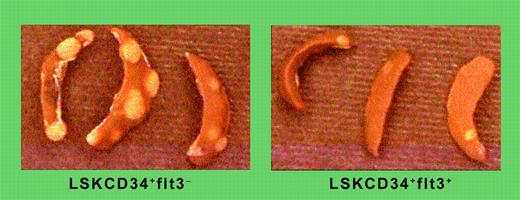
Using the now classical tools of monoclonal antibodies, flow cytometry, murine bone marrow transplantation assays, and the reductionist approach, Yang and colleagues have identified that the murine LSK marrow cells can be further fractionated into functionally distinct subsets of short-term HSCs. Recognizing that long-term HSCs are necessary for sustained hematopoietic reconstitution but generally inadequate to meet the immediate needs for mature myeloerythroid cells for host radioprotection, these investigators searched for short-term HSCs that may be a hierarchical intermediate between long-term HSCs and more differentiated multipotent progenitor cells (MPPs). Yang and colleagues report that the vast majority of bone marrow short-term reconstituting, radioprotective, and spleen colony-forming unit activities (see figure) are derived from LSK cells expressing CD34 but not fetal liver tyrosine kinase 3 (flt3; LSKCD34+flt3–). Evidence is presented that LSKCD34+flt3– cells are descended from transplanted LSKCD34–flt3– long-term HSCs and that the LSKCD34+flt3– short-term HSCs with robust myeloerythroid repopulating potential give rise to LSKCD34+flt3+ MPPs that appear to preferentially replete lymphoid lineages soon after transplantation. These exciting results in the murine model call for identification of their human counterparts, as these progenitors may facilitate more brisk myeloid and lymphoid recovery in patients that received transplants.FIG1
Distinct CFU-S and radioprotective potentials of LSKCD34+flt3–and LSKCD34+flt3+ ST-HSCs. See the complete figure in the article beginning on page 2717.
Distinct CFU-S and radioprotective potentials of LSKCD34+flt3–and LSKCD34+flt3+ ST-HSCs. See the complete figure in the article beginning on page 2717.
HSCs responsible for long-term hematopoiesis are derived from circulating and colonizing embryonic precursors, but defining the exact time and site of the first HSC generation within the embryo has been complicated. In the past, the majority of studies supporting the colonization hypothesis have used the above reductionist approach and classical tools for HSC isolation, enrichment, and adoptive transfer. In this issue, Göthert and colleagues have used the Cre/loxP system to directly trace embryonic HSCs to their adult marrow long-term HSC progeny in vivo. These investigators generated transgenic mice expressing the tamoxifen-inducible Cre-ERT recombinase under control of the stem cell enhancer of the stem cell leukemia (SCL) gene responsible for SCL expression within early hematopoietic progenitors and endothelial cells in vivo. Founder mice that faithfully demonstrated inducible Cre-ERT–mediated recombination were bred to ROSA26-enhanced yellow fluorescence protein (R26R-EYFP) reporter transgenic mice (carrying a floxed transcriptional STOP cassette that blocks EYFP expression unless excised by Cre). In double transgenic fetal mice produced by the genetic cross, in utero exposure to tamoxifen-activated Cre-ERT and EYFP expression was irreversibly turned on, establishing a “mark” that was expressed for the life of the affected HSC and in all of its progeny. Embryonic HSCs could be “marked” as early as days 10 and 11 of gestation and these “marked” HSCs contributed to multilineage hematopoiesis 5 months later in the bone marrow. In a clever experiment, the proportion of “marked” progeny derived from HSCs genetically marked in vivo at day 10.5 and then analyzed in the matured animals 5 months later was compared with HSCs genetically marked in vivo at day 10.5, but 4 days later fetal liver HSCs were isolated and transplanted into congenic myeloablated adult hosts (this approach allowed the investigators to ask if the de novo generation of the majority of marrow HSCs was completed in the midgestation fetus since any HSCs formed in the marrow of the congenic host mice that were not “marked” would decrease the proportion of transplanted “marked” HSCs). Surprisingly, the percentage of EYFP-expressing cells within the recipient mice 5 months after transplantation reflected the proportion of “marked” HSCs at the time of transplantation, suggesting that the generation of HSCs was completed by midgestation in the mouse. What a creative way to assess HSC origins. This method further bolsters the classical approaches to investigating HSC biology and provides us with novel insights into the origins of marrow HSCs. ▪


This feature is available to Subscribers Only
Sign In or Create an Account Close Modal