Abstract
Thrombin activates protease-activated receptor 1 (PAR1) on endothelial cells (ECs) and is critical for angiogenesis and vascular development. However, the mechanism underlying the proangiogenic effect of thrombin has not been elucidated yet. Here, we report the discovery of a novel functional link between thrombin-PAR1 and transforming growth factor-β (TGF-β) signaling pathways. We showed that thrombin via PAR1 induced the internalization of endoglin and type-II TGF-β receptor (TβRII) but not type-I receptors in human ECs. This effect was mediated by protein kinase C-ζ (PKC-ζ) since specific inhibition of PKC-ζ caused an aggregation of endoglin or TβRII on cell surface and blocked their internalization by thrombin. Furthermore, acute and long-term pretreatment of ECs with thrombin or PAR1 peptide agonist suppressed the TGF-β–induced serine phosphorylation of Smad2, a critical mediator of TGF-β signaling. Moreover, activation of PAR1 led to a profound and spread cytosolic clustering formation of Smad2/3 and markedly prevented Smad2/3 nuclear translocation evoked by TGF-β1. Since TGF-β plays a crucial role in the resolution phase of angiogenesis, the down-regulation of TGF-β signaling by thrombin-PAR1 pathway may provide a new insight into the mechanism of the proangiogenic effect of thrombin.
Introduction
Thrombin, a serine protease central to blood coagulation and hemostasis, activates protease-activated receptor 1 (PAR1) on endothelial cells (ECs) and regulates many aspects of EC biology.1 A body of evidence has demonstrated that the thrombin-PAR1 signal pathway is critical for angiogenesis and vascular development.1-3 However, the mechanism underlying the proangiogenic effect of thrombin has not been elucidated yet. Deficiency of PAR1 causes yolk sac defects and bleeding and results in 50% mouse embryonic lethality without altering the plasma coagulation cascade and platelet function.4,5 Reconstitution of a PAR1 transgene driven by an endothelial-specific promoter prevents death of PAR1–/– embryos, indicating that thrombin's action via PAR1 on endothelial rather than other cells contributes to angiogenesis and vascular development.5 Similar defects are observed in mouse embryos deficient in various coagulation factors (eg, factor V, tissue factor, and prothrombin), in which thrombin generation is impaired.6-8 In contrast, deletion of either PAR2, PAR3, or PAR4 is not lethal.2 Furthermore, thrombin induces neoangiogenesis in chick chorioallantoic membrane and the mouse Matrigel system, which requires the activation of PAR1 signal pathways.9,10 Thrombin-PAR1 pathway also promotes tumor progress and metastasis related with angiogenesis.11 However, thrombin (≤ 5 U/mL) itself is not sufficient to induce angiogenesis in vitro.12 In contrast, thrombin at 10 U/mL inhibits EC tube formation.12 It seems likely that thrombin may modulate angiogenesis by regulating functions of growth factors involved in angiogenesis in vivo.
Angiogenesis encompasses activation and resolution phases. In the activation phase, increased vascular permeability and basement membrane degradation allows ECs to proliferate and migrate into the extracellular space and form new capillary sprouts. In the resolution phase, ECs cease proliferation and migration, reconstitute the basement membrane and promote vessel maturation and recruit pericytes. Compelling genetics evidence has demonstrated a pivotal role for transforming growth factor-β (TGF-β) and its receptors (type-I and -II receptors as well as endoglin) in angiogenesis, in particular the resolution phase of angiogenesis such as the establishment and maintenance of vessel wall integrity.13 Deficiency of either TGF-β1, type-I receptor (also known as activin receptor-like kinase [ALK]), type-II receptor (TβRII) as well as endoglin, an ancillary TGF-β receptor, results in mouse embryonic lethality with a similar defect in vascular development, and the human vascular disorder hereditary hemorrhagic telangiectasia has been linked to mutations in ALK1 and endoglin.14-16 However, the mechanism by which TGF-β regulates angiogenesis is still not fully understood, and in particular the functional regulation of TGF-β signaling by other systems during angiogenesis has not been actively explored.
In the present study, we have discovered a new functional link between thrombin and TGF-β signaling pathways. We show that thrombin via PAR1 induces a protein kinase C-ζ (PKC-ζ)–dependent endocytosis of TβRII and endoglin but not type-I receptors and suppresses phosphorylation of the transcriptional activator Smad2 by TGF-β in primary human ECs. Moreover, activation of PAR1 leads to a profound and spread cytosolic clustering formation of Smad2/3 and markedly prevents Smad2/3 nuclear translocation evoked by TGF-β1. These substantial lines of evidence indicate that thrombin-PAR1 pathway is a potent negative regulator of TGF-β signaling in ECs. Our data also indicate that the TGF-β signaling can be modulated by G protein-coupled receptors such as PAR1.
Materials and methods
Reagents
Human α-thrombin, thrombin receptor-activating peptide (TRAP), hirudin, and SB431542 were from Sigma (St Louis, MO). Human TGF-β1 was from R&D Systems (Minneapolis, MN). Short peptide agonists for human PAR1 (SFLLRN), PAR2 (SLIGKV), PAR3 (TFRGAP), and PAR4 (GYPGQV) were from Bachem (Torrance, CA). Myristoylated PKC-ζ peptide inhibitor and other PKC inhibitors were from Calbiochem (La Jolla, CA). Myristoylated PKC-β C2-4 inhibitor was from Biomol (Plymouth Meeting, PA). Reagents for chemiluminescence detection were from Cell Signaling (Beverly, MA).
Antibodies
Antibodies against phospho-nuclear factor (NF-κB) p65 (Ser-536), phospho–PKC-ζ/λ (Thr-410/403), and TβRI (ALK5) were from Cell Signaling. Antibodies against TGF-β1/2/3 (H-112), PAR1 (ATAP2), ALK5 (H-100), TβRII (C-16), and phosphatidylinositol-3-OH kinase (PI3K) were from Santa Cruz Biotechnology (Santa Cruz, CA). Antibodies against extracellular domains of human TβRII and ALK1 were from R&D Systems. Antibody against phospho-Smad2 (Ser-465/467) was from Chemicon (Temecular, CA). Monoclonal antibodies against endoglin and Smad2/3 were from BD Transduction (San Diego, CA). Human endoglin monoclonal antibody (SN6h) was from DakoCytomation (Carpinteria, CA). Smad2 antibody was from Zymed (South San Francisco, CA). Cy™3–conjugated goat antimouse and antirabbit antibodies were from Jackson ImmunoResearch (West Grove, PA).
Cell culture
Human aortic ECs (HAECs) and human umbilical vein ECs (HUVECs) were from Cambrex Bio Science (Walkersville, MD) and cultured in EGM™–2 medium and used for experiments within 10 passages.
Immunoblotting and immunoprecipitation
Immunoblotting and immunoprecipitation were performed as we described previously.17
Identification of the serine-phosphorylated 95-kDa protein
HAECs (20 dishes, 10 cm) were incubated with 1 μM phenylarsine oxide (PAO) for 3 hours. Lysates were prepared and loaded onto a high-Q cartridge column (Bio-Rad, Hercules, CA). Fractions eluted from 350 to 700 mM NaCl, which contain the 95-kDa protein, were combined and concentrated. The concentrated eluates were diluted to a solution of 150 mM NaCl and immunoprecipitated with antibody against phospho–NF-κB p65 (Ser-536). The immune complexes were washed extensively with Nonidet P-40 lysis buffer17 containing 300 mM NaCl and resolved by sodium dodecyl sulfate (SDS)–polyacrylamide gel electrophoresis (PAGE). Proteins were visualized with Coomassie blue staining, and the band corresponding to 95 kDa was excised for in-gel trypsin digestion and analyzed with a matrix-assisted laser desorption delayed-extraction reflectron time of flight (MALDI-TOF) mass spectrometer performed by AmProx (AmProx, Carlsbad, CA).
Subcellular fractionation
EC cytosolic and membrane fractions were prepared as we described previously.17
Potassium depletion
Potassium depletion of ECs was performed essentially as described by Larkin et al.18
Cell surface enzyme-linked immunosorbent assay (ELISA)
Cell surface receptors were determined basically as described previously.19 ECs in 48-well plates were serum-starved for 2 hours and then stimulated with thrombin (3 U/mL) or TRAP (40 μM) for the indicated time periods at 37°C. ECs were washed with phosphate-buffered saline (PBS), fixed with 1% glutaraldehyde for 10 minutes, and blocked with 1% bovine serum albumin (BSA) in PBS for 1 hour. Cells were then incubated at 25°C with antibodies against extracellular domains of endoglin, TβRII, ALK5, ALK1, and PAR1. After 1 hour of incubation, cells were washed 4 times with PBS and incubated with horseradish peroxidase (HRP)–conjugated antimouse or antigoat secondary antibodies (1: 2000 dilution) for 1 hour at 25°C. Cells were again washed with PBS and then incubated in One Step ABTS solution (2,2′-azino-bis-3-ethylbenzthiazoline-6-sulfonic acid; Roche, Indianapolis, IN) for 10 minutes, and the optical density was determined at 405 nm using a universal microplate reader (Elx800; Bio-Tek, Winooski, VT).
Immunofluorescence microscopy
ECs grown on glass coverslips in a 6-well plate were pretreated with or without PKC inhibitors followed by stimulation with thrombin (3 U/mL), TRAP (40 μM), or PAR1-AP (80 μM) for 30 minutes at 37°C, then washed with PBS and fixed in 3.7% formaldehyde solution in PBS for 10 minutes at 25°C. The fixed cells were extracted in ice-cold acetone at –20°C for 5 minutes, washed, and preincubated in PBS containing 1% BSA for 30 minutes at 25°C. Cells were then incubated with monoclonal antibodies (1:100 dilution) against endoglin (SN6h), Smad2/3, and ALK1, or with polyclonal antibodies (1:100 dilution) against ALK5 (H-100) and TβRII (C-16) for 1 hour at 25°C in PBS containing 1% BSA, washed 3 times with PBS, and then incubated with Cy3-conjugated goat antimouse or antirabbit secondary antibodies (1:100 dilution) for 1 hour at 25°C in PBS containing 1% BSA. Control stainings were performed without either primary or secondary antibodies. After washing with PBS, coverslips were mounted on slides with cell-side down with Cytoseal and examined and photographed using a Perkin Elmer Ultra VIEW LCI confocal imaging system (Boston, MA) configured with a Nikon TE2000-S fluorescence microscope (Garden City, NY) fitted with PlanApo 60 × and 100 × oil objectives. Adobe Photoshop 6.0 software was used for image processing.
Results
Identification of the 95-kDa protein as serine-phosphorylated endoglin by a phospho-specific antibody agonist NF-κB p65 (Ser-536) in ECs
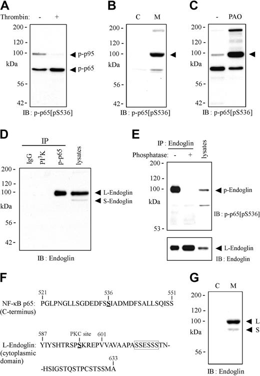
Figure 1A shows that in addition to the phosphorylated NF-κB p65,20 a protein band with 95 kDa was also detected by an antibody against phospho–NF-κB p65 (Ser-536) in HAECs. Interestingly, transient stimulation of HAECs with a physiologic dose of thrombin (3 U/mL) resulted in a profound decrease in the serine phosphorylation of the 95-kDa protein, whereas thrombin promoted the phosphorylation of p65 on Ser-536 (Figure 1A). Figure 1B shows that the 95-kDa protein was merely detected at membrane fraction. To identify the 95-kDa protein by using mass spectrometry, we searched for compounds that could enhance serine phosphorylation of the 95-kDa membrane protein and found that PAO,21,22 a receptor internalization blocker, was the most potent one. As shown in Figure 1C, the serine phosphorylation of the 95-kDa protein was augmented 15-fold by PAO (1 μM) treatment. We then prepared lysates from PAO-treated HAECs, loaded them onto a high-Q cartridge column and then collected fractions containing the phosphorylated 95-kDa membrane protein. The eluates were immunoprecipitated with the phospho-specific antibody, resolved by SDS-PAGE, and analyzed with a MALDI-TOF mass spectrometer as described in “Materials and methods.” Peptide fingerprint analysis identified that the 95-kDa membrane protein was endoglin,23,24 a TGF-β coreceptor highly expressed in proliferating ECs. To confirm this finding, lysates were immunoprecipitated with the Ser-536–phosphorylated p65 antibody, a PI3K antibody, or control IgG from normal rabbit serum and subjected to immnunoblotting with an antibody (clone 35) against NH2-terminus of endoglin (BD Transduction). As shown in Figure 1D, a clear band corresponding to the full-length endoglin (L-endoglin) from the immune complexes of the phospho-specific antibody but not from others was detected. Alternatively, lysates were immunoprecipitated with an antibody against the full-length endoglin (SN6h; DakoCytomation) followed by immunoblotting with the antibody against Ser-536–phosphorylated p65. As shown in Figure 1E, a clear band corresponding to L-endoglin was detected by the phospho-specific antibody. Remarkably, the band was not detected from the endoglin immune complexes treated with alkaline phosphatase to dephosphorylate endoglin. Thus, our findings indicate that the 95-kDa membrane protein detected by the phospho-specific antibody represents the serine-phosphorylated full-length endolin, which accounts for approximately 2% of total endolin in unstimulated HAECs. L-endoglin is composed of 633 amino acids with 47–amino acid residues (residues 587-633) in the cytoplasmic tail, while the S-endoglin consists of 600 amino acids with a 14–amino acid cytoplasmic tail (residues 587-600)25 (Figure 1F). Consistent with previous reports,25-27 90% endoglin was detected as L-form at the membranes of HAECs (Figure 1G). The Ser-536 of NF-κB p65 is surrounded by several Asp and Glu residues (Figure 1F). Since only the L-endoglin is detected by the phospho-specific antibody, a phosphorylated serine within residues 601 to 633 of endoglin, especially within the serine clusters (609SSESSS) adjacent to Glu could be the recognition site of the phospho-specific antibody.
Identification of the 95-kDa protein as the serine-phosphorylated endoglin with an antibody agonist phospho–NF-κB p65 (Ser-536). Lysates from HAECs stimulated with thrombin (3 U/mL) for 30 minutes (A), HAEC cytosolic and membrane fractions (B), or lysates from HAECs treated for 3 hours with phenylarsine oxide (PAO; 1μM) (C) were subjected to immunoblotting (IB) with an antibody against phospho–NF-κB p65 (Ser-536). (D) HAEC lysates were immunoprecipitated (IP) with polyclonal antibodies against phospho–NF-κB p65 (Ser-536) (p-p65) or PI3K, or control immunoglobulin G (IgG) from normal rabbit serum and subjected to immnunoblotting with a monoclonal antibody against NH2-terminus of endoglin. (E) HAEC lysates were immunoprecipitated with a monoclonal antibody (SN6h) against full-length endoglin followed by immunoblotting with the polyclonal antibody against Ser-536–phosphorylated p65. (F) Amino acid sequence of the COOH-terminus of NF-κB p65 or endoglin. (G) Immunoblotting of HAEC cytosolic and membrane fractions with a monoclonal antibody against NH2-terminus of endoglin.
Identification of the 95-kDa protein as the serine-phosphorylated endoglin with an antibody agonist phospho–NF-κB p65 (Ser-536). Lysates from HAECs stimulated with thrombin (3 U/mL) for 30 minutes (A), HAEC cytosolic and membrane fractions (B), or lysates from HAECs treated for 3 hours with phenylarsine oxide (PAO; 1μM) (C) were subjected to immunoblotting (IB) with an antibody against phospho–NF-κB p65 (Ser-536). (D) HAEC lysates were immunoprecipitated (IP) with polyclonal antibodies against phospho–NF-κB p65 (Ser-536) (p-p65) or PI3K, or control immunoglobulin G (IgG) from normal rabbit serum and subjected to immnunoblotting with a monoclonal antibody against NH2-terminus of endoglin. (E) HAEC lysates were immunoprecipitated with a monoclonal antibody (SN6h) against full-length endoglin followed by immunoblotting with the polyclonal antibody against Ser-536–phosphorylated p65. (F) Amino acid sequence of the COOH-terminus of NF-κB p65 or endoglin. (G) Immunoblotting of HAEC cytosolic and membrane fractions with a monoclonal antibody against NH2-terminus of endoglin.
TGF-β1 promotes the serine phosphorylation of endoglin in ECs
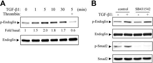
As shown in Figure 2A, TGF-β1 induced a time-dependent serine phosphorylation of endoglin detected by the phospho-specific antibody as described in Figure 1. The stimulatory effect occurred fast (1 minute), reached the maximal level (2-fold) at 5 minutes, and then declined. Interestingly, pretreatment of HAECs with thrombin for 30 minutes suppressed 70% of the serine phosphorylation of endoglin by TGF-β1. The blot was stripped and reprobed with an endoglin antibody (SN6h) to show the equal loading (Figure 2A, lower panel). We next determined the effect of SB431542,28 a specific inhibitor of type-I receptor of TGF-β superfamily, including ALK4, ALK5, and ALK6, on the serine phosphorylation of endoglin by TGF-β in HAECs. As shown in Figure 2B, SB431542 did not affect the TGF-β1–induced serine phosphorylation of endoglin. However, it completely suppressed the basal and TGF-β1–induced serine phosphorylation of Smad2 detected with an antibody against phospho-Smad2 (Ser-465/467; Chemicon). These data suggest that TβRII other than ALK5 may be involved in the serine phosphorylation of endoglin by TGF-β, although other serine/threonine kinases activated by TGF-β could not be excluded. It has been shown that endoglin interacts with and is constitutively phosphorylated by TβRII.29 It appears that the serine-phosphorylated endoglin (2%) detected in HAECs may represent the functional endoglin associated with and phosphorylated by TβRII.
TGF-β1 promotes the serine phosphorylation of endoglin in ECs. (A) HAECs were serum-starved for 2 hours, then pretreated with (+) or without thrombin (3 U/mL) for 30 minutes followed by stimulation with TGF-β1 (2 ng/mL) for various time periods as indicated. Lysates were immunoblotted with the antibody against phospho–NF-κB p65 (Ser-536) that also detects the serine-phosphorylated endoglin as described in Figure 1. The serine phosphorylation of endoglin by TGF-β1 was shown as fold increase relative to control cells by densitometric analysis. The same blot was stripped and reprobed with an endoglin antibody (SN6h) to show the equal loading of lysates (lower panel). (B) HAECs were left untreated (control) or pretreated with SB431542 (10 μM) for 30 minutes, then stimulated with TGF-β1 (2 ng/mL) for 5 minutes. Lysates were subjected to immunoblotting with indicated antibodies against the serine-phosphorylated endoglin as described in Figure 1, endoglin (SN6h), phospho-Smad2 (Ser-465/467), or Smad2. Results shown are representative immunoblots of 3 independent experiments.
TGF-β1 promotes the serine phosphorylation of endoglin in ECs. (A) HAECs were serum-starved for 2 hours, then pretreated with (+) or without thrombin (3 U/mL) for 30 minutes followed by stimulation with TGF-β1 (2 ng/mL) for various time periods as indicated. Lysates were immunoblotted with the antibody against phospho–NF-κB p65 (Ser-536) that also detects the serine-phosphorylated endoglin as described in Figure 1. The serine phosphorylation of endoglin by TGF-β1 was shown as fold increase relative to control cells by densitometric analysis. The same blot was stripped and reprobed with an endoglin antibody (SN6h) to show the equal loading of lysates (lower panel). (B) HAECs were left untreated (control) or pretreated with SB431542 (10 μM) for 30 minutes, then stimulated with TGF-β1 (2 ng/mL) for 5 minutes. Lysates were subjected to immunoblotting with indicated antibodies against the serine-phosphorylated endoglin as described in Figure 1, endoglin (SN6h), phospho-Smad2 (Ser-465/467), or Smad2. Results shown are representative immunoblots of 3 independent experiments.
Thrombin down-regulates the serine-phosphorylated endoglin through PAR1 in ECs
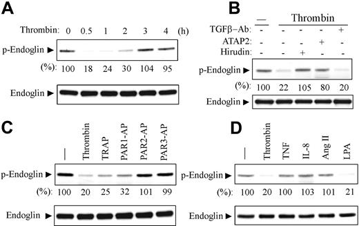
PAR1 is the major receptor for thrombin in ECs in mice and humans.1 As shown in Figure 3A, thrombin markedly down-regulated the serine-phosphorylated endoglin in a time-dependent manner in HAECs detected with the antibody against the serine-phosphorylated endoglin associated with, and phosphorylated by, TGF-β receptor complexes as described in Figures 1 and 2. The down-regulatory effect occurred fast (< 5 minutes, data not shown), reached a maximal level at 30 minutes (82% down-regulation), and gradually returned to basal level by 3 hours after thrombin treatment. Hirudin (6 U/mL), a specific inhibitor of thrombin, and an antibody (ATAP2) that inhibits PAR1 cleavage and activation30 markedly prevented the down-regulatory effect of thrombin (Figure 3B). Whereas pretreatment of HAECs with an antibody against TGF-β1/2/3 had virtually no effect, indicating a mechanism independent of TGF-β secretion (Figure 3B). Moreover, TRAP (SFLLRNPNDKYEPF), a 14–amino acid peptide agonist for human PAR1, and a short peptide agonist for human PAR1 (PAR1-AP; SFLLRN)31 mimicked the effect of thrombin in HAECs. In contrast, selective peptide agonists for human PAR2 and PAR3 had no effect (Figure 3C). Thus, thrombin down-regulates the phosphorylated and functional endoglin through PAR1 in HAECs. Interestingly, lysophosphatidic acid (LPA) also down-regulated the serine-phosphorylated endoglin. In contrast, angiotensin II (Ang II), tumor necrosis factor (TNF), and interlukin-8 (IL-8) had no effect (Figure 3D). These findings were reproduced in HUVECs (data not shown).
Thrombin down-regulates the serine-phosphorylated endoglin through PAR1 in ECs. (A) HAECs were serum-started for 2 hours and then stimulated with thrombin (3 U/mL) for the indicated time periods. (B) HAECs were left untreated (–) or preincubated with hirudin (6 U/mL), a monoclonal PAR1 antibody (ATAP2, 10 μg/mL), or an antibody against TGF-β1/2/3 (10 μg/mL) for 30 minutes, then stimulated with thrombin (3 U/mL) for 30 minutes. (C) HAECs were stimulated with thrombin (3 U/mL), thrombin receptor-activating peptide (TRAP, 40 μM), peptide agonists (100 μM) for human PAR1 (PAR1-AP), human PAR2 (PAR2-AP), or PAR3 (PAR3-AP). (D) HAECs were stimulated for 30 minutes with thrombin (3 U/mL), tumor necrosis factor (TNF; 10 ng/mL), interleukin 8 (IL-8; 2 nM), angiotensin II (Ang II; 100 nM), or lysophosphatidic acid (LPA; 10 μM). Lysates were subjected to immunoblotting with the antibody against the serine-phosphorylated endoglin as described in Figures 1 and 2. The serine phosphorylation of endoglin was shown as percentage relative to untreated control cells by densitometric analysis. The same blot was stripped and reprobed with an endoglin antibody (SN6h) to show the equal loading. Results shown are representative immunoblots of 3 independent experiments.
Thrombin down-regulates the serine-phosphorylated endoglin through PAR1 in ECs. (A) HAECs were serum-started for 2 hours and then stimulated with thrombin (3 U/mL) for the indicated time periods. (B) HAECs were left untreated (–) or preincubated with hirudin (6 U/mL), a monoclonal PAR1 antibody (ATAP2, 10 μg/mL), or an antibody against TGF-β1/2/3 (10 μg/mL) for 30 minutes, then stimulated with thrombin (3 U/mL) for 30 minutes. (C) HAECs were stimulated with thrombin (3 U/mL), thrombin receptor-activating peptide (TRAP, 40 μM), peptide agonists (100 μM) for human PAR1 (PAR1-AP), human PAR2 (PAR2-AP), or PAR3 (PAR3-AP). (D) HAECs were stimulated for 30 minutes with thrombin (3 U/mL), tumor necrosis factor (TNF; 10 ng/mL), interleukin 8 (IL-8; 2 nM), angiotensin II (Ang II; 100 nM), or lysophosphatidic acid (LPA; 10 μM). Lysates were subjected to immunoblotting with the antibody against the serine-phosphorylated endoglin as described in Figures 1 and 2. The serine phosphorylation of endoglin was shown as percentage relative to untreated control cells by densitometric analysis. The same blot was stripped and reprobed with an endoglin antibody (SN6h) to show the equal loading. Results shown are representative immunoblots of 3 independent experiments.
Blockage of receptor internalization prevents the thrombin-induced down-regulation of the serine-phosphorylated endoglin in ECs
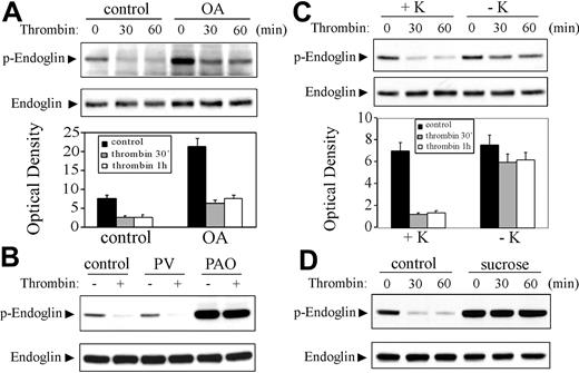
As shown in Figure 4A, thrombin down-regulated the serine-phosphorylated endoglin by TβRII in a similar efficiency in both control HAECs and HAECs treated with okadaic acid (100 nM),32 a serine/threonine phosphatase inhibitor, although the basal serine phosphorylation of endoglin was markedly enhanced by okadaic acid. This finding suggests that endocytosis of endoglin other than the serine/threonine phosphatases may be involved in the thrombin-induced down-regulation of the serine-phosphorylated endoglin in ECs.
Blockage of receptor internalization prevents the thrombin-induced down-regulation of the serine-phosphorylated endoglin by TβRII in ECs. (A, top) HAECs were left untreated (control) or pretreated with okadaic acid (OA; 100 nM) for 30 minutes, then stimulated with thrombin (3 U/mL) for the indicated time periods. (Bottom) Optical density. ▪ represents control (no thrombin stimulation); ▦, thrombin 30 min; □ thrombin 1 h. Error bars indicate standard deviation. (B) HAECs were left untreated (control) or pretreated with pervanadate (PV; 50 μM) for 30 minutes or phenylarsine oxide (PAO; 1 μM) for 3 hours, then stimulated with thrombin for 30 minutes. (C, top) Control cells (+K) or HAECs depleted of potassium (–K) were stimulated with thrombin (3 U/mL) for the indicated time periods. (Bottom) Optical density. Shading indicates same treatment types as in panel A. Error bars indicate standard deviation. (D) HUVECs were left untreated (control) or pretreated with 0.45 M sucrose for 10 minutes then stimulated with thrombin for the indicated time periods. Lysates were subjected to immunoblotting with the antibody against the serine-phosphorylated endoglin as described in Figures 1 and 2. The same blot was stripped and reprobed with an endoglin antibody (SN6h) to show the equal loading. Results shown are representative immunoblots and densitometric analyses of 3 independent experiments.
Blockage of receptor internalization prevents the thrombin-induced down-regulation of the serine-phosphorylated endoglin by TβRII in ECs. (A, top) HAECs were left untreated (control) or pretreated with okadaic acid (OA; 100 nM) for 30 minutes, then stimulated with thrombin (3 U/mL) for the indicated time periods. (Bottom) Optical density. ▪ represents control (no thrombin stimulation); ▦, thrombin 30 min; □ thrombin 1 h. Error bars indicate standard deviation. (B) HAECs were left untreated (control) or pretreated with pervanadate (PV; 50 μM) for 30 minutes or phenylarsine oxide (PAO; 1 μM) for 3 hours, then stimulated with thrombin for 30 minutes. (C, top) Control cells (+K) or HAECs depleted of potassium (–K) were stimulated with thrombin (3 U/mL) for the indicated time periods. (Bottom) Optical density. Shading indicates same treatment types as in panel A. Error bars indicate standard deviation. (D) HUVECs were left untreated (control) or pretreated with 0.45 M sucrose for 10 minutes then stimulated with thrombin for the indicated time periods. Lysates were subjected to immunoblotting with the antibody against the serine-phosphorylated endoglin as described in Figures 1 and 2. The same blot was stripped and reprobed with an endoglin antibody (SN6h) to show the equal loading. Results shown are representative immunoblots and densitometric analyses of 3 independent experiments.
PAO is a blocker of clathrin-mediated receptor endocytosis.21,22 As shown in Figure 4B, pretreatment of HAECs with PAO (1 μM) resulted in a profound increase in the serine phosphorylation of endoglin and abolished the thrombin-induced down-regulation of the serine-phosphorylated endoglin. In addition, PAO is also an inhibitor of protein tyrosine phosphatase.33 To determine the possible involvement of tyrosine phosphatase, we treated HAECs with pervanadate,34 a potent inhibitor of tyrosine phosphatase, and found that pervanadate affected neither basal level nor thrombin-mediated down-regulation of the functional endoglin, indicating a tyrosine phosphatase–independent mechanism. We next determined the effect of inhibiting clathrin-mediated endocytosis by hypertonic sucrose35 or potassium depletion.18 As shown in Figure 4C-D, the thrombin-induced down-regulation of the serine-phosphorylated endoglin was markedly prevented by potassium depletion and abolished by sucrose treatment. The basal phosphorylation of endoglin was markedly enhanced by sucrose but slightly increased by potassium depletion. Thus, these data can be interpreted to mean that endoglin may undergo constitutive clathrin-dependent internalization in unstimulated ECs. The thrombin-induced down-regulation of the serine-phosphorylated endoglin may result from endoglin internalization by thrombin.
Thrombin induces a PKC-ζ–dependent internalization of endoglin and TβRII in ECs
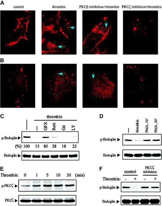
To determine whether thrombin induces internalization of endoglin, we performed immunofluorescence microscopy using a confocal microscope to access the localization of endoglin in HUVECs. In unstimulated HUVECs, endoglin was localized primarily at the plasma membrane detected with a monoclonal antibody (SN6h) against L-endoglin (Figure 5A). Stimulation of HUVECs with thrombin for 30 minutes led to the progressive appearance of a vesicular pattern typical of endocytic vesicles. Serial laser scans ranged from the top surface levels and into the cell interior showed that the thrombin-induced endocytic vesicles of endoglin was only observed inside the cell, indicating the internalization of endoglin by thrombin (Figure 5A, 2nd panel). Since TβRII interacts constitutively with the cytoplasmic domain of endoglin,29 we assessed the effect of thrombin on the localization of TβRII. Serial laser scans ranged from the top surface levels and into the cell interior revealed that TβRII was stained primarily at the cell surface detected with a TβRII antibody29 that recognizes the COOH-terminal domain of human TβRII (Figure 5B). Stimulation of HUVECs with thrombin for 30 minutes induced a progressive appearance of the endocytic vesicles containing TβRII that was only observed inside the cell, indicating the internalization of TβRII by thrombin. Similar effect of thrombin or PAR1-AP on the internalization of endoglin and TβRII was observed in HAECs (data not shown). In contrast, we found that the localization of ALK5 and ALK1 was not altered by thrombin, and there is no appearance of the endocytic vesicles formation by thrombin in ECs (Figure S1 at the Blood website; see the Supplemental Figure link at the top of the online article). Thus, thrombin induces the internalization of endoglin and TβRII but not ALK5 and ALK1 in ECs, which is distinct from TGF-β–induced internalization of TGF-β receptors.36
Thrombin induces a PKC-ζ–dependent internalization of endoglin and TβRII in ECs. (A-B) HUVECs grown on glass coverslips were left untreated (control) or pretreated with myristoylated PKC-β or PKC-ζ peptide inhibitors, then stimulated for 30 minutes with thrombin (3 U/mL), washed twice, and fixed. Endoglin (A) and TβRII (B) were viewed (10 × 100) by staining the fixed cells with antibodies against L-endoglin (SN6h) or TβRII (C-16). Arrows indicate the internalized endoglin or TβRII. (C) HUVECs were left untreated (–) or pretreated for 30 minutes with GF109203X (GFX; 15 μM), rottlerin (Rott; 10 μM), Gö6976 (Gö; 300 nM), or LY294002 (LY; 20 μM), then stimulated with thrombin (3 U/mL) for 30 minutes. (D) HUVECs were stimulated with thrombin (3 U/mL) for 30 minutes or phorbol 12-myristate 13-acetate (PMA; 100 nM) for 10 or 30 minutes. Lysates were subjected to immunoblotting with the antibody against the serine-phosphorylated endoglin as described in Figures 1 and 2. (E) HUVECs were stimulated with thrombin (3 U/mL) for the indicated time periods. Lysates were immunoblotted with an antibody against phospho–PKC-ζ/λ (Thr-410/403). (F) HUVECs were left untreated (control) or pretreated with a myristoylated PKC-ζ peptide inhibitor for 30 minutes, then stimulated with thrombin (3 U/mL) for 30 minutes. The serine phosphorylation of endoglin was determined as described in Figures 1 and 2. Results shown are representative of 3 independent experiments.
Thrombin induces a PKC-ζ–dependent internalization of endoglin and TβRII in ECs. (A-B) HUVECs grown on glass coverslips were left untreated (control) or pretreated with myristoylated PKC-β or PKC-ζ peptide inhibitors, then stimulated for 30 minutes with thrombin (3 U/mL), washed twice, and fixed. Endoglin (A) and TβRII (B) were viewed (10 × 100) by staining the fixed cells with antibodies against L-endoglin (SN6h) or TβRII (C-16). Arrows indicate the internalized endoglin or TβRII. (C) HUVECs were left untreated (–) or pretreated for 30 minutes with GF109203X (GFX; 15 μM), rottlerin (Rott; 10 μM), Gö6976 (Gö; 300 nM), or LY294002 (LY; 20 μM), then stimulated with thrombin (3 U/mL) for 30 minutes. (D) HUVECs were stimulated with thrombin (3 U/mL) for 30 minutes or phorbol 12-myristate 13-acetate (PMA; 100 nM) for 10 or 30 minutes. Lysates were subjected to immunoblotting with the antibody against the serine-phosphorylated endoglin as described in Figures 1 and 2. (E) HUVECs were stimulated with thrombin (3 U/mL) for the indicated time periods. Lysates were immunoblotted with an antibody against phospho–PKC-ζ/λ (Thr-410/403). (F) HUVECs were left untreated (control) or pretreated with a myristoylated PKC-ζ peptide inhibitor for 30 minutes, then stimulated with thrombin (3 U/mL) for 30 minutes. The serine phosphorylation of endoglin was determined as described in Figures 1 and 2. Results shown are representative of 3 independent experiments.
We next determined the mechanism of thrombin-induced endocytosis of endoglin and TβRII in ECs. Using the phospho-specific antibody that recognizes the serine-phosphorylated endoglin by TβRII as described in Figures 1 and 2, we found that the thrombin-induced down-regulation of the serine-phosphorylated endoglin (ie, endoglin internalization) was markedly prevented by a general PKC inhibitor GF109203X37 (Figure 5C). Rottlerin,38 a selective PKCδ inhibitor, or LY294002,39 a selective inhibitor of PI3K, had a minor effect. In contrast, Gö6976,40 a selective conventional PKC inhibitor, had virtually no effect. Moreover, phorbol-12-myristate-13-acetate (PMA),41 a potent activator of conventional and novel PKCs, did not affect endoglin phosphorylation (Figure 5D). These data were highly reproducible in HAECs (data not shown). These findings suggest that the atypical PKCs may be involved in the thrombin-induced endocytosis of endoglin in ECs. It has been shown that human ECs, including HUVECs, express PKC-α, -β, -δ, -ϵ, -η, -θ, and -ζ isoforms but not PKC-ι/λ.42-44 Consistent with a previous report,45 we found that thrombin induced a rapid activation of PKC-ζ detected with an antibody against phospho–PKC-ζ (Thr-410) (Figure 5E). The activation of PKC-ζ requires the phosphorylation of Thr-410 on the activation loop by phosphoinositide-dependent kinase.46 To assess the role of PKC-ζ in the endocytosis of endoglin, HUVECs were pretreated with a myristoylated PKC-ζ peptide inhibitor, and lysates were immunoblotted with the phospho-specific antibody that detects the functional endoglin associated with and phopshorylated by TβRII. As shown in Figure 5F, specific inhibition of PKC-ζ markedly prevented down-regulation of the functional endoglin or endoglin internalization by thrombin. We then performed confocal immunofluorescence microscopy to confirm the role of PKC-ζ in the endocytosis of endoglin and TβRII by thrombin. Remarkably, specific inhibition of PKC-ζ with the myristoylated PKC-ζ peptide inhibitor led to the aggregation or cluster formation of endoglin or TβRII on cell surface and blocked their endocytosis by thrombin (Figure 5A-B), suggesting that an overlap or similar pathway may be involved in the thrombin-induced endocytosis of TβRII and endoglin in ECs. In contrast, the thrombin-induced endocytosis of TβRII and endoglin was not affected by a myristoylated PKC-β peptide inhibitor (Figure 5A-B) and a conventional PKC inhibitor Gö6976 in HUVECs (data not shown). Taken together, these findings indicate that PKC-ζ plays a critical role in the internalization of TβRII and endoglin evoked by thrombin in ECs.
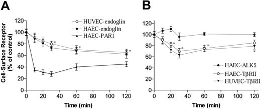
Finally, we performed a cell surface ELISA assay19 with monoclonal antibodies against the extracellular domains of endoglin or TβRII to quantify the internalization of endoglin and TβRII by thrombin in ECs. We found that thrombin induced a time-dependent internalization of endoglin, with a maximal level of 38% or 35% internalization at 2 hours in HAECs and HUVECs, respectively (Figure 6A). As expected, thrombin induced a rapid and profound internalization of PAR1, with a maximal effect at 30 minutes (72% internalization) detected with an antibody (ATAP2)47 that recognizes both intact and cleaved PAR1 (Figure 6A). Figure 6B shows that thrombin induces a time-dependent internalization of TβRII, with a maximal level of 30% or 37% internalization at 30 minutes in HAECs and HUVECs, respectively. Similar to the pattern of thrombin-induced down-regulation of the serine-phosphorylated endoglin (Figure 3A), the cell surface TβRII tended to recover after 30 minutes of treatment with thrombin (Figure 6B). In addition, consistent with the observation by immunofluorescence microscopy, the level of cell surface ALK5 was not significantly altered by thrombin (Figure 6B).
Quantification of the cell-surface endoglin and TβRII using cell ELISA. HAECs and HUVECs were stimulated with thrombin (3 U/mL) for indicated time periods. Cell-surface receptors were measured by using cell ELISA with antibodies against the NH2-terminus of endoglin, PAR1, ALK5, and TβRII. (A) ○ indicates HUVEC-endoglin; •, HAEC-endoglin; ▴, HAEC-PAR1. (B) • indicates HAEC-ALK5; ○, HAEC-TβRII; ▾, HUVEC-TβRII. Results shown are representative of 3 independent experiments. *P < .05. Error bars indicate standard deviation.
Quantification of the cell-surface endoglin and TβRII using cell ELISA. HAECs and HUVECs were stimulated with thrombin (3 U/mL) for indicated time periods. Cell-surface receptors were measured by using cell ELISA with antibodies against the NH2-terminus of endoglin, PAR1, ALK5, and TβRII. (A) ○ indicates HUVEC-endoglin; •, HAEC-endoglin; ▴, HAEC-PAR1. (B) • indicates HAEC-ALK5; ○, HAEC-TβRII; ▾, HUVEC-TβRII. Results shown are representative of 3 independent experiments. *P < .05. Error bars indicate standard deviation.
Thrombin down-regulates TGF-β signaling in ECs
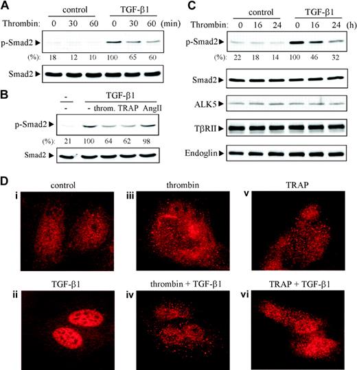
Since TGF-β binds directly to TβRII, which is essential for the downstream signal transduction of TGF-β,48-50 we determined effects of thrombin on the proximal signaling events upon activation of TGF-β receptors such as phosphorylation of the transcriptional activators Smad2 and Smad3 in ECs. As shown in Figure 7A-B, the TGF-β1–induced phosphorylation of Smad2, which was detected with a phospho-Smad2 (Ser-465/467) antibody, was suppressed 35% to 40% by acute treatment of HUVECs with thrombin or TRAP. In contrast, Ang II did not affect the phosphorylation of Smad2 by TGF-β1. Remarkably, the TGF-β1–induced Smad2 phosphorylation was suppressed 54% and 68% by 16 or 24 hours of treatment of HUVECs with thrombin, respectively. The protein levels of Smad2, ALK5, TβRII, and endoglin were not altered by long-term thrombin treatment (Figure 7C). In addition, we found that long-term Ang II treatment did not affect the Smad2 phosphorylation by TGF-β1 (data not shown). Moreover, we found that the cell surface receptor levels of ALK5, endoglin, and TβRII were down-regulated 15% ± 3%, 12% ± 2%, and 20% ± 4% by 24-hour treatment of HUVECs with thrombin, respectively. It seems that the profound decrease (68%) in the TGF-β1–induced Smad2 phosphorylation by long-term thrombin treatment may not merely result from the small amount of down-regulation (10%-20%) of the cell surface TGF-β receptors, suggesting the involvement of other mechanisms.
Thrombin down-regulates TGF-β signaling in ECs. (A-C) HUVECs were left untreated (control) or pretreated with thrombin (3 U/mL), TRAP (40 μM), or Ang II (100 nM) for 30 minutes or as indicated or preincubated with thrombin (3 U/mL) for 16 or 24 hours, then stimulated with TGF-β1 (2 ng/mL) for 25 minutes. Lysates were subjected to immunoblotting with antibodies against phospho-Smad2 (Ser-465/467), Smad2, ALK5, TβRII, or endoglin. The serine phosphorylation of Smad2 by TGF-β1 was shown as the percentage relative to control cells stimulated with TGF-β1 alone by densitometric analysis from 3 independent experiments. (D) HUVECs grown on glass coverslips were left untreated (control) or pretreated 30 minutes with thrombin (3 U/mL) or TRAP (40 μM), then stimulated with TGF-β1 (2 ng/mL) for 20 minutes. Cells were fixed and stained with a monoclonal antibody to Smad2/3 (10 × 100). Results shown are representative of 3 independent experiments.
Thrombin down-regulates TGF-β signaling in ECs. (A-C) HUVECs were left untreated (control) or pretreated with thrombin (3 U/mL), TRAP (40 μM), or Ang II (100 nM) for 30 minutes or as indicated or preincubated with thrombin (3 U/mL) for 16 or 24 hours, then stimulated with TGF-β1 (2 ng/mL) for 25 minutes. Lysates were subjected to immunoblotting with antibodies against phospho-Smad2 (Ser-465/467), Smad2, ALK5, TβRII, or endoglin. The serine phosphorylation of Smad2 by TGF-β1 was shown as the percentage relative to control cells stimulated with TGF-β1 alone by densitometric analysis from 3 independent experiments. (D) HUVECs grown on glass coverslips were left untreated (control) or pretreated 30 minutes with thrombin (3 U/mL) or TRAP (40 μM), then stimulated with TGF-β1 (2 ng/mL) for 20 minutes. Cells were fixed and stained with a monoclonal antibody to Smad2/3 (10 × 100). Results shown are representative of 3 independent experiments.
Upon serine phosphorylation by ALK5, Smad2 and Smad3 form a heterotrimeric complex with the common Smad4 and translocate to the nucleus where they regulate the transcriptional response to TGF-β.48-50 We assessed effects of thrombin and TRAP on the TGF-β1–induced nuclear translocation of Smad2 and Smad3 using immunofluorescence microscopy. In unstimulated HUVECs, Smad2 and Smad3 were predominantly cytoplasmic detected with a monoclonal antibody against both Smad2 and Smad3 (BD Transduction). Some bright dots, which may represent the oligomers of Smad2/3, were seen around the nucleus (Figure 7Di). Stimulation of HUVECs with TGF-β1 activated Smad2/3 by inducing their nuclear translocation. We found that 85% of the cells were positive with a profound nuclear staining of Smad2/3 (Figure 7Dii). Remarkably, acute treatment of HUVECs with thrombin or TRAP alone led to a profound appearance of the bright and spread Smad2/3 clusters in cytosol (Figure 7Diii,v). Furthermore, thrombin or TRAP markedly suppressed the nuclear translocation of Sma2/3 evoked by TGF-β1. We found that about 30% of cells pretreated with thrombin or TRAP were stained positively with the nuclear translocation of Smad2/3 but in a much less density compared with that induced by TGF-β1 alone (Figure 7Dii,iv,vi). We also noticed that cells with a more cytosolic clustering formation of Smad2/3 had a less nuclear translocation of Smad2/3 by TGF-β1. Similar phenomena were observed in HAECs (data not shown). Taken together, the thrombin-PAR1 pathway is a potent negative-regulator of TGF-β signaling in ECs.
Discussion
In the present study, we report the discovery of a novel functional link between thrombin-PAR1 and TGF-β signaling pathways. We have demonstrated that thrombin-PAR1 pathway down-regulates TGF-β signaling at least through the PKC-ζ–dependent internalization of TβRII and endoglin and the sequestering of Smad2/3 in cytosol using primary human ECs. Since TGF-β plays a pivotal role in the resolution phase of angiogenesis such as the establishment and maintenance of vessel wall integrity,13-16 our findings that the down-regulation of TGF-β signaling by thrombin-PAR1 pathway, which facilitates the transition of the angiogenic resolution phase to activation phase, may provide new insights into the mechanism of the proangiogenic effect of thrombin.
The first finding obtained from this study is that thrombin via PAR1 induces the internalization of endoglin and TβRII but not the type I receptors (ALK1 and ALK5) in human ECs, which has not been reported to date, as far as we know. TGF-β binds directly to TβRII.51 Upon ligand binding, TβRII recruits and transphosphorylates type I receptor in ECs, which in turn phosphorylates and activates Smads to modulate gene transcription.48-50 Endoglin (CD105), an ancillary TGF-β receptor, is an 180-kDa homodimeric membrane glycoprotein primarily expressed on ECs.23 Endoglin antagonizes TGF-β signaling.26,52 Endoglin cannot bind ligands on its own and does not alter ligand-binding to TβRII, but it interacts with ALK5 and TβRII independent of TGF-β.29,51,53 Both ALK5 and TβRII interact with the cytoplasmic domain of endoglin, but ALK5 only interacts when the kinase domain is inactive, whereas TβRII remains associated with endoglin regardless of its kinase activity.29 We found that thrombin via PAR1 induced a co-internalization of endoglin and TβRII but not ALK1 and ALK5 in ECs, suggesting a tight interaction between endoglin and TβRII. It has been shown that phosphorylation of the cytoplasmic tail of betaglycan, an endoglin homologue normally not expressed in ECs,54 causes a co-internalization of betaglycan and TβRII but not ALK5, leading to the down-regulation of TGF-β signaling in HEK-293 cells.55 Furthermore, we found that the amount of the internalized endoglin (30%-40%) is clearly larger than the estimated amount of endoglin (1%) associated with TβRII in ECs.53 This suggests that thrombin may promote the internalization of endoglin free from, or associated with, TβRII. Since endoglin associates constitutively with TβRII, it appears likely that the internalization of TβRII may result from the endocytosis of endoglin by thrombin. However, we found that more than 80% of the serine-phosphorylated endoglin associated with TβRII was internalized by thrombin in ECs (Figures 2, 3). These findings suggest that the TβRII-associated endoglin may be preferably internalized by thrombin.
We further showed that the PKC-ζ activity was required to drive the endocytosis of TβRII and endoglin by thrombin since specific inhibition of PKC-ζ with a myristoylated PKC-ζ peptide inhibitor caused the aggregation or cluster formation of TβRII or endoglin on cell surface and blocked their internalization by thrombin. These findings also suggest that an overlap or similar pathway may be involved in the thrombin-induced endocytosis of TβRII and endoglin in ECs. The cytoplasmic domain of endoglin contains a putative PKC phosphorylation site at Ser-596 (Figure 1F). On the basis of our data, the serine phosphorylation sites of endoglin phosphorylated by TβRII probably locate within a sequence (609SSESSS614) (Figures 1F and 2). The cytoplasmic domains of endoglin and its homologue betaglycan share a serine- and threonine-rich sequence (HSIGSTQSTPCS) that resembles β-arrestin–binding domain in G protein-coupled receptors.56 Thr-841 of betaglycan within the sequence has been shown to be phosphorylated by TβRII in HEK-293 cells.55 Studies to assess roles of the direct phosphorylation of endoglin by PKC-ζ or TβRII in the thrombin-induced endocytosis of endoglin and TβRII are underway.
The second finding from this study is that activation of PAR1 leads to a profound and spread cytosolic clustering formation of Smad2/3, and markedly suppresses Smad2/3 phosphorylation and nuclear translocation evoked by TGF-β1 in ECs. We also noticed that ECs with a more cytosolic clustering formation of Smad2/3 had a less nuclear translocation of Smad2/3 by TGF-β1. The nuclear translocation of Smad2/3 is initiated through their phosphorylation by ALK5, which promotes formation of a heterotrimer consisting of Smad2, Smad3, and Smad4.48-50 Two groups recently reported that cytosolic sequestering of Smad3 by Akt/PKB (protein kinase B) prevented Smad3 phosphorylation, nuclear translocation, and transcriptional activity.57,58 It appears likely that the cytosolic sequestering of Smad2/3 upon activation of PAR1, in addition to the endocytosis of TβRII from the cell surface, may play an important role in the down-regulation of TGF-β signaling by thrombin in ECs. The interaction of Akt with Smad3 is positively regulated by the phosphorylation of Akt at Thr-308 and Ser-473.57,58 We found that the phosphorylation of Akt at Thr-308 and Ser-473 was not significantly altered by thrombin (data not shown), suggesting involvement of an Akt-independent mechanism in the cytosolic sequestering of Smad2/3 by thrombin. The mechanism underlying the thrombin-induced cytosolic clustering formation or sequestering of Smad2/3 merits further studies. In addition, we found that the TGF-β1–induced nuclear translocation of Smad2/3 was inhibited more than 65% by acute thrombin treatment, while the Smad2 phosphorylation was only inhibited 35% to 40% by thrombin. Thus, besides the reduced phosphorylation of Smad2/3, the sequestering of Smad2/3 or other mechanism may also be involved in preventing Smad2/3 nuclear translocation evoked by TGF-β in ECs. It has been shown that phosphorylation of Smad2 and Smad3 by extracellular signal–regulated kinase and Ca2+/calmodulin-dependent protein kinase II impairs their nuclear translocation,59,60 whereas the PKC activator PMA causes phosphorylation of Smad2 and Smad3, which abrogates DNA binding but not nuclear translocation of Smad3.61
In summary, we have obtained substantial evidence indicating that thrombin-PAR1 pathway down-regulates TGF-β signaling in primary human ECs, which may offer new insights into the mechanism of the proangiogenic effect of thrombin in vivo.
Prepublished online as Blood First Edition Paper, November 2, 2004; DOI 10.1182/blood-2004-08-3308.
Supported in part by grants from the National Institutes of Health (HL-69806) (H.T.) and from the American Heart Association (0130038N) (H.T.).
The online version of the article contains a data supplement.
The publication costs of this article were defrayed in part by page charge payment. Therefore, and solely to indicate this fact, this article is hereby marked “advertisement” in accordance with 18 U.S.C. section 1734.

This feature is available to Subscribers Only
Sign In or Create an Account Close Modal