Abstract
Treatment for 14 to 24 hours with low concentrations of arsenic trioxide (As2O3, 1-4 μM) caused apoptosis in U-937 promonocytes and other human myeloid leukemia cell lines (HL-60, NB4). This effect was potentiated by cotreatment with the phosphatidylinositol 3-kinase (PI3K) inhibitors LY294002 and wortmannin, and the Akt inhibitor Akti5. However, the inhibitors did not increase the toxicity of the mitochondria-targeting drug lonidamine, and the DNA-specific drugs camptothecin and cisplatin, when used under similar experimental conditions as As2O3. The potentiation of As2O3-provoked apoptosis involved the increased disruption of mitochondrial transmembrane potential, increased caspase-3 activation and cytochrome c release from mitochondria, increased Bax and Bid activation, and attenuation of 27-kDa heat shock protein (HSP27) expression; the potentiation was prevented by Bcl-2 overexpression. The PI3K/Akt inhibitors decreased the intracellular glutathione content, and caused intracellular oxidation, as measured by peroxide accumulation. Cotreatment with subcytotoxic concentrations of hydrogen peroxide increased apoptosis induction by As2O3. On the other hand, the treatments did not significantly affect glutathione S-transferase π expression and activity. These results, which indicate that glutathione is a target of PI3K/Akt in myeloid leukemia cells, may partially explain the selective increase of As2O3 toxicity by PI3K/Akt inhibitors, and may provide a rationale to improve the efficacy of these inhibitors as therapeutic agents.
Introduction
Arsenic trioxide (As2O3, ATO) is a clinically effective agent in the treatment of acute promyelocytic leukemia (APL).1 At physiologically tolerable concentrations (< 5 μM in plasma), As2O3 causes the degradation of the promyelocytic leukemia-retinoic acid receptor α (PML-RARα) fusion oncoprotein, expressed in the vast majority of APLs, forcing the cells to terminal differentiation and/or apoptosis.1,2 Moreover, albeit with lower efficacy, As2O3 also induces apoptosis in tumor cells lacking PML-RARα, including other types of leukemia and multiple myeloma, which opens the possibility for broader clinical application of this compound.1,2 Nevertheless, to be effective, this possibility requires the elaboration of strategies to increase the apoptotic action of As2O3 and to reduce the drug dosage to physiologically tolerable concentrations.
To date, the glutathione (GSH)-based redox system is the best known determinant of As2O3 sensitivity.2,3 Thus, the apoptotic action of As2O3 inversely correlates with the endogenous levels of GSH or GSH-associated enzymes in different leukemia cell types4-6 ; and treatments that experimentally deplete or enhance the GSH content exacerbate or decrease, respectively, the As2O3 toxicity.4,7 Due to the multiplicity of functions of glutathione,8 this molecule may modulate drug toxicity by different mechanisms. This includes the scavenging of reactive oxygen species (ROSs), the production of which may be stimulated by As2O3,5,9 and the inactivation/detoxification of As2O3, either by direct binding to GSH10 or through a reaction catalysed by glutathione S-transferases.11,12 Hence, the modulation of intracellular GSH level appears as a promissory tool to regulate the clinical efficacy of As2O3.
An important aspect of apoptosis regulation is the signaling by serine/threonine kinases, a broad category of kinases that includes, among others, the mitogen-activated protein kinases (MAPKs) and the protein kinase B (PKB, Akt).13 Among the 3 main members of the MAPK family in mammalian cells, the extracellular signal-regulated kinases (ERK1/2) are associated to mitogenesis, and as such are generally considered as antiapoptotic.13,14 In a similar manner, the phosphatidylinositol 3-kinase (PI3K)/Akt pathway is considered as a critical survival-signaling pathway.15,16 Akt-mediated phosphorylation may alter the activity of proteins such as caspase-9, some Bcl-2 family members, and nuclear factor κB (NF-κB) and other transcription factors, which trigger or restrain apoptosis; and PI3K/Akt deregulation may contribute to tumorigenesis, metastasis, and resistance to chemotherapy.15,16 For this reason, the PI3K/Akt signaling pathway represents a promissory target of therapeutic intervention. Actually, the PI3K inhibitors LY294002 and wortmannin were observed to exert antitumor activity in animal cell models15 ; and phase 1 and 2 clinical trials using rapamycin and rapamycin derivatives, which target the Akt downstream kinase mammalian target of rapamycin (mTOR), are ongoing.17
Of note, protein kinases and intracellular GSH are not unrelated factors. Thus, Sordet et al18 reported that the protein kinase C activator 12-O-tetradecanoylphorbol-13-acetate (TPA) potentiated the As2O3 toxicity by decreasing GSH content in U-937 human promonocytic leukemia cells. These observations were corroborated by us, and we also demonstrated that the TPA-provoked GSH decrease was apparently mediated by ERK activation.19 In addition, other authors demonstrated that PI3K inhibitors abrogated the insulin-provoked increase in GSH levels in rat cardiac myocytes20 and hepatocytes.21 These findings led us to examine whether GSH may represent a target of PI3K/Akt in myeloid cells, and if so, whether GSH mediates the changes in viability derived of PI3K/Akt disruption. The obtained results indicate that pharmacologic inhibitors of PI3K/Akt cause intracellular GSH depletion, increase peroxide accumulation, and potentiate the apoptotic action of As2O3 in a GSH-dependent manner, in U-937 promonocytes and other myeloid leukemia cell lines.
Materials and methods
Chemicals
All components for cell culture were obtained from Invitrogen (Carlsbad, CA). Monochlorobimane, dichlorodihydrofluorescein diacetate (H2DCFDA), and rhodamine 123 (R123) were obtained from Molecular Probes (Eugene, OR). DAPI (4,6-diamino-2-phenylindole) was obtained from Serva (Heidelberg, Germany). The kinase inhibitors PD98059, U0126, SB203580, SP600125, LY294002, wortmannin, and 1L-6-hydroxymethyl-chiroinositol 2-(R)-2-O-methyl-3-O-octadecylcarbonate (Akti5); the caspase-3-specific substrate N-acetyl-Asp-Glu-Val-Asp-P-nitroanilide (Ac-DEVD-pNA); the caspase inhibitor benzyloxy-carbonyl-Val-Ala-Asp-fluoromethylketone (Z-VAD-Fmk); and rabbit anti-human glutathione S-transferase P1-1 polyclonal antibody (pAb) were obtained from Calbiochem (Darmstad, Germany). Rabbit polyclonal antibodies against human Akt, phospho-Akt (Ser473), p44/42 MAPK, and phospho-p44/42 MAPK (Thr202/Tyr204) were obtained from Cell Signaling Technology (Beverly, MA). Mouse anti-pigeon cytochrome c monoclonal antibody (mAb) clone 7H8.2C12, mouse anti-Bax mAb clone 6A7, and rabbit anti-rat Bcl-X pAb were obtained from BD PharMingen (San Diego, CA). Mouse anti-human Bcl-2 (100) mAb and rabbit anti-human Bax (N-20) pAb were from Santa Cruz Biotechnology (Santa Cruz, CA). Mouse anti-human 70-kDa heat shock protein (HSP70) mAb (clone C92F3A-5, which specifically recognizes the stress-inducible form of HSP70) and mouse anti-human HSP27 mAb were obtained from StressGen Biotechnologies (Victoria, BC). All peroxidase- and fluorescein isothiocyanate (FITC)-conjugated immunoglobulin G (IgG) antibodies were obtained from DAKO Diagnósticos (Barcelona, Spain). All other reagents were from Sigma (Madrid, Spain).
Cells and treatments
The human leukemia cell lines U-937 (promonocytic),22 HL-60 (myelomonocytic),23 and NB4 (acute promyelocytic)24 ; and Bcl-2-transfected U-937 cells (U4 clone, kindly provided by Dr Jacqueline Bréard, Institut National de la Santé et de la Recherche Médicale [INSERM] 461, Chatenay Malabry, France)25 were routinely grown in RPMI 1640 supplemented with 10% (vol/vol) heat-inactivated calf serum, 0.2% sodium bicarbonate, and antibiotics in a humidified 5% CO2 atmosphere at 37°C. For experiments, 16 to 24 hours before the initiation of the treatments the cell concentration was adjusted at approximately 105 cells/mL. Stock solutions of TPA (20 mM), camptothecin (10 mM), PD98059 (20 mM), U0126 (2.63 mM), SB203580 (20 mM), SP600125 (20 mM), LY294002 (20 mM), wortmannin (1 mM), Akti5 (20 mM), Z-VAD-Fmk (25 mM), monochlorobimane (200 mM), Ac-DEVD-pNA (5 mM), N-acetyl-l-cysteine (NAC, 3 M), and lonidamine (100 mM) were prepared in dimethyl sulfoxide; a stock solution of cis-platinum(II)-diammine dichloride (cisplatin, 3.3 mM) was prepared in distilled water; and a stock solution of H2DCFDA (5 mM) was prepared in ethanol. All of these solutions were stored at -20°C. Stock solutions of DAPI (10 μg/mL), propidium iodide (PI, 1 mg/mL), and R123 (1 mg/mL) were prepared in phosphate-buffered saline (PBS); and a stock solution of As2O3 (100 mM) was prepared in distilled water. These solutions were stored at 4°C. dl-buthionine-S, R-sulfoximine (BSO), and ascorbic acid (AA) were dissolved in distilled water at 50 and 10 mM, respectively, just before application.
Flow cytometry
The analysis of samples was carried out using an EPICS XL flow cytometer (Coulter, Hialeah, FL) equipped with an air-cooled argon laser tuned to 488 nm. The specific fluorescence signals corresponding to FITC, H2DCFDA, and R123 were collected with a 525-nm band pass filter and the signal corresponding to PI, with a 620-nm band pass filter.
Determination of apoptosis
Distinctive characteristics of apoptotic cells were the presence of chromatin condensation/fragmentation and the acquisition of sub-G1 DNA content. To examine chromatin structure, cells were fixed with ethanol, stained with DAPI, and examined by fluorescence microscopy. To measure DNA content, cells were permeabilized, stained with PI, and examined by flow cytometry. These procedures were described in detail elsewhere.26
Measurement of caspase-3 activity
Samples of 4 × 10⁶ cells were collected by centrifugation, washed twice with ice-cold PBS, resuspended in 50 μL ice-cold lysis buffer (1 mM dithiothreitol, 0.03% nonidet P-40 [vol/vol] in 50 mM Tris [tris(hydroxymethyl)aminomethane, pH 7.5]), kept on ice for 30 minutes, and finally centrifuged at 14000g for 15 minutes at 4°C. Samples containing aliquots of the supernatants (corresponding to 10 μg total protein), 8 μL caspase substrate (Ac-DEVD-pNA), and PBS to complete 200 μL were prepared in triplicate in 96-well microtiter plates and incubated for one hour at 37°C. The absorption was measured by spectrometry at 405 nm.
Determination of active Bax
Cells were fixed with 0.35% (vol/vol) formaldehyde and permeabilized with 0.1% (wt/vol) saponin in PBS for 5 minutes on ice. After incubation for 30 minutes at 4°C with anti-Bax antibody clone 6A7, and for 30 minutes at 4°C with FITC-conjugated anti-IgG antibody, the fluorescence was estimated by flow cytometry. The 6A7 clone recognizes a NH2-terminal region of Bax that is occluded under normal conditions, but that is exposed as a consequence of changes in conformation associated to Bax translocation to mitochondria in stressed cells.27
Measurement of peroxide accumulation and mitochondrial transmembrane potential
Intracellular oxidation was measured using the fluorescent probe H2DCFDA, which is preferentially sensitive to peroxides.28 With this aim, the cells were collected by centrifugation and incubated for one hour at 37°C in red phenol-lacking RPMI medium containing 5 μM H2DCFDA, centrifuged again, and resuspended in PBS. The fluorescence was then measured by flow cytometry.
To measure the mitochondrial transmembrane potential (ΔΨm), the cells were incubated for 20 minutes at 37°C with PBS containing 1 μg/mL R123. After washing with PBS, the cells were resuspended in PBS and the fluorescence was measured by flow cytometry. Under these conditions, incubation with the depolarizing agent carbonyl cyanide P-(trifluoromethoxy) phenylhydrazone (100 μM) greatly decreased ΔΨm.
Measurement of glutathione content and glutathione S-transferase π activity
The intracellular reduced glutathione (GSH) content was currently determined by fluorometry after cell loading with monochlorobimane, following the previously described procedure.29 In some experiments, the content of both reduced glutathione (GSH) and glutathione disulfide (GSSG) was determined by high-pressure liquid chromatography (HPLC), according to the procedure described by Fariss and Reed,30 using a 5-μM Spherisorb NH2 analytical column (Water, Milford, MA).
Immunoblot assays
To obtain total cellular protein extracts, cells were collected by centrifugation, washed with PBS, and lysed by 5-minute heating at 100°C followed by sonication in Laemmli buffer containing a protease inhibitor cocktail, 10 mM sodium fluoride, and 1 mM sodium orthovanadate. To obtain cytosolic extracts (aimed at determining cytochrome c release from mitochondria), cells were collected for centrifugation; resuspended in 100 μL ice-cold PBS containing 80 mM KCl, 250 mM sucrose, and 200 μg/mL digitonin; and kept on ice for 5 minutes. After centrifugation (10 000g for 15 minutes at 4°C) the pellet was discarded. Fractions of the total or cytosolic extracts, containing equal protein amounts, were analyzed by sodium dodecyl sulfate (SDS)-polyacrylamide gel electrophoresis, blotted onto membranes, and immunodetected, as previously described.32
Results
Apoptosis induction by As2O3 and other antitumor drugs, and its modulation by PI3K/Akt inhibitors
First we analyzed the capacity of As2O3, alone or in combination with the PI3K inhibitors LY294002 and wortmannin, to induce apoptosis in U-937 cells. LY294002 and wortmannin, which are known to inhibit PI3K activity with different action mechanisms,15 were used at 30 and 0.5 μM, respectively. These concentrations were adopted on the grounds of earlier publications, and their efficacy was here corroborated by measuring the level of phosphorylated Akt (Figure 1A, inset). It was observed that As2O3 caused a concentration-dependent (Figure 1A) and time-dependent (Figure 1B) increase in the frequency of cells with fragmented chromatin, which is characteristic of apoptosis. Some accumulation of cells at the G2 phase of the growth cycle was also detected at treatments longer than 24 hours (result not shown). Treatments for 24 hours with the PI3K inhibitors alone were innocuous (Figure 1A). Longer treatments (48-72 hours) did not cause cell death, but decreased cell proliferation by accumulating cells at the G1 phase of the growth cycle (result not shown). When used in combination, the PI3K inhibitors greatly potentiated the generation of apoptosis by As2O3, with maximum efficacy in the case of LY294002 (Figure 1A-B). These conclusions were confirmed by measuring the frequency of cells with sub-G1 DNA content (Figure 1C, and results not shown), which is also an indicator of apoptosis. Moreover, the pan-caspase inhibitor Z-VAD-Fmk abrogated the toxicity of As2O3 alone or with LY294002 (Figure 1D), corroborating that the measured cell death is a bona fide caspase-dependent, typical apoptosis. The potentiation of As2O3-provoked apoptosis was also observed using 30 μM Akti5, a novel inhibitor that is known to directly prevent Akt phosphorylation33 (Figure 1A).
To determine whether the potentiation of As2O3 toxicity by PI3K/Akt inhibitors was a cell line-specific response, experiments were carried out using human leukemia HL-60 (myelomonocytic) and NB4 (acute promyelocytic) cells. It was observed that LY294002 also potentiated the apoptotic action of As2O3 in both cell lines (Figure 1E). NB4 cells exhibited a higher sensitivity to As2O3 than U-937 and HL-60 cells, as earlier reported.4,5
Generation of apoptosis by antitumor drugs and PI3K/Akt inhibitors in human myeloid leukemia cell lines. (A) Frequency of apoptotic cells, as determined by chromatin fragmentation, in untreated U-937 cell cultures (Cont); in cultures treated for 24 hours with LY294002 (LY), wortmannin (Wt), or Akti5 alone; and in cultures treated for 24 hours with the indicated concentrations of As2O3, either alone (As) or in combination with LY294002 (LY/As), wortmannin (Wt/As), or Akti5 (Akti5/As). The inset represents the relative level of total and phosphorylated Akt (t-Akt and p-Akt, respectively) in total cellular extracts obtained from untreated cells (Cont) and cells treated for 24 hours with LY294002 and wortmannin, as determined by immunoblot. (B) Frequency of apoptosis in U-937 cell cultures treated for the indicated time periods with 4 μMAs2O3, alone or in combination with LY294002. (C) Cell distribution according to their DNA content in untreated U-937 cell cultures (Cont) and in cultures treated for 24 hours with 4 μM As2O3 and LY294002, alone and in combination. The fraction of cells with sub-g1 DNA content (Ap) is considered as apoptotic. (D) Frequency of apoptosis in U-937 cell cultures treated for 24 hours with 4 μMAs2O3, alone or with LY294002, either in the absence (-) or the presence (+)of 50 μM Z-VAD-FMK (Z-VAD). (E) Frequency of apoptosis in human NB4 and HL-60 cell cultures, treated for 24 hours with LY294002 and the indicated concentrations of As2O3, alone or in combination. (F) Frequency of apoptosis in U-937 cell cultures treated for 24 hours with the indicated concentrations of lonidamine (Lon), 20 nM camptothecin (Cpt), and 4 μM cisplatin (CDDP), alone (-) and in combination with LY294002 or wortmannin. The asterisks indicate significant differences (*P < .02, **P < .0005, Student t test), in relation to cells treated with the correspondent antitumor drug alone. LY294002, wortmannin, and Akti5 were used at 30, 0.5, and 30 μM, respectively. All agents (antitumor drugs, kinase inhibitors, and Z-VAD-FMK) were applied simultaneously. The data in panels A-B and D-F represent the mean ± SD of at least 3 determinations. The data in panel C and the inset in panel A are representative of 2 determinations with similar results.
Generation of apoptosis by antitumor drugs and PI3K/Akt inhibitors in human myeloid leukemia cell lines. (A) Frequency of apoptotic cells, as determined by chromatin fragmentation, in untreated U-937 cell cultures (Cont); in cultures treated for 24 hours with LY294002 (LY), wortmannin (Wt), or Akti5 alone; and in cultures treated for 24 hours with the indicated concentrations of As2O3, either alone (As) or in combination with LY294002 (LY/As), wortmannin (Wt/As), or Akti5 (Akti5/As). The inset represents the relative level of total and phosphorylated Akt (t-Akt and p-Akt, respectively) in total cellular extracts obtained from untreated cells (Cont) and cells treated for 24 hours with LY294002 and wortmannin, as determined by immunoblot. (B) Frequency of apoptosis in U-937 cell cultures treated for the indicated time periods with 4 μMAs2O3, alone or in combination with LY294002. (C) Cell distribution according to their DNA content in untreated U-937 cell cultures (Cont) and in cultures treated for 24 hours with 4 μM As2O3 and LY294002, alone and in combination. The fraction of cells with sub-g1 DNA content (Ap) is considered as apoptotic. (D) Frequency of apoptosis in U-937 cell cultures treated for 24 hours with 4 μMAs2O3, alone or with LY294002, either in the absence (-) or the presence (+)of 50 μM Z-VAD-FMK (Z-VAD). (E) Frequency of apoptosis in human NB4 and HL-60 cell cultures, treated for 24 hours with LY294002 and the indicated concentrations of As2O3, alone or in combination. (F) Frequency of apoptosis in U-937 cell cultures treated for 24 hours with the indicated concentrations of lonidamine (Lon), 20 nM camptothecin (Cpt), and 4 μM cisplatin (CDDP), alone (-) and in combination with LY294002 or wortmannin. The asterisks indicate significant differences (*P < .02, **P < .0005, Student t test), in relation to cells treated with the correspondent antitumor drug alone. LY294002, wortmannin, and Akti5 were used at 30, 0.5, and 30 μM, respectively. All agents (antitumor drugs, kinase inhibitors, and Z-VAD-FMK) were applied simultaneously. The data in panels A-B and D-F represent the mean ± SD of at least 3 determinations. The data in panel C and the inset in panel A are representative of 2 determinations with similar results.
To determine whether the potentiation of As2O3 toxicity was a drug-specific response, experiments were carried out using lonidamine—an agent that, as As2O3, directly targets the mitochondria34 —and the DNA-specific antitumor drugs camptothecin and cisplatin. For homogeneity, we adopted a similar experimental design as in the case of As2O3, namely prolonged treatments (24 hours) with relatively low drug concentrations. It was found that, by contrast to As2O3, the PI3K inhibitors did not modify the apoptotic action of lonidamine, and they even decreased the apoptotic action of camptothecin and cisplatin, as measured by chromatin fragmentation (Figure 1F). This conclusion was corroborated by measuring the frequency of cells with decreased (sub-G1) DNA content by flow cytometry assays, which also indicated that camptothecin and cisplatin provoked G2 arrest, while lonidamine did not cause significant phase-specific blockade (results not shown).
Protein kinase activation
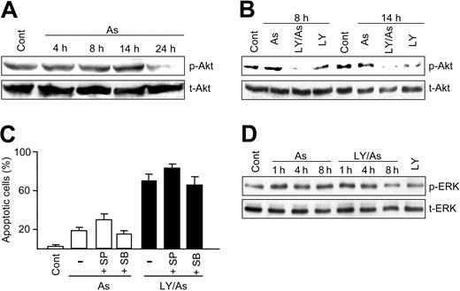
It was reported that cytotoxic agents may affect Akt phosphorylation/activation in U-937 cells.35,36 For this reason, we wanted to measure Akt phosphorylation upon treatment with As2O3, in the absence and presence of LY294002. Treatment with As2O3 alone caused a late decrease (24 hours) in Akt phosphorylation (Figure 2A). On the other hand, treatment with As2O3 plus LY294002 caused an earlier decrease in phosphorylation (8 hours and thereafter), with higher efficacy than treatment with LY294002 alone (Figure 2B).
Earlier publications indicated that PI3K/Akt may negatively regulate MAPK pathways.37-39 Hence, we queried whether the potentiation of As2O3 toxicity by PI3K/Akt inhibitors could be mediated by MAPK activation. To analyze this possibility, experiments were carried out using appropriate MAPK inhibitors, namely 10 μM PD98059 and 2.5 μM U0126 (specific for mitogen-induced extracellular kinase [MEK]/ERK), 10 μM SB20358 (specific for p38), and 10 μM SP600125 (specific for Jun N-terminal kinase [JNK]). These concentrations proved to efficaciously block kinase activation in a preceding work.19 It was found that SB20358 and SP20358, which were nontoxic when used alone or in combination with LY294002 (results not shown), did not affect or only minimally modified the generation of apoptosis by As2O3 plus LY294002 (Figure 2C), indicating that the potentiation of As2O3 toxicity may not be explained by a possible p38 or JNK activation. Experiments with MEK/ERK inhibitors could not be carried out since PD98059 and U0126, although nontoxic when used alone, were extremely toxic in combination with LY294002, even in the absence of As2O3 (result not shown). Nonetheless, immunoblot assays revealed that ERK phosphorylation was not affected by LY294002, either alone or in combination with As2O3 (Figure 2D), a result that apparently excludes a possible regulation via the MEK/ERK pathway.
Akt and ERK activation, and effect of MAPK inhibitors. (A-B) Relative level of total and phosphorylated Akt in extracts (40 μg protein per lane) obtained from untreated U-937 cells (Cont) and cells treated for the indicated time periods with 4 μMAs2O3 (A); and from cells treated for the indicated time periods with As2O3 and LY294002, alone and in combination (B). (C) Frequency of apoptotic cells (mean ± standard deviation of 4 determinations) in cultures treated for 24 hours with 4 μM As2O3, alone or with LY294002, either in the absence (-) or the presence of 10 μM SP600125 (SP) or 10 μM SB203580 (SB). (D) Relative level of total (t-ERK) and phosphorylated (p-ERK) ERKs in extracts (10 μg protein per lane) obtained from untreated cells and cells treated for the indicated time periods with As2O3 and LY294002, alone and in combination. All drugs were applied simultaneously. All other conditions were as in Figure 1.
Akt and ERK activation, and effect of MAPK inhibitors. (A-B) Relative level of total and phosphorylated Akt in extracts (40 μg protein per lane) obtained from untreated U-937 cells (Cont) and cells treated for the indicated time periods with 4 μMAs2O3 (A); and from cells treated for the indicated time periods with As2O3 and LY294002, alone and in combination (B). (C) Frequency of apoptotic cells (mean ± standard deviation of 4 determinations) in cultures treated for 24 hours with 4 μM As2O3, alone or with LY294002, either in the absence (-) or the presence of 10 μM SP600125 (SP) or 10 μM SB203580 (SB). (D) Relative level of total (t-ERK) and phosphorylated (p-ERK) ERKs in extracts (10 μg protein per lane) obtained from untreated cells and cells treated for the indicated time periods with As2O3 and LY294002, alone and in combination. All drugs were applied simultaneously. All other conditions were as in Figure 1.
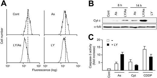
Disruption of mitochondrial transmembrane potential (ΔΨm), cytochrome c release, and caspase-3 activation. (A) The figure shows the changes in ΔΨm in U-937 cells treated for 14 hours with 4 μMAs2O3 and LY294002, alone or in combination, as determined by flow cytometry after cell loading with R123. The vertical, dotted lines represent the mean fluorescence value in untreated cells (Cont), to better discern the displacement caused by the treatments. (B) The blot shows the presence of cytochrome c (Cyt c) in cytosolic extracts (25 μg protein per lane) obtained from untreated cells (Cont), from cells treated for 14 hours with LY291002 alone, and from cells treated for the indicated time periods with 4 μMAs2O3, alone or in combination with LY294002. The level of α-tubulin (α-tub) was also measured as a control. (C) The figure shows the relative caspase-3 activity, as determined using Ac-DEVD-pNA as substrate, in extracts from untreated cells (Cont) and cells treated for 24 hours with 4 μMAs2O3, 20 nM camptothecin, and 4 μM cisplatin, alone (-)orin combination with LY294002 (+LY). The results (mean ± standard deviation of 3 determinations) are represented in relation to the control, which was given the arbitrary value of one. All other conditions were as in Figure 1.
Disruption of mitochondrial transmembrane potential (ΔΨm), cytochrome c release, and caspase-3 activation. (A) The figure shows the changes in ΔΨm in U-937 cells treated for 14 hours with 4 μMAs2O3 and LY294002, alone or in combination, as determined by flow cytometry after cell loading with R123. The vertical, dotted lines represent the mean fluorescence value in untreated cells (Cont), to better discern the displacement caused by the treatments. (B) The blot shows the presence of cytochrome c (Cyt c) in cytosolic extracts (25 μg protein per lane) obtained from untreated cells (Cont), from cells treated for 14 hours with LY291002 alone, and from cells treated for the indicated time periods with 4 μMAs2O3, alone or in combination with LY294002. The level of α-tubulin (α-tub) was also measured as a control. (C) The figure shows the relative caspase-3 activity, as determined using Ac-DEVD-pNA as substrate, in extracts from untreated cells (Cont) and cells treated for 24 hours with 4 μMAs2O3, 20 nM camptothecin, and 4 μM cisplatin, alone (-)orin combination with LY294002 (+LY). The results (mean ± standard deviation of 3 determinations) are represented in relation to the control, which was given the arbitrary value of one. All other conditions were as in Figure 1.
Modulation of mitochondria-related events
It is known that As2O3 directly targets the mitochondria by interacting with the permeability transition pore.40 This led us to examine 2 critical mitochondria-associated regulatory events, namely the dissipation of mitochondrial transmembrane potential (ΔΨm) and the release of cytochrome c, which initiates apoptosis execution along the “intrinsic” pathway.41 Figure 3A shows the alteration in ΔΨm, as measured by flow cytometry using the cationic dye R123. Treatment for 14 hours with As2O3 alone caused a slight ΔΨm decrease, which was potentiated by cotreatment with LY294002. Of note, treatment with the inhibitor alone, although nontoxic (Figure 1), sufficed to slightly reduce ΔΨm. Qualitatively similar results were obtained using the cationic dye 5,5′,6,6′-tetrachloro-1,1′,3,3′-tetraethylbenzimidazolcarbocyanine iodide (JC-1) instead of R123 (results not shown). Figure 3B shows the release of cytochrome c from mitochondria to the cytosol, as revealed by immunoblot using cytosolic extracts. As2O3 slightly induced cytochrome c release, which was greatly potentiated by cotreatment with LY294002. By contrast to ΔΨm dissipation, no cytochrome c release could be detected in cells treated with LY294002 alone. The changes in cytochrome c location were followed by the activation of the executioner caspase-3, as determined by measuring DEVDase activity in cellular extracts. Thus, extracts from cells treated with As2O3 alone exhibited a moderate increase in DEVDase activity, which was higher in cells treated with As2O3 plus LY294002 (Figure 3C). In contrast, the PI3K inhibitor did not potentiate, and instead decreased, DEVDase activity in combination with camptothecin or cisplatin, which agrees with the changes in apoptosis (indicated in Figure 1).
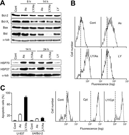
The release of cytochrome c is regulated by proteins of the Bcl-2 family, which may either inhibit (eg, the antiapoptotic proteins Bcl-2 and Bcl-XL) or promote (eg, the proapoptotic proteins Bax and Bid) the process.41 Bax remains inactive in the cytosol, and to be functional requires changes in conformation and translocation to the mitochondrial membrane.27 Bid is activated by cleavage followed by translocation to the mitochondrial membrane.42 In addition, the “heat-shock” proteins HSP27 and HSP70 also inhibit apoptosis by blocking cytochrome c and/or other elements of the intrinsic pathway.43 Hence, experiments were carried out to measure the expression and modification of these proteins. The results, represented in Figure 4, were as follows: (1) Treatment with As2O3, with or without LY294002, did not significantly modify the total Bcl-2 and Bax levels, but caused a slight decrease in Bcl-XL (Figure 4A). (2) As2O3 caused Bax activation, and this activation was further enhanced by cotreatment with LY294002, as determined by flow cytometry using the 6A7 antibody. By contrast, the PI3K inhibitor did not potentiate the camptothecin-provoked Bax activation (Figure 4B). (3) LY294002 also potentiated the As2O3-provoked Bid cleavage, using as criterion the loss of the 21-kDa proform26 (Figure 4A). (4) As2O3 increased HSP70 and HSP27 expression. However, while HSP70 was not affected by LY294002, the PI3K inhibitor reduced the basal HSP27 level as well as the increase caused by As2O3 (Figure 4A). Hence, Bax and Bid activation, and HSP27 decrease, may contribute to the increased cytochrome c release and apoptosis execution in As2O3 plus LY294002-treated cells. The possible contribution of Bad, another proapoptotic member of the Bcl-2 family (the activity of which is regulated by Akt-mediated changes in phosphorylation),16 could not be determined since phosphorylated Bad was below detection level in our immunoblot assays. Finally, it was observed that the frequency of apoptosis was greatly reduced in Bcl-2-transfected U-937 cells (which, according to our control observations, possess an 8-fold increase in Bcl-2 content in relation to the nontransfected cells) (Figure 4C). This indicates that the generation of apoptosis by As2O3 and its potentiation by LY294002 is Bcl-2 regulated, as expected in the intrinsic pathway.
Expression and modification of Bcl-2 and HSP family members, and effect of Bcl-2 overexpression. (A) Relative levels of Bcl-2, Bcl-XL, Bax, Bid (21-kDa proform), HSP70, HSP27, and α-tubulin (used as a control) in total cellular extracts (15 μg per lane) obtained from untreated U-937 cells (Cont), from cells treated for 14 hours with LY294002 alone, and from cells treated for the indicated time periods with 4 μMAs2O3, alone or in combination with LY294002, as determined by immunoblot. (B) Relative level of activated Bax in untreated cells (Cont), in cells treated for 14 hours with LY294002 alone, and in cells treated for 14 hours with As2O3 and camptothecin, alone or in the presence of LY294002, as determined by flow cytometry using the 6A7 antibody. The vertical, dotted lines represent the mean fluorescence value in the control, to better discern the displacement caused by the treatments. (C) Frequency of apoptotic cells (mean ± standard deviation of 3 determinations) in nontransfected (U-937) and Bcl-2-transfected (U4/Bcl-2) U-937 cell cultures at 24 hours of treatment with 4 μMAs2O3, alone or in combination with LY294002. All other conditions were as in Figure 1.
Expression and modification of Bcl-2 and HSP family members, and effect of Bcl-2 overexpression. (A) Relative levels of Bcl-2, Bcl-XL, Bax, Bid (21-kDa proform), HSP70, HSP27, and α-tubulin (used as a control) in total cellular extracts (15 μg per lane) obtained from untreated U-937 cells (Cont), from cells treated for 14 hours with LY294002 alone, and from cells treated for the indicated time periods with 4 μMAs2O3, alone or in combination with LY294002, as determined by immunoblot. (B) Relative level of activated Bax in untreated cells (Cont), in cells treated for 14 hours with LY294002 alone, and in cells treated for 14 hours with As2O3 and camptothecin, alone or in the presence of LY294002, as determined by flow cytometry using the 6A7 antibody. The vertical, dotted lines represent the mean fluorescence value in the control, to better discern the displacement caused by the treatments. (C) Frequency of apoptotic cells (mean ± standard deviation of 3 determinations) in nontransfected (U-937) and Bcl-2-transfected (U4/Bcl-2) U-937 cell cultures at 24 hours of treatment with 4 μMAs2O3, alone or in combination with LY294002. All other conditions were as in Figure 1.
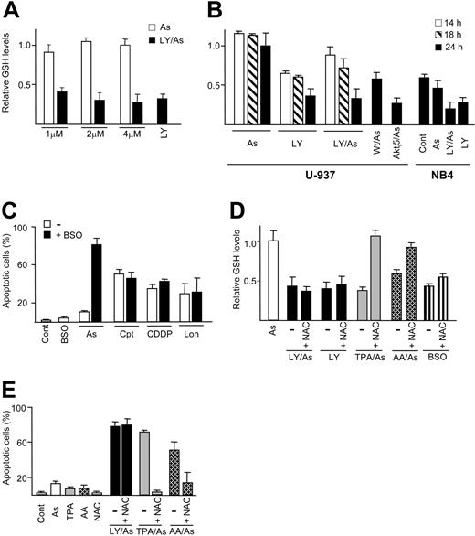
Modulation of GSH level and its relationship with apoptosis. (A-B) Relative GSH levels, as determined by monochlorobimane derivatization, in U-937 cell cultures treated for 24 hours with LY294002 and the indicated concentrations of As2O3, alone and in combination (A); and in U-937 and NB4 cell cultures treated for the indicated time periods with 4 μM (U-937) and 2 μM (NB4) As2O3 alone, with LY294002 alone, and with As2O3 plus either LY294002, wortmannin, or Akti5 (B). All results are expressed in relation to untreated U-937 cell cultures (approximate GSH content, 9 nmol/106 cells), which received the arbitrary value of one. (C) Frequency of apoptotic cells in U-937 cell cultures treated for 24 hours with BSO alone, and with As2O3, camptothecin, cisplatin, and lonidamine, either alone (-) or in combination with BSO. (D) Relative GSH levels in U-937 cells treated for 24 hours with LY294002, with BSO, and with the combination of As2O3 plus either LY294002, TPA, or ascorbic acid (AA), in the absence (-) or the presence of NAC (+NAC). (E) Frequency of apoptotic cells in U-937 cell cultures treated for 24 hours with As2O3 alone, TPA alone, AA alone, and NAC alone, and with the combinations of As2O3 plus either LY294002, TPA, or AA, in the absence (-) or the presence of NAC. BSO, TPA, AA, and NAC were used at 1 mM, 20 nM, 150 μM, and 10 mM, respectively. All drugs were applied simultaneously. The data represent the means ± standard deviation of at least 3 determinations. All other conditions were as in Figure 1.
Modulation of GSH level and its relationship with apoptosis. (A-B) Relative GSH levels, as determined by monochlorobimane derivatization, in U-937 cell cultures treated for 24 hours with LY294002 and the indicated concentrations of As2O3, alone and in combination (A); and in U-937 and NB4 cell cultures treated for the indicated time periods with 4 μM (U-937) and 2 μM (NB4) As2O3 alone, with LY294002 alone, and with As2O3 plus either LY294002, wortmannin, or Akti5 (B). All results are expressed in relation to untreated U-937 cell cultures (approximate GSH content, 9 nmol/106 cells), which received the arbitrary value of one. (C) Frequency of apoptotic cells in U-937 cell cultures treated for 24 hours with BSO alone, and with As2O3, camptothecin, cisplatin, and lonidamine, either alone (-) or in combination with BSO. (D) Relative GSH levels in U-937 cells treated for 24 hours with LY294002, with BSO, and with the combination of As2O3 plus either LY294002, TPA, or ascorbic acid (AA), in the absence (-) or the presence of NAC (+NAC). (E) Frequency of apoptotic cells in U-937 cell cultures treated for 24 hours with As2O3 alone, TPA alone, AA alone, and NAC alone, and with the combinations of As2O3 plus either LY294002, TPA, or AA, in the absence (-) or the presence of NAC. BSO, TPA, AA, and NAC were used at 1 mM, 20 nM, 150 μM, and 10 mM, respectively. All drugs were applied simultaneously. The data represent the means ± standard deviation of at least 3 determinations. All other conditions were as in Figure 1.
Changes in glutathione content
As mentioned in the “Introduction,” the toxicity of As2O3 is greatly dependent on the intracellular GSH content, which in turn may be affected by changes in protein kinase activities. For these reasons, determinations using the GSH-sensitive fluorescent probe monoclorobimane were carried out to analyze the possible effects of As2O3 and PI3K/Akt inhibitors on intracellular GSH in U-937 cells. As indicated in Figure 5A-B, the GSH content was not reduced by treatment with 1 to 4 μM As2O3 alone, but was considerably decreased by treatment with LY294002, either alone or in combination with As2O3. This response was corroborated using combinations of As2O3 plus wortmannin or Akti5, and using NB4 and HL-60 cells instead of U-937 cells (Figure 5B, and results not shown). The importance of GSH depletion as mediator of apoptosis induction in our experimental conditions was confirmed using BSO, a specific inhibitor of γ-glutamylcysteine synthetase (γ-GCS) activity, the rate-limiting enzyme of GSH biosynthesis. Preliminary determinations indicated that a 24-hour treatment with 1 mM BSO caused approximately a 60% inhibition in the intracellular GSH content (result not shown). It was observed that treatment with BSO alone was nontoxic, but as expected cotreatment with BSO increased the apoptotic action of As2O3, in the same manner as the PI3K/Akt inhibitors. By contrast, BSO failed to increase the apoptotic action of cisplatin, camptothecin, and lonidamine (Figure 5C). This result indicates that under the conditions used here, cisplatin, camptothecin, and lonidamine behave as GSH-insensitive drugs, and may therefore explain the inability of the PI3K inhibitors to potentiate their toxicity (Figure 1F).
To shed some light on the mechanisms by which PI3K/Akt inhibition could deplete GSH, determinations were carried out using NAC, an agent earlier used as a cysteine donor for GSH biosynthesis in experiments with As2O3.4,7 In addition to LY294002 and BSO, in these assays we used TPA and AA, which were also reported to reduce GSH in myeloid cells and other cell systems.4,7,18,19 The results are indicated in Figure 5D. NAC was unable to restore the GSH content in cells treated with BSO (which, as indicated above, blocks enzyme activity); and it was similarly ineffective in cells treated with LY294002, either alone or with As2O3. However, NAC restored the GSH content in TPA plus As2O3- and AA plus As2O3-treated cells. This suggests that, in contrast to TPA and AA, and in the same manner as BSO, PI3K/Akt inhibitors modulate GSH biosynthesis at levels other than substrate availability. Moreover, in good parallelism with these results, NAC abrogated the increase in toxicity in the case of TPA and AA, but not in the case of LY294009 (Figure 5E), confirming again the importance of GSH as a mediator of the increased apoptosis in cells treated with As2O3 plus PI3K inhibitors.
Final information was obtained by means of HPLC. As indicated in Table 1, this technique allowed us to corroborate the capacity of LY294002 to cause GSH depletion, and also proved that the GSH decrease provoked by the PI3K inhibitor was not adequately compensated by an increase in GSSG. Nonetheless, the ratio of GSH to GSSG was diminished, especially in As2O3 plus LY294002-treated cells, indicating that the treatments caused GSH oxidation to some degree.
Effect of treatment with LY-294002 alone (LY) and LY294002 plus As2O3 (LY/As) on the intracellular GSH and GSSG content in U-937 cells
. | Control . | LY . | LY/As . |
|---|---|---|---|
| No. | 6 | 6 | 3 |
| GSH, nmol/mg protein | 75.1 ± 11.7 | 45.7 ± 6.1 | 37.1 ± 6.6 |
| GSSG, nmol/mg protein | 1.5 ± 0.4 | 1.6 ± 0.6 | 3.1 ± 0.8 |
| GSH/GSSG, nmol/mg protein | 50.5 | 28.6 | 12.0 |
. | Control . | LY . | LY/As . |
|---|---|---|---|
| No. | 6 | 6 | 3 |
| GSH, nmol/mg protein | 75.1 ± 11.7 | 45.7 ± 6.1 | 37.1 ± 6.6 |
| GSSG, nmol/mg protein | 1.5 ± 0.4 | 1.6 ± 0.6 | 3.1 ± 0.8 |
| GSH/GSSG, nmol/mg protein | 50.5 | 28.6 | 12.0 |
The values (nmol/mg protein) were obtained at 24 hours of treatment. For other conditions, see Figure 1 legend.
ROS accumulation and GSTπ expression and activity
One of the roles of GSH is the scavenging of ROSs.8 Hence, we queried whether the GSH depletion derived from PI3K/Akt inhibition could result in ROS overaccumulation, which might in turn explain the increase in As2O3 toxicity. To answer this question, flow cytometry determinations were carried out using the peroxide-sensitive fluorescent probe H2DCFDA. The results are represented in Figure 6A: (1) treatment with As2O3 alone, which as indicated in Figure 5 did not decrease GSH, did not affect peroxide accumulation; (2) treatment with LY294002 alone, which as indicated in Figures 5 and 1 sufficed to decrease GSH but was not toxic per se, increased the intracellular peroxide accumulation; and (3) a similar increase was obtained by treatment with As2O3 plus LY294002, which as indicated in Figures 5 and 1 decreased GSH and was highly toxic.
Peroxide accumulation and effects of hydrogen peroxide. (A) Intracellular peroxide accumulation in untreated U-937 cells (Cont), in cells treated for 14 hours with LY294002 alone, and in cells treated for 14 hours with 4 μMAs2O3, alone or in combination with LY294002, as determined by flow cytometry after cell loading with H2DCFDA. The vertical, dotted lines represent the mean fluorescence value in the control, to better discern the displacement caused by the treatments. (B) Frequency of apoptotic cells (mean ± standard deviation of at least 4 determinations) in U-937 cell cultures treated for 24 hours with 20 and 40 μM H2O2 alone, and with 1 to 4 μ As2O3, camptothecin, and cisplatin, either alone (-) or in combination with H2O2. All other conditions were as in Figure 1.
Peroxide accumulation and effects of hydrogen peroxide. (A) Intracellular peroxide accumulation in untreated U-937 cells (Cont), in cells treated for 14 hours with LY294002 alone, and in cells treated for 14 hours with 4 μMAs2O3, alone or in combination with LY294002, as determined by flow cytometry after cell loading with H2DCFDA. The vertical, dotted lines represent the mean fluorescence value in the control, to better discern the displacement caused by the treatments. (B) Frequency of apoptotic cells (mean ± standard deviation of at least 4 determinations) in U-937 cell cultures treated for 24 hours with 20 and 40 μM H2O2 alone, and with 1 to 4 μ As2O3, camptothecin, and cisplatin, either alone (-) or in combination with H2O2. All other conditions were as in Figure 1.
The preceding observations could indicate that while the cells tolerate a limited ROS overaccumulation without concomitant toxicity, such overaccumulation may facilitate the generation of apoptosis by As2O3. To investigate this possibility, experiments were carried out in which cells were cotreated with As2O3 plus low H2O2 concentrations (20-40 μM), which were per se innocuous or slightly toxic. In agreement with our hypothesis, H2O2 potentiated or exerted a synergic effect with As2O3, at the concentrations of 2 to 4 μM. By contrast, H2O2 did not modify or exhibited only additive effects in combination with camptothecin and cisplatin (Figure 6B).
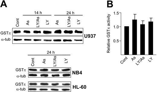
It has been indicated that GSTπ is key element in the As2O3 detoxification machinery.11,12 For this reason, we asked whether the potentiation of As2O3 toxicity by PI3K/Akt inhibitors involved an alteration in GSTπ expression and/or activity. The results in Figure 7 indicate that both the enzyme content (as measured by immunoblot assays) and activity remained unaltered or were slightly increased upon treatment with As2O3 and LY294002, alone and in combination, when compared with the values in untreated cells.
Discussion
The results in this work indicate that the generation of apoptosis by low concentrations of As2O3 is potentiated by cotreatment with PI3K/Akt inhibitors (LY294002, wortmannin, Akti5) in U-937 promonocytic and other myeloid leukemia cells. In spite of the importance of PI3K/Akt as a survival-promoting pathway, treatment with the inhibitors alone did not significantly cause cell death, in contrast with the results sometimes obtained in other cell models.44,45 A possible explanation is that the intact MEK/ERK pathway provides enough survival signals to keep the cell viability. In fact, the simultaneous inactivation of PI3K/Akt and MEK/ERKs by cotreatment with LY294002 plus either PD98059 or U0126 drastically induced apoptosis in U-937 cells. The potentiation by PI3K/Akt inhibitors of the As2O3-provoked apoptosis represented a drug-specific response, which could not be reproduced using lonidamine, camptothecin, and cisplatin. This result was unexpected, since with few exceptions PI3K/Akt inhibition was earlier reported to potentiate the lethality of cytotoxic drugs in myeloid cells.35,46,47 Such discrepancy might be explained by differences in the experimental designs used. While other works used elevated drug concentrations for short time periods (typically 4-6 hours),35,46,47 in the present study we used relatively low concentrations during a longer time period (24 hours). Under these conditions, the alterations of cell cycle regulatory events derived of PI3K/Akt inhibition may considerably affect the action of the antitumor drugs, which often behave as cycle phase-specific drugs (eg, G2-arrest by As2O3, camptothecin, and cisplatin in U-937 cells, which are p53-null cells48 ). Actually, this argument was earlier used to explain the LY294002-provoked attenuation of toxicity upon long-term cisplatin treatment in 32D murine myeloid cells.49 However, while such explanation might account for the discrepancies between our and other studies, it does not satisfactorily explain the different modulation by PI3K inhibitors of apoptosis induction by As2O3 and the other assayed drugs, observed in the present work. In accordance with its property as a mitochondria-targeting drug, the action of As2O3 and the potentiation by LY294002 exhibited typical characteristics of the “intrinsic” pathway—namely increased cytochrome c release, negative regulation by Bcl-2, and positive regulation by Bax. Moreover, while the mechanism by which LY294002 inhibits HSP27 expression is still unknown, such inhibition may also contribute to the increase in toxicity, since HSP27 is an antiapoptotic protein.43 Nonetheless, the possibility that the “extrinsic” (receptor-mediated) pathway could also participate in the process may not be excluded, since As2O3 was reported to activate this pathway in some cell types.50,51 This possibility is presently under investigation in our laboratory.
GSTπ expression and activity. (A) Relative GSTπ levels in extracts (10μg protein per lane) obtained from untreated cells (Cont) and cells treated for the indicated time periods with LY294002 and As2O3, alone and in combination. As2O3 was used at 4 μM in the case of U-937 and HL-60, and at 2 μM in the case of NB4 cell cultures. (B) GSTπ activity in untreated U-937 cells and cells subjected for 24 hours to the same treatments as in panel A. The results (mean ± standard deviation of 3 determinations) are expressed in relation to untreated cells (main value, 44.5 ± 6.9 nmol/mg protein/min), which received the arbitrary value of one. All other conditions were as in Figure 1.
GSTπ expression and activity. (A) Relative GSTπ levels in extracts (10μg protein per lane) obtained from untreated cells (Cont) and cells treated for the indicated time periods with LY294002 and As2O3, alone and in combination. As2O3 was used at 4 μM in the case of U-937 and HL-60, and at 2 μM in the case of NB4 cell cultures. (B) GSTπ activity in untreated U-937 cells and cells subjected for 24 hours to the same treatments as in panel A. The results (mean ± standard deviation of 3 determinations) are expressed in relation to untreated cells (main value, 44.5 ± 6.9 nmol/mg protein/min), which received the arbitrary value of one. All other conditions were as in Figure 1.
In addition, the present results demonstrate that the PI3K/Akt signaling pathway is required to maintain the normal GSH metabolism in myeloid cells, since the basal GSH content was reduced upon treatment with PI3K/Akt inhibitors, either alone or in combination with As2O3. Although our mechanistic studies on GSH regulation by PI3K/Akt are still preliminary, it may be concluded that (1) GSH depletion was not a mere consequence of cell death, since a 24-hour treatment with LY294002 alone, which was nontoxic, sufficed to considerably decrease GSH; (2) the GSH depletion was not adequately compensated by the appearance of the oxidized form, GSSG; and (3) GSH was not regulated at the level of substrate availability since (in contrast to TPA and AA, and in the same manner as BSO) the LY294002-provoked GSH depletion was not prevented by NAC. Therefore, a possible explanation is that PI3K/Akt may directly regulate γ-GCS, either at the level of enzyme synthesis or activity. Actually, PI3K inhibitors were recently reported to abrogate the insulin-mediated increase in γ-GCS catalytic subunit synthesis in rat hepatocytes,21 and γ-GCS activity is susceptible to modulation by changes in phosphorylation.52 Whatever the mechanism, GSH depletion appears to be an important factor in explaining the selective potentiation by PI3K/Akt inhibitors of apoptosis induction by As2O3, but not by other antitumor drugs. In fact, in contrast to As2O3, under the hereby assayed conditions, camptothecin, cisplatin, and lonidamine behaved as GSH-insensitive drugs, as indicated by the inability of the GSH-specific agent BSO to increase their toxicity. Due to the multiplicity of functions of glutathione, it is conceivable that different mechanisms may participate in potentiating As2O3 toxicity under GSH-depleting conditions. Thus, in our experiments the LY294002-provoked GSH decrease was paralleled by an increase in intracellular peroxides. This is important since ROS elevation has been reported to sensitize leukemic cells to As2O3-provoked apoptosis53 —a response here corroborated by us by cotreatment with low H2O2 concentrations. In addition, since GSH directly reacts with As2O3,10 GSH depletion may result in an increase in free, acting intracellular As2O3 concentration and hence in toxicity, even in the absence of significant alterations in GSTπ expression and activity. Of course, the GSH-based regulation does not exclude the concurrence of additional mechanisms (eg, alterations of critical cell-cycle regulatory events), which as indicated earlier in this section might explain the unusual attenuation by PI3K inhibitors of camptothecin and cisplatin toxicity, observed in this and other reports.49
In summary, the present study provides the first demonstration that GSH is a target of the PI3K/Akt in myeloid leukemia cells, and as such it may partially explain the capacity of PI3K/Akt inhibitors to selectively potentiate the apoptotic action of the antileukemic drug As2O3. We believe that this property might provide a rationale to improve the efficacy of therapies based on the use of pharmacologic inhibitors of the PI3K/Akt pathway.
Prepublished online as Blood First Edition Paper, January 21, 2005; DOI 10.1182/blood-2004-07-2802.
Supported by grant SAF2004-01250 from the Plan Nacional de Investigación Científica, Desarrollo e Innovación Tecnológica, Dirección General de Investigación, Ministerio de Educación y Ciencia, Spain; grant GR/SAL/0639/2004 from the Dirección General de Universidades e Investigación, Consejería de Educación, Comunidad de Madrid, Spain; and by grant 2001-592 from the INTAS program (European Union). A.M.R. was a recipient of a postdoctoral fellowship from the Fundación Carolina, Spain. P.S. and C.F. were recipients of predoctoral fellowships from the Ministerio de Educación, Cultura y Deporte and the Ministerio de Ciencia y Tecnología, Spain, respectively.
The publication costs of this article were defrayed in part by page charge payment. Therefore, and solely to indicate this fact, this article is hereby marked ”advertisement” in accordance with 18 U.S.C. section 1734.
We thank Dr J. Bréard for providing Bcl-2-transfected U-937 cells.



This feature is available to Subscribers Only
Sign In or Create an Account Close Modal