Abstract
Despite the rapid and widespread integration of chimeric CD20 monoclonal antibody (mAb), rituximab, into the management of non-Hodgkin lymphoma, its efficacy remains variable and often modest when used as a single agent. To develop more potent reagents, human immunoglobulin transgenic mice were used to generate a panel of immunoglobulin G1κ (IgG1κ) CD20 mAbs. All reagents bound strongly to CD20+ cells and recruited mononuclear cells for the lysis of malignant B cells. However, 2 mAbs, 2F2 and 7D8, were exceptionally active in complement-dependent cytotoxicity (CDC), being able to lyse a range of rituximab-resistant targets, such as CD20-low chronic lymphocytic leukemia (CLL), in the presence of human plasma or unfractionated blood. Further analysis showed that 2F2 and 7D8, like rituximab, redistributed CD20 into Triton X-100-insoluble regions of the plasma membrane, but that they had markedly slower off-rates. To determine whether off-rate influenced CDC, a non-complement activating F(ab′)2 antihuman κ reagent was used. This reagent markedly slowed the off-rate of rituximab and increased its CDC activity to that of 2F2 and 7D8. Thus, with increasing evidence that mAb therapeutic activity in vivo depends on complement activation, these new CD20 reagents with their slow off-rates and increased potency in CDC hold considerable promise for improved clinical activity. (Blood. 2004;104:1793-1800)
Introduction
The growing success of monoclonal antibodies (mAbs) in the treatment of human diseases continues to focus attention on their mechanisms of action in vivo.1-4 In the field of oncology, the human/mouse chimeric CD20 mAb, rituximab, is rapidly becoming integrated into the treatment of B-cell malignancies.5-8 It is particularly effective when combined with conventional chemotherapies such as cyclophosphamide, doxorubicin, vincristine, and prednisone (CHOP).9 Despite such success, we still have a relatively poor understanding of how rituximab mediates its activity or even the best regimen under which it should be used. It is apparent that anticancer mAbs can use a wide range of effector mechanisms in vivo, such as recruitment of Fc-bearing effectors,10-13 activation of complement,14-19 and induction of apoptosis,18,20-23 and that the target antigen itself is critical in determining which of these predominates. For example, complement is likely to operate only when an immunoglobulin G (IgG) antibody (Ab) binds to an antigen that is highly expressed.24 In the case of CD20, most evidence suggests that Fc-Fc receptor (FcR) interactions are critical in both animal models13 and humans.10,12 However, determining whether such interactions are required for classical antibody-dependent cellular cytotoxicity (ADCC) mediated by natural killer (NK) or myeloid cells, or whether they provide cross-linking, which promotes apoptosis, has been difficult to resolve.18,22
The role of complement in the depletion of malignant and normal cells is less convincing, and a number of antitumor mAbs appear to operate in the absence of lytic complement.25-27 Complement-dependent cytotoxicity (CDC) requires at least 10 times more Abs at the cell surface than does ADCC,24 and consequently while most mAbs will mediate ADCC, very few reach the surface density necessary to activate the classical complement pathway.28,29 A number of laboratories, including our own, have recently reexamined the role of complement in Ab immunotherapy, and, in the case of CD20 mAb have found support for its involvement in both animals14,19 and humans.16-18 Furthermore, there is a strong correlation between the level of CD20 expression and therapeutic outcome for rituximab,17 which matches the known dependence of CDC on antigen density.30 However, there is also contrary evidence, and Weng and Levy26 have shown no correlation between expression of complement regulatory proteins (CD55 and CD59) and clinical response to rituximab.
To further complicate the situation, we recently defined CD20 mAbs as either type I or II, based upon their efficacy in various in vitro assays.28 Type I mAbs (rituximab and most CD20 mAbs) are potent in CDC assays and less effective inducers of apoptosis, whereas type II mAbs (B1 and 11B8) are ineffective in CDC but potent at inducing homotypic adhesion and apoptosis.21 Both types of mAbs are equally effective in ADCC. The ability of type I mAbs to induce CDC appears dependent on their ability to translocate CD20 into Triton X-100 (Tx100)-insoluble “lipid raft” areas of the plasma membrane, a property that type II mAbs lack.28,31
In the current work we describe a novel set of CD20 mAbs, derived from human immunoglobulin (Ig) transgenic mice, that have been fully characterized into type I and II reagents. The type II mAb (11B8) mediates ADCC but not CDC, in keeping with its inability to redistribute CD20 into lipid rafts. Despite having the same IgG1 isotype as rituximab and behaving similarly in ADCC, 2 type I reagents (2F2 and 7D8) are strikingly more active in CDC assays allowing lysis of a range of B-cell lines and fresh tumor isolates that were resistant to rituximab-mediated CDC. This correlates with a superior binding activity, which manifests itself as a slower off-rate in all 3 human mAbs.
Materials and methods
Cell lines
Cells were from the European Collection of Cell Cultures (ECACC) or Deutsche Sammlung von Microorganismen und Zellkulturen (DSMZ) and maintained in supplemented RPMI 1640.
Antibody production and labeling
The mAbs used are detailed in Table 1. Hybridomas were expanded in tissue culture (HyQ medium; Perbio Sciences, Helsingborg, Sweden) and purified on Protein A columns (Amersham Biosciences, Buckinghamshire, United Kingdom). F(ab′)2 fragments were produced by pepsin digestion,32 and Fab′, by reduction with 2 mM dithiothreitol for 30 minutes at 25°C, followed by alkylation with 4 mM N-ethyl maleimide.
Details of mAbs used in this study
mAb . | Specificity . | Isotype . | Source . |
|---|---|---|---|
| B1 | CD20 | IgG2a | Coulter, Miami, FL |
| Rituximab | CD20 | Chimeric Hu Fc | Roche, Basel, Switzerland |
| 1F5 | CD20 | IgG1 | ATCC, Manassas, VA |
| 2F2 | CD20 | Hu IgG1, hybridoma | Genmab, Utrecht, the Netherlands |
| 7D8 | CD20 | Hu IgG1, hybridoma | Genmab |
| 11B8 | CD20 | Hu IgG1, transfectoma | Genmab |
| HuMab | KLH | Hu IgG1, hybridoma | Genmab |
ATCC indicates American Type Culture Collection.
Generation of human CD20 mAbs
HCo7 and KM mice (Genmab)33 were immunized with human CD20-transfected NS/0 cells. For priming, mice received 1 × 10⁷ CD20-transfected cells in complete Freund adjuvant (CFA) intraperitoneally and were boosted with cells in phosphate-buffered saline (PBS). After development of a positive CD20 Ab titer, mice received a final intravenous boost of 0.5 × 10⁷ cells 3 days prior to fusion.33 Hybridomas making IgG1 Ab that recognized CD20-transfected but not untransfected NS-0 cells were sub-cloned by limiting dilution.
Measurement of 125I-labeled CD20 mAb binding to CD20-expressing cells
IgG, F(ab′)2, or Fab′ fragments were iodinated using Iodobeads (Pierce Chemical, Rockford, IL). 125I-labeled mAb binding was performed as described previously.34 Briefly, labeled mAbs were serially diluted and incubated with cells for 2 hours at 37°C in the presence of sodium azide and 2-deoxyglucose to prevent endocytosis. The cells were then centrifuged through phthalate oils, allowing rapid separation of bound and free 125I-labeled mAbs. The pelleted cells were then counted using a gamma counter (Wallac, Milton Keynes, United Kingdom).
Dissociation of labeled CD20 mAbs in the absence and presence of cross-linking antibody
To determine the dissociation rate of mAbs we followed methods described by Mason and Williams.35 Briefly, cells were pelleted and resuspended in medium containing 2 μg/mL 125I-labeled CD20 mAb IgG. Cells were incubated for 2 hours at room temperature, pelleted, and resuspended in medium containing 1 mg/mL of the unlabeled IgG. An aliquot was taken and layered onto phthalate oil to determine the starting level of mAb binding. Similar aliquots were taken over 3 hours, and the amount of mAb remaining on the cell was determined and expressed as a percentage of the initial count. To determine the effect of cross-linking on the dissociation of CD20 mAbs, DOHH cells (λ positive) were incubated with labeled CD20 mAbs, the cells were pelleted by centrifugation, and the supernatant was removed. The cells were then resuspended in cold medium, split into 2 aliquots, and a polyclonal sheep F(ab′)2 antihuman κ reagent (10 μg/mL) was added to one and medium to the second. After incubating on ice for 30 minutes, 1F5 IgG (final concentration, 1 mg/mL) was added to each aliquot, and over 3 hours samples were taken to determine the level of mAbs remaining on the cell surface. In these cross-linking experiments, the mouse CD20 mAb 1F5 was used as a cold competitor to ensure that it did not interfere with the binding of the cross-linking sheep Ab, which recognized only human κ chains.
Cytotoxicity assays
ADCC assays were performed as described.36 To determine the CDC activity of mAbs, elevated membrane permeability was assessed using 51Cr-release or propidium iodide (PI) exclusion assays.36 When required, plasma was obtained for cytotoxicity assays by centrifugation of citrate anticoagulated whole blood and was recalcified by adding complete medium (1 part plasma + 3 parts medium). To assess the longevity of the CDC-inducing effect of the mAbs, cells were labeled with mAbs for 10 to 15 minutes at room temperature, washed twice, and resuspended in RPMI 1640/10% fetal calf serum (FCS) at 37°C. Samples were taken at various time points, and human serum (20% vol/vol) was added as a source of complement. In one set of experiments, CDC was performed in the presence of a cross-linking F(ab′)2 reagent that prevented mAb dissociation. This was the sheep antihuman κ reagent used in the off-rate study, which lacked Fc and was unable to activate CDC. In these experiments DOHH cells were labeled with CD20 mAbs for 15 minutes at room temperature, washed, and various concentrations of cross-linker added, followed by human serum (20% vol/vol). After incubation for 45 minutes at 37°C, cells were harvested and lysis was detected by flow cytometry.
Assessment of raft-associated CD20
As a rapid assessment of the presence of CD20 in raft microdomains, we used a flow cytometry method based on their Triton X-100 (Tx100) insolubility at low temperatures, as described previously.28 To assess fluorescence resonance energy transfer (FRET), mAbs were directly conjugated to bifunctional N-hydroxysuccinimide (NHS)-ester derivates of cyanin 3 (Cy3) and Cy5 (Amersham Biosciences) as described in the manufacturer's instructions and analyzed as described previously.28,37
Deposition of C1q and C4c
To assess the extent of cellular C1q and C4c deposition, various concentrations of mAbs were added to cells (1 × 10⁵/well in a 96-well plate) and allowed to bind for 10 to 15 minutes at room temperature. Serum was then added to a final concentration of 1% (vol/vol) followed by incubation at 37°C for 10 minutes. After washing, fluorescein isothiocyanate (FITC)-conjugated polyclonal antibodies against C1q (F0254) or C4c (F0169) (DakoCytomation, Glostrup, Denmark) were added (1:100) and samples incubated for 30 minutes at 4°C and analyzed by flow cytometry.
C1q binding to plate-bound human mAbs was also assessed as determined by enzyme-linked immunosorbent assay (ELISA). Briefly, plates were coated with varying concentrations of control and CD20 mAbs (10 μg/mL to 0.06 μg/mL). After blocking, pure human C1q (Sigma, St Louis, MO) at 2 μg/mL was added and then detected with rabbit antihuman C1q polyclonal Ab (DakoCytomation), followed by peroxidase-conjugated swine anti-rabbit IgG-Fc Ab (DakoCytomation) and ABTS (2,2′-azino-bis[3-ethylbenzthiazoline-6-sulfonic acid]) solution (1 mg/mL). To confirm that the level of each of the CD20 mAbs bound to the plates was similar, IgG was measured on the same plates (after washing and blocking) using alkaline-phosphatase-conjugated goat anti-human IgG (Jackson, Brunswick, NJ) and p-nitrophenylphosphate (pNNp) solution (KPL, Gaithersburg, MD) as substrate. Optical densities (ODs) were read at 405 nm in a Biotek plate reader (Biotek Instruments, Winooski, VT).
Results
Characterization of human CD20 mAbs
Following immunization of human Ig transgenic mice with CD20-transfected cells, 3 new human mAbs, 2F2, 7D8, and 11B8, were raised. All 3 reagents showed strong staining of B-cell areas in immunohistochemistry, similar to rituximab, and bound to a wide range of CD20-expressing B-cell tumors. CD20 specificity was confirmed by binding to CD20-expressing NS-0 and SKBR3 cells, but not to the nontransfected parent cells and by cross blocking of a range of known CD20 mAbs (not shown). Thus by all criteria tested, these human mAbs were specific for human CD20.
We next assessed the ability of these new mAbs to translocate CD20 into detergent-insoluble fractions of the plasma membrane, a property that separates mAbs into type I (rituximab-like) or type II (B1-like) reagents.28 The most straightforward technique for assessing this uses flow cytometry to measure binding of CD20 mAbs before and after treatment with Tx100 detergent at 4°C. Once an mAb (type I) has moved CD20 into the Tx100-insoluble fraction, the cells remain stained with FITC-labeled CD20 mAbs. Figure 1A-B shows results for 2 typical B-cell lines, Daudi and DOHH, which clearly revealed that rituximab, 2F2, and 7D8 translocated CD20 into the Tx100-insoluble fraction, but that B1 and 11B8 did not.
Comparison of the ability of CD20 mAbs to redistribute antigens on the cell surface. (A-B) Translocation of CD20 into the Tx100-insoluble fraction of the cell. Daudi (A) or DOHH (B) cells were incubated with FITC-labeled CD20 mAb (10 μg/mL) for 15 minutes at 37°C and washed. After chilling on ice, half of each sample was treated with 0.5% Tx100 for 15 minutes on ice. Both samples (lysed, dark gray bars; and not lysed, light gray bars) were washed and analyzed by flow cytometry (mean fluorescence intensity [MFI]) to assess bound FITC-CD20 mAbs. (C) FRET determination: Daudi cells were labeled with an equimolar mixture of Cy3 (donor)- and Cy5 (acceptor)-conjugated CD20 mAbs at 37°C with or without C8-deficient (C8-def) serum. FRET, which measures the proximity of neighboring mAb molecules, was assessed by flow cytometric analysis. These graphs are representative of at least 3 experiments, each showing similar results.
Comparison of the ability of CD20 mAbs to redistribute antigens on the cell surface. (A-B) Translocation of CD20 into the Tx100-insoluble fraction of the cell. Daudi (A) or DOHH (B) cells were incubated with FITC-labeled CD20 mAb (10 μg/mL) for 15 minutes at 37°C and washed. After chilling on ice, half of each sample was treated with 0.5% Tx100 for 15 minutes on ice. Both samples (lysed, dark gray bars; and not lysed, light gray bars) were washed and analyzed by flow cytometry (mean fluorescence intensity [MFI]) to assess bound FITC-CD20 mAbs. (C) FRET determination: Daudi cells were labeled with an equimolar mixture of Cy3 (donor)- and Cy5 (acceptor)-conjugated CD20 mAbs at 37°C with or without C8-deficient (C8-def) serum. FRET, which measures the proximity of neighboring mAb molecules, was assessed by flow cytometric analysis. These graphs are representative of at least 3 experiments, each showing similar results.
The relative distribution of mAbs on cell membranes can also be followed using fluorescence resonance energy transfer (FRET). Figure 1C shows that the type I mAbs (rituximab, 2F2, and 7D8) gave clear increases in FRET following binding either in the presence or absence of serum, whereas B1 and 11B8 did not. C8-deficient serum was used in these experiments to engage early complement components, such as C1 and C3, without lysis. It is clear that any interaction with complement components did not markedly change the redistribution of CD20-mAb complexes. These results were consistent with the concept that type I mAbs cause coalescence of dispersed CD20 molecules into localized Tx100-insoluble lipid rafts, resulting in energy transfer from the Cy3 to Cy5 mAb (Figure 1A-B). In contrast, B1 and 11B8 did not induce reorganization, hence the lack of energy transfer.
In initial functional experiments, the cytotoxic activity of these mAbs was assessed against ARH-77 B cells, using unfractionated blood as a source of natural effectors. Blood contains complement in the plasma, together with FcR-expressing cellular effectors, such as polymorphonuclear cells (PMNs) and mononuclear cells (MNCs). In Figure 2A, rituximab was unable to mediate detectable lysis with unfractionated blood, even when used at 10 μg/mL, a concentration that would saturate binding sites (below). In contrast, 7D8 and 2F2 gave up to 60% specific 51Cr release. 11B8 was less active and gave only 20% to 30% specific 51Cr-release. Thus, despite the fact that these reagents were all IgG1 isotype and cross blocked each other, they displayed very distinct cytotoxic activity with fresh blood, presumably as a result of differences in type (Figure 1), epitope recognition, or binding characteristics.
Killing of ARH B-cells and fresh tumors (CLL and HCL) by CD20 mAbs with human effectors. (A-B) Lysis of ARH-77 lymphoma cells was measured in 3-hour 51Cr release assays. (A) The influence of antibody concentration was investigated using unfractionated blood from healthy donors as effector source. Symbols: rituximab, ▪; 7D8, ▴; 2F2, ▾ ; and 11B8, •. This figure shows the mean ± SEM of 4 separate experiments determining the percent specific 51Cr-release. (B) To identify individual effector mechanisms, human blood (whole blood) was fractionated into polymorphonuclear (PMN) or mononuclear (MNC) cells, or into complement-containing plasma (plasma). Antibodies were used at 10 μg/mL, and isolated PMNs and MNCs at an effector-to-target cell ratio of 40:1. Data represent the mean ± SEM of 8 separate experiments determining percent specific 51Cr-release. (C-D) Similar experiments were performed against 51Cr-labeled, freshly isolated lymphoma cells. As effector source, we used unfractionated blood from healthy volunteers, which was fractionated into polymorphonuclear (PMN) or mononuclear (MNC) cells, or into complement-containing plasma (plasma). (C) Mean ± SEM of experiments determining percent specific 51Cr-release from 8 primary hairy cell leukemias. (D) Data from 16 primary B-CLLs. All mAbs were used at 10 μg/mL, and isolated PMNs or MNCs at an effector-to-target cell ratio of 40:1. Data are presented as the mean ± SEM of the percent specific 51Cr-release.
Killing of ARH B-cells and fresh tumors (CLL and HCL) by CD20 mAbs with human effectors. (A-B) Lysis of ARH-77 lymphoma cells was measured in 3-hour 51Cr release assays. (A) The influence of antibody concentration was investigated using unfractionated blood from healthy donors as effector source. Symbols: rituximab, ▪; 7D8, ▴; 2F2, ▾ ; and 11B8, •. This figure shows the mean ± SEM of 4 separate experiments determining the percent specific 51Cr-release. (B) To identify individual effector mechanisms, human blood (whole blood) was fractionated into polymorphonuclear (PMN) or mononuclear (MNC) cells, or into complement-containing plasma (plasma). Antibodies were used at 10 μg/mL, and isolated PMNs and MNCs at an effector-to-target cell ratio of 40:1. Data represent the mean ± SEM of 8 separate experiments determining percent specific 51Cr-release. (C-D) Similar experiments were performed against 51Cr-labeled, freshly isolated lymphoma cells. As effector source, we used unfractionated blood from healthy volunteers, which was fractionated into polymorphonuclear (PMN) or mononuclear (MNC) cells, or into complement-containing plasma (plasma). (C) Mean ± SEM of experiments determining percent specific 51Cr-release from 8 primary hairy cell leukemias. (D) Data from 16 primary B-CLLs. All mAbs were used at 10 μg/mL, and isolated PMNs or MNCs at an effector-to-target cell ratio of 40:1. Data are presented as the mean ± SEM of the percent specific 51Cr-release.
To investigate which component in blood was responsible for cytotoxic activity, further assays were performed using various “effector-fractions,” including plasma, MNCs, and PMNs. The results (Figure 2B) confirmed that rituximab was unable to mediate cytotoxicity of ARH-77 with unfractionated blood, but gave good killing with the purified MNC fraction as a source of effectors in conventional ADCC. Further work showed that this activity resides in the NK fraction within the MNCs (data not shown), and that killing was evident only when these cells are enriched in the MNC fraction. Again, all 3 human mAbs, 11B8, 7D8, and 2F2, mediated killing with unfractionated blood (20%-40% specific 51Cr release). In the case of 11B8, killing, like that for rituximab, was restricted to the MNC fraction, with no activity in the plasma or PMN fractions. It is not clear why 11B8 killed with unfractionated blood while rituximab did not, especially as both appeared to use the same MNC fraction to mediate killing. Finally, in addition to showing killing with unfractionated blood and MNCs, 7D8 and 2F2 were able to use plasma to lyse ARH-77 cells, suggesting that these 2 mAbs were perhaps unusually active in recruiting complement and evoking CDC. Evidence that this cytotoxicity was likely to be mediated by complement is presented below where we show that heat inactivation, which destroys complement, also wipes out such activity.
We next investigated whether the new CD20 mAbs would also kill fresh tumor. Hairy cell leukemia (HCL) and B-chronic lymphocytic leukemia (B-CLL) were chosen as examples of malignancies that are relatively sensitive and resistant, respectively, to the therapeutic activity of rituximab.38-40 Against HCL, rituximab, 7D8, and 2F2 were highly cytotoxic with either unfractionated blood or plasma. Figure 2C shows results for 8 cases of HCL. In contrast, 11B8 showed measurable activity with the MNC fractions, but nothing with plasma or unfractionated blood. None of the mAbs were active with nonstimulated PMNs.
B-CLL cells generally express lower levels of CD20 than many other B-cell malignancies and are also less amenable to rituximab treatment. Interestingly, we found lysis with unfractionated blood only with 7D8 or 2F2, whereas both rituximab and 11B8 were inactive (Figure 2D). As noted with the ARH-77 cells (2A/B), this unusual activity of 7D8 and 2F2 was explained by their ability to use plasma. With B-CLL, all the mAbs gave a low level of ADCC when used with the MNC fraction.
Potency of different CD20 mAbs in CDC and complement fixation
As the killing activity of 7D8 and 2F2 with fresh plasma was likely to be due to complement activation, we next explored the potency of the CD20 mAbs in mediating CDC with a range of B-cell targets showing various levels of complement resistance due to differential expression of the complement defense molecules CD55 and CD59 (Table 2). The SU-DHL-4 line has unusually high levels of CD20 and is very sensitive to CDC (Figure 3). These results clearly showed that 2F2, 7D8, and rituximab were all able to mediate potent CDC against these cells and that CDC activity was completely lost when complement was heat inactivated (not shown). Similar results were obtained with Daudi cells, which are also relatively sensitive to CDC. In both cases, the 2 human mAbs (7D8 and 2F2) were more potent than rituximab with higher levels of lysis at low concentrations. As expected given its type II status, 11B8 was unable to mediate CDC, even against these complement-sensitive targets. The third line used was a complement-resistant variant of Raji, which expresses modest levels of CD20 and high levels of both complement defense molecules, CD55 and CD59 (Table 2). In this case only the 2 human CD20 mAbs 7D8 and 2F2 gave appreciable lysis.
Expression levels of CD20, CD55, and CD59 on different B-cell lines
Cell lines . | CD20 . | CD55 . | CD59 . |
|---|---|---|---|
| Daudi | 682 ± 129 | 22 ± 3 | 9 ± 2 |
| DOHH | 853 ± 99 | 54 ± 6 | 71 ± 9 |
| ARH-77 | 747 ± 154 | 99 ± 12 | 124 ± 54 |
| Raji | 481 ± 96 | 45 ± 4 | 154 ± 39 |
| SU-DHL-4 | 1295 ± 272 | 40 ± 18 | 94 ± 26 |
Cell lines . | CD20 . | CD55 . | CD59 . |
|---|---|---|---|
| Daudi | 682 ± 129 | 22 ± 3 | 9 ± 2 |
| DOHH | 853 ± 99 | 54 ± 6 | 71 ± 9 |
| ARH-77 | 747 ± 154 | 99 ± 12 | 124 ± 54 |
| Raji | 481 ± 96 | 45 ± 4 | 154 ± 39 |
| SU-DHL-4 | 1295 ± 272 | 40 ± 18 | 94 ± 26 |
Expression levels of CD20, CD55, and CD59 were assessed by flow cytometry and are shown as MFI ± SEM from at least 5 experiments.
CDC induced by CD20 mAbs on B-cell lines.SU-DHL-4 (A), Daudi (B), or Raji (C) cells were incubated with a range of CD20 mAbs followed by addition of normal human serum (NHS, 20% vol/vol) as a source of complement and incubation at 37°C for 45 minutes. CDC activity was determined by PI exclusion assay as described in “Materials and methods.” The results show a representative experiment of 5 separate experiments. Symbols: rituximab, ▪; 7D8, ▴; 2F2, ▾ ; and 11B8, •.
CDC induced by CD20 mAbs on B-cell lines.SU-DHL-4 (A), Daudi (B), or Raji (C) cells were incubated with a range of CD20 mAbs followed by addition of normal human serum (NHS, 20% vol/vol) as a source of complement and incubation at 37°C for 45 minutes. CDC activity was determined by PI exclusion assay as described in “Materials and methods.” The results show a representative experiment of 5 separate experiments. Symbols: rituximab, ▪; 7D8, ▴; 2F2, ▾ ; and 11B8, •.
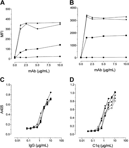
To further assess the potent complement-activating capacity of the human mAbs, we measured their ability to fix complement components to cells. Figure 4A shows that the binding of C1q, measured using FITC-labeled anti-C1q, closely paralleled lytic activity in that the type I human mAb bound more C1q than rituximab. Thus C1q binding was in the order 7D8 = 2F2 > rituximab > > 11B8, with 11B8 mAb giving no detectable C1q binding. The hierarchy of activity was equally pronounced for the deposition of C4c (Figure 4B).
Complement deposition by CD20 mAbs. (A-B) To detect C1q and C4c binding to CD20 mAbs coated on the cell surface, DOHH cells were incubated with CD20 mAbs at 10 μg/mL for 15 minutes at room temperature, followed by the addition of NHS (1% vol/vol) as a source of complement and incubation at 37°C for 10 minutes. Deposition of complement components was assessed by flow cytometry after incubation of the cells with FITC-labeled antibody against C1q (A) or C4c (B). (C-D) To assess C1q binding to CD20 mAbs coated onto ELISA plates, plates were coated with CD20 or control IgG1 mAb. Human C1q (2 μg/mL) was added to IgG-coated wells and detected with a rabbit antihuman C1q antibody followed by a peroxidase-conjugated anti-rabbit IgG-Fc antibody (D). To confirm that similar levels of CD20 mAbs were bound to the plates, they were then washed, and bound IgG was detected with an alkaline-phosphatase-conjugated goat anti-human IgG (C). Symbols: KLH (control), ○; rituximab, ▪; 7D8, ▾ ; 2F2, ▴; and 11B8, •. Data show OD values in the ELISA systems and represent 1 of 3 separate experiments yielding similar results.
Complement deposition by CD20 mAbs. (A-B) To detect C1q and C4c binding to CD20 mAbs coated on the cell surface, DOHH cells were incubated with CD20 mAbs at 10 μg/mL for 15 minutes at room temperature, followed by the addition of NHS (1% vol/vol) as a source of complement and incubation at 37°C for 10 minutes. Deposition of complement components was assessed by flow cytometry after incubation of the cells with FITC-labeled antibody against C1q (A) or C4c (B). (C-D) To assess C1q binding to CD20 mAbs coated onto ELISA plates, plates were coated with CD20 or control IgG1 mAb. Human C1q (2 μg/mL) was added to IgG-coated wells and detected with a rabbit antihuman C1q antibody followed by a peroxidase-conjugated anti-rabbit IgG-Fc antibody (D). To confirm that similar levels of CD20 mAbs were bound to the plates, they were then washed, and bound IgG was detected with an alkaline-phosphatase-conjugated goat anti-human IgG (C). Symbols: KLH (control), ○; rituximab, ▪; 7D8, ▾ ; 2F2, ▴; and 11B8, •. Data show OD values in the ELISA systems and represent 1 of 3 separate experiments yielding similar results.
As the differences between the mAbs in CDC may reflect small differences in Fc structure, we analyzed complement fixation in a system independent of CD20 binding. Human CD20 mAbs were first bound to ELISA plates before being incubated with purified C1q, and then the level of C1q binding was detected (Figure 4D). To confirm that the CD20 mAb IgG all bound at a similar level, the plates were washed and the binding of anti-human IgG was detected (Figure 4C). The results clearly show that the CD20 mAbs bound equally well to the plates and were all, including 11B8, equally efficient at binding C1q. Thus, they appeared to have equal intrinsic capacity to bind C1q and potentially activate complement.
Binding properties of CD20 mAbs on B-cell lines
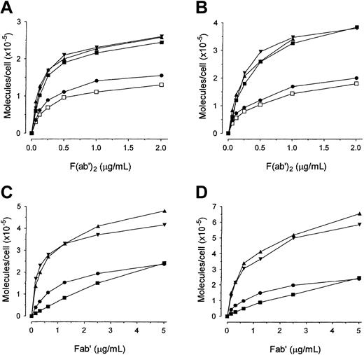
Given that 2F2 and 7D8 were very similar to rituximab in their ability to move CD20 into the Tx100-insoluble fraction (Figure 1), and yet were more potent in recruiting complement, we next addressed whether they had similar binding properties. Using conventional binding assays with radiolabeled IgG and F(ab′)2, we found that 2F2, 7D8, and rituximab gave almost identical binding curves to each other on both cell lines tested and with both IgG (not shown) and F(ab′)2 (Figure 5, upper panels). Furthermore, attempts to demonstrate differences in rates of association showed that all IgG mAbs had very similar on-rates, which reached maximal levels of binding in fewer than 15 minutes at both room temperature and 37°C (data not shown). Therefore, these results did not explain the unusual ability of 2F2 or 7D8 to recruit complement. These binding studies also confirmed that 11B8 was a type II mAb in that it, like B1, bound at approximately half the level seen with the type I reagents. This 2:1 ratio has been noted in our previous work21 and is currently undergoing further investigation.
Binding of radiolabeled CD20 mAb F(ab′)2 and Fab′ fragments to Daudi and EHRB cells.125I-labeled F(ab′) 2 (A-B) or Fab′ fragments (C-D) of CD20 mAbs were incubated with EHRB (A,C) or Daudi (B,D) cells for 2 hours at 37°C. The cell-bound and free 125I-labeled mAbs were then separated by centrifugation through phthalate oils, and the cell pellets together with bound antibody were counted for radioactivity. Symbols: rituximab, ▪; 7D8, ▴; 2F2, ▾ ; 11B8, •; and B1, □. The results show 1 of at least 2 separate experiments with similar results.
Binding of radiolabeled CD20 mAb F(ab′)2 and Fab′ fragments to Daudi and EHRB cells.125I-labeled F(ab′) 2 (A-B) or Fab′ fragments (C-D) of CD20 mAbs were incubated with EHRB (A,C) or Daudi (B,D) cells for 2 hours at 37°C. The cell-bound and free 125I-labeled mAbs were then separated by centrifugation through phthalate oils, and the cell pellets together with bound antibody were counted for radioactivity. Symbols: rituximab, ▪; 7D8, ▴; 2F2, ▾ ; 11B8, •; and B1, □. The results show 1 of at least 2 separate experiments with similar results.
We next investigated the binding activity of these mAbs using univalent Fab′ fragments. In contrast to the bivalent molecules, the Fab′ binding curves revealed a distinct difference between rituximab and the human mAb. Figure 5 (lower panels) shows that on both cell lines the human mAbs gave binding curves that appeared to saturate at the expected level, indicating the number of CD20 molecules on each cell to be more than 400 000 with EHRB (Ramos) and more than 600 000 with Daudi. Again these curves, like those of the IgG and F(ab′)2 fragments, showed approximately a 2:1 ratio of binding between types I and II CD20 mAbs. However, in contrast, the 125I-Fab′ of rituximab gave a very low level of binding to both lines, with “flatter,” nonsaturating curves, indicative of low-affinity binding and that rituximab has a weaker binding interaction with CD20 than the new human mAbs.
Ab off-rate and CDC
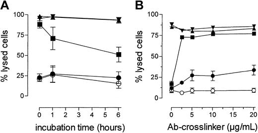
To investigate whether the difference in Ab binding activity could account for the observed differences in CDC, we next developed a CDC assay to investigate mAb dissociation. Cells were coated with CD20 mAbs, washed, and then placed in culture with CD20 mAb-free medium. At various times, samples were taken, serum was added, and lysis was assessed after incubation at 37°C. Daudi cells were selected as suitable targets due to their high sensitivity to CDC (Figure 3). This assay showed a relatively rapid disappearance of CDC mediated by rituximab compared with 2F2 and 7D8 (Figure 6A). Thus by 6 hours the CDC mediated by rituximab had dropped to around 50%, while the 2 human type I mAbs were still giving more than 90% CDC. Flow cytometry confirmed that the rituximab-treated cells still expressed unchanged levels of surface CD20.41 Similar results were obtained with other sensitive lines such as SU-DHL4 (not shown).
CDC activity over time.(A) Loss of CDC activity over time. Daudi cells were incubated with CD20 mAbs (10 μg/mL) for 15 minutes at room temperature to allow binding to cells. Cells were then washed to remove unbound CD20 mAbs, resuspended in 100 μL RPMI/10% FCS, and then incubated at 37°C. At various time points aliquots were taken, NHS (20% vol/vol) was added, and the cells were incubated at 37°C for 45 minutes. Cell lysis was assessed by flow cytometry using a PI exclusion assay, and the level of CDC expressed as PI-positive cells as percentage of total cells. Symbols: rituximab, ▪; 7D8, ▴; 2F2, ▾ ; 11B8, •, and KLH (control), ○. The results show mean ± SEM for 3 separate experiments. (B) CDC by CD20 mAbs following cross-linking. DOHH cells were incubated with CD20 mAbs (10 μg/mL) for 15 minutes at room temperature to allow binding. After washing away unbound antibody, various concentrations of sheep antihuman-κ F(ab′)2 fragments (20, 10, 5, and 0 μg/mL) were added as cross-linker, followed by the addition of normal human serum (NHS, 20% vol/vol) as a source of complement. Cell lysis was assessed by flow cytometry using a PI exclusion assay. The level of CDC is expressed as PI-positive cells as percentage of total cells. Groups: rituximab, ▪; 7D8, ▴; 2F2, ▾ ; 11B8, •; and KLH (control), ○. The figure shows the mean (± SEM) of 3 separate experiments.
CDC activity over time.(A) Loss of CDC activity over time. Daudi cells were incubated with CD20 mAbs (10 μg/mL) for 15 minutes at room temperature to allow binding to cells. Cells were then washed to remove unbound CD20 mAbs, resuspended in 100 μL RPMI/10% FCS, and then incubated at 37°C. At various time points aliquots were taken, NHS (20% vol/vol) was added, and the cells were incubated at 37°C for 45 minutes. Cell lysis was assessed by flow cytometry using a PI exclusion assay, and the level of CDC expressed as PI-positive cells as percentage of total cells. Symbols: rituximab, ▪; 7D8, ▴; 2F2, ▾ ; 11B8, •, and KLH (control), ○. The results show mean ± SEM for 3 separate experiments. (B) CDC by CD20 mAbs following cross-linking. DOHH cells were incubated with CD20 mAbs (10 μg/mL) for 15 minutes at room temperature to allow binding. After washing away unbound antibody, various concentrations of sheep antihuman-κ F(ab′)2 fragments (20, 10, 5, and 0 μg/mL) were added as cross-linker, followed by the addition of normal human serum (NHS, 20% vol/vol) as a source of complement. Cell lysis was assessed by flow cytometry using a PI exclusion assay. The level of CDC is expressed as PI-positive cells as percentage of total cells. Groups: rituximab, ▪; 7D8, ▴; 2F2, ▾ ; 11B8, •; and KLH (control), ○. The figure shows the mean (± SEM) of 3 separate experiments.
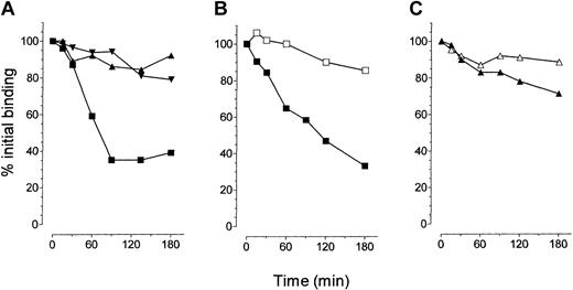
To confirm that loss of CDC might result from Ab dissociation, we performed conventional binding “off-rate” experiments using radiolabeled IgG. Figure 7A shows that compared with 2F2 and 7D8, rituximab had a markedly faster off-rate. Thus, when DOHH cells were coated with 125I-labeled mAb, approximately 30% of the rituximab, and more than 70% of 2F2 and 7D8, remained bound to the cells after 3 hours. In all cases, the culture medium contained 1 mg/mL cold IgG as a competitor to prevent rebinding of the radiolabeled Ab. As expected, similar results were achieved with F(ab′)2 (not shown) excluding an interaction with FcγR on target cells influencing mAb dissociation.
Effect of crosslinking on dissociation of radiolabeled CD20 mAb.Comparison of the dissociation of radiolabeled CD20 mAb IgG (A) and the effect of cross-linking on dissociation (B-C). (A) 125I-labeled CD20 mAbs (2 μg/mL) were added to DOHH cells, incubated for 2 hours at room temperature, and then pelleted and resuspended in 200 μL medium containing 1 mg/mL unlabeled mAbs. Cells were then cultured at room temperature and at the times indicated aliquots were taken to determine the level of cell-bound mAbs over time. Counts remaining at each time point were expressed as a percentage of the initial count. Symbols: rituximab, ▪; 7D8, ▴; and 2F2, ▾. This figure shows 1 of 3 experiments that gave similar results. (B-C) To determine the effect of cross-linking on mAb dissociation, DOHH cells were incubated with labeled rituximab (B) or 2F2 (C) as described in “Materials and methods,” pelleted, and the supernatant was removed. Cells were then resuspended and treated with sheep antihuman κ F(ab′)2 fragment (open symbols) or not (filled symbols) for 30 minutes on ice. Excess mouse CD20 mAb, 1F5 (1 mg/mL), was added to act as a cold competitor that would not compete for binding to the sheep cross-linking Ab. At the times shown, aliquots of cells were taken and bound radiation was determined as for panel A. These results are representative of 2 experiments each showing the same outcome.
Effect of crosslinking on dissociation of radiolabeled CD20 mAb.Comparison of the dissociation of radiolabeled CD20 mAb IgG (A) and the effect of cross-linking on dissociation (B-C). (A) 125I-labeled CD20 mAbs (2 μg/mL) were added to DOHH cells, incubated for 2 hours at room temperature, and then pelleted and resuspended in 200 μL medium containing 1 mg/mL unlabeled mAbs. Cells were then cultured at room temperature and at the times indicated aliquots were taken to determine the level of cell-bound mAbs over time. Counts remaining at each time point were expressed as a percentage of the initial count. Symbols: rituximab, ▪; 7D8, ▴; and 2F2, ▾. This figure shows 1 of 3 experiments that gave similar results. (B-C) To determine the effect of cross-linking on mAb dissociation, DOHH cells were incubated with labeled rituximab (B) or 2F2 (C) as described in “Materials and methods,” pelleted, and the supernatant was removed. Cells were then resuspended and treated with sheep antihuman κ F(ab′)2 fragment (open symbols) or not (filled symbols) for 30 minutes on ice. Excess mouse CD20 mAb, 1F5 (1 mg/mL), was added to act as a cold competitor that would not compete for binding to the sheep cross-linking Ab. At the times shown, aliquots of cells were taken and bound radiation was determined as for panel A. These results are representative of 2 experiments each showing the same outcome.
Subsequently, we investigated the dissociation of these mAbs in the presence of F(ab′)2 fragments from a sheep antihuman κ Ab, the rationale being to assess how effectively a “second-layer” of cross-linking Ab (bound to the κ chain of the CD20 mAb) could prevent dissociation. If successful, this approach would allow us to determine the effect of manipulating the rituximab off-rate in relation to CDC efficacy. DOHH cells were chosen for these experiments due to their relative resistance to CDC (Figure 6B) and their λ expression, which ensured that the cross-linking F(ab′)2 anti-κ Ab bound only to the κ + CD20 mAb. Figure 7B shows that adding the cross-linking F(ab′)2 arrested the loss of radiolabeled rituximab from the cell surface resulting in more than 80% retention after 3 hours (Figure 7C). Thus, cross-linking with this polyclonal F(ab′)2 reagent provided a very efficient means of maintaining the primary Ab at the cell surface, greatly reducing off-rate. It is important to note that because excess CD20 mAb had been removed from the supernatant before adding the sheep anti-κ Ab, cross-linking prevented only dissociation without capturing additional mAb from the supernatant.
Having established that the cross-linking agent could retain rituximab at the cell surface, we repeated the CDC assay against DOHH cells to address whether tethering it to the cell surface would augment its lytic activity. Figure 6B shows quite clearly that adding the F(ab′)2 anti-κ cross-linker increased the CDC activity of rituximab to that of the 2F2 and 7D8 mAbs. These results confirmed that the lower CDC activity of rituximab resulted from its relatively rapid off-rate. Interestingly, the lack of CDC activity of the 11B8 mAb could not be fully restored by cross-linking and only increased by about 20%.
Discussion
There is growing support for an important role for complement in rituximab immunotherapy, either through direct lysis of tumor cells or though its ability to activate effectors and opsonize targets.14,16-19 Here we describe 3 novel human mAbs directed at CD20 that offer the potential of improved therapeutic activity, especially via the classical complement pathway. Of these reagents, 2F2 and 7D8 were classified as type I CD20 mAbs as they were capable of moving CD20 into the Tx100-insoluble fraction of the plasma membrane. Perhaps surprisingly, they were able to lyse B-cell lines and fresh tumors that were resistant to rituximab, despite having the same human γ1 heavy-chain constant regions. This was most evident when comparing the lysis of Raji cells, which express modest levels of CD20 and high levels of CD55 and CD59 (Figure 3). Similarly, we also found that unlike rituximab, 2F2 and 7D8 could mediate lysis of fresh B-CLL cells either with unfractionated blood or plasma (Figure 2C and D), thus confirming their ability to engage complement efficiently. When testing these mAbs for C1q binding in a system that is independent of CD20 binding (mAbs bound to plastic), all exhibited similar activity (Figure 4A-B). This virtually rules out that the differences in CDC were due to minor variations in amino acid sequence of the constant domains.
One possible explanation for the potent CDC of CD20 mAbs relates to the nature of the CD20 target. It has been suggested that effective CDC is critically dependent on the distance between the plasma membrane and the constant parts of the sensitizing antibody.29,42,43 This may help explain why mAbs directed at membrane-proximal targets, such as CD20 and CD52, are highly active in C recruitment, while those binding to equally expressed but more extended molecules, such as CD45, are often not.44 Thus, the closer to a cell membrane a mAb binds, the more efficiently it coats the target cell with active complement components for lysis. Xia et al29,43 pointed out that a range of small glycolipids, such as CD24 and CD52, have the property of being “good” targets for Ab-mediated CDC. These workers also suggest that because the labile thioester bond that is exposed upon activation of C4 or C3 has a short half-life, CDC may require that complement activation occurs proximal to the membrane. Interestingly, because the half-life of the nascent thioester bond in C4b is only 25 to 30 μs,45 it will survive on average long enough to diffuse only 6 nm from its site of generation. This is rather less than the length of an Ab Fab arm.42,43 Perhaps the most persuasive evidence to support these suggestions comes from class-switch variants of alemtuzumab (anti-CD52), showing that while an IgG3 isoform binds C1q more efficiently than IgG1, it is less active in lysis.42 Furthermore, when the long hinge of IgG3 was mutated to the shorter length of that in IgG1, lysis efficiency was improved up to 10 times.46 Collectively, such work makes a strong case for membrane proximal activation of complement delivering more membrane damage.
Almost all CD20 mAbs raised to date appear to bind to the same 44-amino acid extracellular loop, causing substantial cross blocking, indicating that the binding sites are not sufficiently different to influence the CDC efficacy of the mAbs. Indeed, Polyak and Deans47 have shown that the AxP motif at position 170-172 in the extracellular loop is by far the dominant epitope. One property that can substantially affect the ability of a mAb to induce potent CDC is whether it is able to redistribute the target antigen into Tx100-insoluble areas of the plasma membrane. This is perhaps most clearly demonstrated for the B1 and 11B8 mAbs that bind to CD20, have good complement activating isotypes, but do not redistribute into lipid rafts and show very little efficacy in CDC. However, differences in redistribution do not explain the superior lytic activity of 2F2 and 7D8 over rituximab, as these mAbs all show equivalent performance in the Tx100 insolubility assay.
The most apparent difference between the new human mAbs and rituximab is the speed at which they dissociate from the cells. 2F2 and 7D8 have much slower off-rates than rituximab such that over 3 hours, the dissociation for the human mAbs is only 20% to 30%, yet is 70% to 80% for rituximab. Similarly, when dissociation was measured using a CDC assay, lysis mediated via rituximab was poor after washing and incubation for 6 hours, while 2F2 and 7D8 retained full activity. Interestingly, when an F(ab′)2 cross-linking anti-κ polyclonal reagent was added, the CDC activity of rituximab was comparable with that of 7D8 and 2F2.
Given this striking observation regarding off-rate and CDC, next we must consider why off-rate influences CDC so profoundly. One likely possibility is that the mAb off-rate influences the amount of C1q bound at the cell surface and the locality at which complement is activated (Figure 4). For example, a mAb that dissociates rapidly may not allow full engagement of C1q, even when the Ab is present at saturating concentrations. Although the off-rates we have measured are relatively slow and could not directly account for the marked differences in C1q binding and CDC activity, it is likely that these off-rates instead reflect binding stability. As such, while the complete dissociation of a bivalent mAb such as rituximab might be a relatively slow event, requiring the release of both Fab arms, it may continually engage and disengage individual Fab arms without being lost from the cell. The instability of rituximab binding relative to the human CD20 mAbs is most clearly shown by the inability of its Fab′ fragments to give saturation of binding curves (Figure 5). So how might the stability of binding influence binding and activation of C1? The C1q component of C1 is a hexamer containing 6 globular heads, which provide IgG binding sites, attached to 6 collagen-like tails. Activation of C1 requires fixation of the globular heads of C1q at an angle favoring autoactivation of the C1rC1s tetramer.48 Therefore, perhaps when Ab is less firmly attached and is resonating between univalent and bivalent binding, it is less able to capture C1 via multiple globular heads, and hence cannot hold it long enough in a conformation required for autoactivation.
If C1 does become activated at the cell surface, but theAb to which it is bound disengages, then this could favor fluid phase activation and effectively take the activated C1 complex away from the cell surface, which enhances susceptibility for inactivation by C1-inhibitor.49 In addition, not only C1 but subsequent stages in the complement cascade could also be influenced by the longevity of Ab binding at the cell surface. For example, the ability of complement components (C4 and C3) to covalently fix to the activating mAb and generate C3- and C5-convertases can influence the membrane stages of complement activation. Interestingly, complement activation itself may influence antigen binding, a phenomenon known as solubilization,50 and we may speculate that those Abs that tend toward dissociation may be more easily dissociated upon complement activation. If fixing complement components to the mAb itself is beneficial to lysis, then dissociation will effectively halt, or at least severely limit, activation of complement at the membrane (C5 and beyond). Hence there are numerous potential mechanisms that would allow Ab off-rates to influence lysis. We are currently undertaking further investigations to determine which of these is active with CD20 mAbs.
In conclusion, the new CD20 mAbs presented here offer exciting reagents for studying the critical in vivo mechanisms involved with CD20 mAb immunotherapy. In addition, their extremely slow off-rates will provide advantages for their use, not only as “naked” Abs where they should function at lower concentrations than currently used reagents, but also as radioconjugates where their lack of dissociation will ensure that a high proportion remains bound to the target cells. Finally, as the complete protein structure of these compounds is of human origin, they are likely to be the least immunogenic form of mAbs currently available. It now remains to be seen how these interesting and potent reagents perform in the clinic.
Prepublished online as Blood First Edition Paper, June 1, 2004; DOI 10.1182/blood-2004-01-0039.
Supported by Genmab, the Biotechnology and Biosciences Research Council (BBSRC), Tenovus of Cardiff, the Cancer Research United Kingdom, and the Leukaemia Research Fund.
Several of the authors (J.L.T, J. van den B., M.P, H.H., P.W.H.I.P, J.G.J. van de W., and M.J.G.) have declared a financial interest in Genmab and Medarex, whose products were studied in the present work.
J.L.T. and R.R.F. contributed equally to this study.
An Inside Blood analysis of this article appears in the front of this issue.
The publication costs of this article were defrayed in part by page charge payment. Therefore, and solely to indicate this fact, this article is hereby marked “advertisement” in accordance with 18 U.S.C. section 1734.
We wish to express our gratitude to all members of the Genmab and Tenovus laboratories who provided expert technical support and valuable discussion. We are also particularly indebted to Jeffrey Beekman, Peter Johnson, Ron Taylor, Mike Clark, and Geoff Hale for helpful discussion.

![Figure 1. Comparison of the ability of CD20 mAbs to redistribute antigens on the cell surface. (A-B) Translocation of CD20 into the Tx100-insoluble fraction of the cell. Daudi (A) or DOHH (B) cells were incubated with FITC-labeled CD20 mAb (10 μg/mL) for 15 minutes at 37°C and washed. After chilling on ice, half of each sample was treated with 0.5% Tx100 for 15 minutes on ice. Both samples (lysed, dark gray bars; and not lysed, light gray bars) were washed and analyzed by flow cytometry (mean fluorescence intensity [MFI]) to assess bound FITC-CD20 mAbs. (C) FRET determination: Daudi cells were labeled with an equimolar mixture of Cy3 (donor)- and Cy5 (acceptor)-conjugated CD20 mAbs at 37°C with or without C8-deficient (C8-def) serum. FRET, which measures the proximity of neighboring mAb molecules, was assessed by flow cytometric analysis. These graphs are representative of at least 3 experiments, each showing similar results.](https://ash.silverchair-cdn.com/ash/content_public/journal/blood/104/6/10.1182_blood-2004-01-0039/6/m_zh80180466650001.jpeg?Expires=1767712754&Signature=Jwkxi0i520nCKr5tWX1Atta2DtjF9v7RoOaB0iESYKYUX1XbyEV2GatlA1-PpTsN68PcLajUxUvSMakSDKTg29YP4ggbNiykvYgfOo4wYdcE6y9KATSVkIzdcVj7shdJxo8iPu9k~4fzEv1JKA1Cy5O6C59BZV6qrQ7GEk9BGoa86vuuUge-p2zweLqtZonnJeBSoNbBcWxBdAI2AGBdfXGxtW20ly2Z8~8efncC-~rAnCjNKv6J1L7ibHLA11ttDIbyo3yVCnWznsQwu0EDwTjTfkO8E2RdjfJB25EE8NHCwMoB6clhUqbxLHjpQryoYqhq3vJLYOdKrVhiXJrFyA__&Key-Pair-Id=APKAIE5G5CRDK6RD3PGA)


This feature is available to Subscribers Only
Sign In or Create an Account Close Modal