The mechanisms that dictate the evolution of tumor heterogeneity are not well understood, but the findings of 2 articles suggest a dynamic interplay between subpopulations of multiple myeloma cells, both with each other and with the tumor microenvironment.
There are 2 articles published in this issue of Blood that describe approaches and techniques (Kumar and colleagues [Figure 1] and Maxwell and colleagues [Figure 2C]) that address distinct mechanisms important (among many others) in the pathobiology of multiple myeloma (MM), enhanced angiogenesis in bone marrow, and altered signaling from the microenvironment. The investigators use an in vitro human angiogenesis model or single-cell reverse transcriptase–polymerase chain reaction (RT-PCR) that detects attograms of amplified product and make contrasting conclusions about the roles of population dynamics versus clonal overgrowth in determining malignant potential of MM. Both studies are timely, since the concept that malignant human tumors arise primarily from clonal expansion of dominant mutant cells is increasingly challenged. Certain leukemias1 and breast tumors2 contain subpopulations of poorly proliferative but plastic progenitor cells that are responsible for the majority of tumor-initiating activity. These cells are thought to serve as “tumor-initiating stem cells” that give rise to aggressive tumors. It has been shown clearly that the microenvironment can be dominant over mutations in determining tumorigenic potential, emphasizing the importance of epigenetic influences in tumor progression.3,4
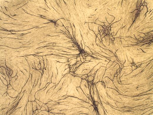
Increased bone marrow microvessel density in MM patients is an important prognostic factor. Using a unique human in vitro angiogenesis assay, Kumar and colleagues have identified inhibitory factor(s) in normal and monoclonal gammopathy of unknown significance (MGUS) bone marrow that cannot be overcome by the addition of the strongly proangiogenic vascular endothelial growth factor (VEGF). The authors have also confirmed a correlation between expression of angiogenic stimulatory factors and disease severity in myeloma patients, but the percentage of plasma cells expressing angiogenic factors and their receptors was not significantly different between early and late stages of the disease. Several important conclusions were made from this study. Enhanced production of angiogenic factors such as VEGF and basic fibroblast growth factor by “aggressive” plasma cell clones is likely not a dominant factor in induction of angiogenesis. Rather, a balance between inhibitory bone marrow factors versus stimulatory plasma cell factors from the total tumor population determines the extent of progression-associated bone marrow angiogenesis. This study clearly illustrates an example where the interplay between specific microenvironmental factors and tumor cells alters the course of the disease.FIG1
Human in vitro angiogenesis assay. See the complete figure in the article beginning on page 1159.
Human in vitro angiogenesis assay. See the complete figure in the article beginning on page 1159.
By contrast, Maxwell and colleagues conclude that population dynamics may be a major contributing factor to disease progression. In their article, they demonstrate that elevated levels of the hyaluronan-binding protein receptor for hyaluronan-mediated motility (RHAMM) correlate with progression of MM. Elevated RHAMM levels were associated with osteolytic bone lesions independently of other implicated factors (eg, dickkopf1 [DKK]5 ). Several isoforms of RHAMM are expressed in MM, particularly a full-length (FL) and an alternatively spliced form lacking exon 4 (exon4del). Using single-cell RT-PCR, the authors find that RHAMMFL was rarely expressed in the same plasma cell as RHAMM (exon4del). Analysis of the ratio of the exon4del variant/full-length forms indicated a positive correlation of this ratio with disease severity. One conclusion made from this study was that clones expressing the exon4del variant of RHAMM have a growth advantage, leading to gradual overgrowth of this subpopulation in patients with advanced disease.
Further studies addressing these complex interactions may lead to important new insights into the way in which tumors are formed (eg, via tumor-initiating stem cells) versus how they progress to form malignant disease (eg, interplay between tumor subpopulations and the microenvironment). The studies have important implications for understanding why current therapies fail, and studies such as these will hopefully lead to new strategies for the improved management of patients with malignant tumors such as multiple myelomas.


This feature is available to Subscribers Only
Sign In or Create an Account Close Modal