Abstract
Idiopathic CD4 lymphocytopenia (ICL) is characterized by reduced CD4+ T cell counts in the absence of HIV infection or other defined causes. Patients with ICL generally have CD4+ T cell counts < 100/μL and are at risk for infections. Administration of IL-2 has been reported to reduce the risk of infections in some patients, but CD4+ T cell counts remain low. Currently, there is no available therapy to increase the number of CD4+ T cells in these patients.
We have developed the Xcellerate™ Technology, in which T cells are activated and expanded ex vivo from peripheral blood mononuclear cells (PBMC) using microscopic paramagnetic beads conjugated with anti-CD3 and anti-CD28 monoclonal antibodies (Xcyte™Dynabeads®). In patients with low CD4 counts due to HIV or cancer chemotherapy, administration of T cells activated and expanded using the Xcellerate Technology or similar process leads to significant increases in CD4+ T cell counts to normal levels that are sustained over several months.
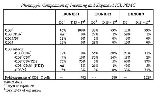
In the current study, we tested the ability of the Xcellerate Technology to expand T cells from patients with ICL. We collected data on T cell phenotype, cell expansion, activation marker expression, cytokine secretion, T cell receptor (TCR) repertoire diversity, functional potential and response to restimulation. T cells expanded a median of 902 fold (range 281–1529; n=3 patients), and demonstrated typical induction of surface-activation marker expression, including upregulation of CD25 (IL-2R) and CD154 (CD40L). Phenotypic compositions of the ICL donor PBMC were heterogenous both before and after T cell expansion. Two patients had very high levels of CD4−CD8− T cells, and one of these two patients also had very high levels of γδ+ T cells. Each of these populations was maintained throughout expansion. The other patient displayed high levels of circulating CD3+CD56+ NKT cells that did not proliferate as robustly as other T cells during the expansion process. For two patients, flow cytometric analysis revealed a normal pattern of TCR Vβ surface expression while the remaining tissue had a skewed TCR Vβ distribution pattern. Following initial expansion and subsequent restimulation, T cells rapidly re-expressed surface activation markers and cytokines. In these preliminary studies, T cells from patients with ICL were expanded successfully using the Xcellerate Technology, suggesting that this approach might be used for clinical application in this disease.
Author notes
Corresponding author


This feature is available to Subscribers Only
Sign In or Create an Account Close Modal