Abstract
Adult bone marrow contains a population of hematopoietic stem cells (HSCs) that can give rise to cells capable of targeting sites of neovascularization in the peripheral or retinal vasculature. However, relatively little is known about the mechanism of targeting of these cells to sites of neovascularization. We have analyzed subpopulations of HSCs for the expression of a variety of cell surface adhesion molecules and found that R-cadherin, a calcium-dependent cell-cell adhesion molecule important for normal retinal endothelial cell guidance, was preferentially expressed by functionally targeting HSCs. Preincubation of HSCs with function-blocking anti-R-cadherin antibodies or novel R-cadherin-specific peptide antagonists effectively prevented targeting of bone marrow-derived cells to the developing retinal vasculature in vivo. Whereas control-injected HSCs targeted to all 3 normal developing retinal vascular layers, blocking R-cadherin-mediated adhesion resulted in mistargeting of the HSCs to the normally avascular outer retina. Our results suggest that vascular targeting of bone marrow-derived HSCs is dependent on mechanisms similar to those used by endogenous retinal vascular endothelial cells. Thus, R-cadherin antagonists may be useful in the treatment of neovascular diseases in which circulating HSCs contribute to abnormal angiogenesis. (Blood. 2004;103:3420-3427)
Introduction
Normal adult bone marrow contains a population of hematopoietic stem cells (HSCs) that can be divided into lineage positive (Lin+) and lineage negative (Lin-) subpopulations, depending on whether the cells differentiate into formed elements of the blood (Lin+) or not (Lin-). In addition, it has been shown that cells capable of forming blood vessels in vitro and in vivo are contained within the Lin- subpopulation.1,2 These vascular targeting cells can be mobilized from the bone marrow into the circulation, where they target sites of ischemia in the peripheral3 or central4 circulation. More recently it has also been demonstrated that these cells, when injected systemically, will participate in hypoxia-induced retinal neovascularization.5 We have demonstrated that Lin- HSCs can target and stabilize degenerating retinal vasculature after intraocular injection in a model of retinal degeneration.2 While it is becoming increasingly clear that these adult bone marrow-derived progenitor cells may prove useful in treating a variety of vascular disorders, the mechanism by which they target to sites of ischemia or vascular injury remains poorly understood.
The retina consists of well-defined and highly organized layers of neuronal, glial, and vascular elements, and disruption of this ordered array can lead to neuronal degeneration and significant visual loss. Development of the mouse retinal vasculature occurs during the first 3 postnatal weeks in a manner similar to that observed in human fetuses during the last trimester of gestation.6-8 During retinal vascular development, endothelial cells migrate from the optic nerve head toward the retinal periphery, forming a network of interconnecting superficial retinal vessels during the first week of life in the rodent. Subsequently, between postnatal day 7 (P7) and P12, these vessels branch laterally, extending endothelial cells into the retina, where they migrate to the outer edge of the inner nuclear layer, forming a vascular plexus parallel to the previously formed superficial layer. Finally, between P14 and P21 a third, intermediate vascular plexus is formed, followed by vascular remodeling and maturation of the retinal vasculature. The superficial, or primary, network of retinal vessels is formed as endothelial cells migrating from the optic nerve head target a preexisting astrocytic template spread over the surface of the retina. This model not only has been useful in the understanding of mechanisms of developmental angiogenesis,8,9 but also has applications to the study of pathological neovascularization.10-12
Using specific, function-blocking antibodies, we found that R-cadherin adhesion plays a role in developmental retinal vascularization.8 Disruption of R-cadherin adhesion during formation of the superficial vascular plexus results in a loss of the complex vascular interconnections observed during normal vascular patterning. When R-cadherin adhesion is blocked during the subsequent formation of deep vascular layers, key guidance cues are lost, causing the vessels to migrate past the normal deep vascular plexuses into the normally avascular photoreceptor layer.8
The use of, and targeting to, a preexisting retinal astrocytic template is specific for endogenous retinal vascular endothelial cells; nonretinal endothelial cells do not target, but we found that intravitreally injected Lin- HSCs also will target astrocytes in the superficial retinal vascular layer ahead of the newly forming superficial vascular layer.2 As the endogenous vasculature reaches the periphery containing the astrocyte-targeted HSCs, Lin- HSCs are incorporated into the newly forming vessels forming functional mosaic vessels with mixed populations of injected HSCs and endogenous endothelial cells. Lin- HSCs also can target to the deep and intermediate vascular plexuses before normal vascularization of these layers. Incorporation of these cells rescues the deep vasculature of rd/rd mice when compared to untreated or control Lin+ HSC-injected mice.2
The targeting of Lin- HSCs to astrocytes and deep vascular regions prior to endogenous developmental vascularization suggests that these cells express cell-surface adhesion molecules that serve a function similar to those involved in the targeting of the endogenous endothelial cells. Since R-cadherin adhesion is required for targeting of endogenous developing endothelial cells to astrocytes as well as the correct deep vascular plexuses during normal developmental retinal angiogenesis, we tested the role of cadherin-mediated adhesion for targeting of Lin- HSCs as well. Here we demonstrate, using both function-blocking anti-R-cadherin antibodies and novel peptide antagonists against specific cadherin family members, that R-cadherin adhesion also is required for targeting of bone marrow-derived HSCs to the developing retinal vasculature.
Materials and methods
Hematopoietic stem cell isolation
Hematopoietic stem cells (HSCs) were isolated and enriched for a subpopulation of vascular targeting cells as described.2 Briefly, bone marrow cells were isolated from ACTbEGFP transgenic mice (The Jackson Laboratory, Bar Harbor, ME), where a green fluorescent protein (GFP) marker was fused to the actin promoter, causing all cells to express GFP. Mononuclear cells were collected by density gradient separation using Fico/Lite-LM (Atlanta Biologicals, Norcross, GA), and these cells were labeled with biotin-conjugated lineage panel antibodies (CD45, CD3, Ly-6G, Mac1, and TER-119, Pharmingen, San Diego, CA). Using these markers, cells that had already begun to differentiate into formed cells of the peripheral blood (Lin+ HSCs) were separated using a magnetic separation column (MACS, Miltenyi Biotech, Auburn, CA), leaving the Lin- cells to be collected by the flow-through. CD31-, CD34-, and Mac1- cells also were isolated from mononuclear cells and used as a negative control for the functional Lin- HSCs.
Various subpopulations of HSCs were analyzed for R-cadherin expression using anti-R-cadherin antibodies (sc-6456, Santa Cruz Biotechnology, Santa Cruz, CA), fluorescently labeled donkey anti-goat secondaries (Molecular Probes, Eugene, OR), and a FACSCalibur flow cytometer (Becton Dickinson, Franklin Lakes, NJ). Preimmune goat IgGs were used to establish a background baseline for flow cytometry analysis.
Peptide design and synthesis
Peptides were designed based on structural,13 biochemical,14 and similar peptide mimetic studies of N-cadherin-mediated neurite outgrowth.15,16 All peptides were synthesized using the solid phase synthesis method and were purified to the highest grade possible (> 95% pure) as analyzed by high-performance liquid chromatography (HPLC) analysis. The sequences were analyzed by mass spectrometry to ensure correct synthesis. All peptides were amide blocked at the amino terminus and acetylated at the carboxy terminus. Cysteine residues were generated at the amino and carboxy terminal ends for formation of cyclic peptide rings by disulfide tethering.
L-cell transfections
The mouse R-cadherin and N-cadherin plasmids were generous gifts from Dr Masatoshi Takeichi (Kyoto University, Japan).17,18 R-cadherin or N-cadherin sequences were subcloned into pDsRed2 N1 vectors (Clontech). L-cells (mouse fibroblast L929 cells, ATCC #CRL-2148) were stably transfected with either R- or N-cadherin pDsRed2 N1 using the Calcium Phosphate Transfection System (Life Technologies, Rockville, MD) according to the manufacturer's protocol. After screening by growth in media supplemented with 700 μg/mL G418 (Geneticin, Gibco Life Technologies, Rockville, MD), positive clones were selected. Cells were examined for expression of red fluorescent protein (pDSRed2 N1) and were tested for cadherin expression by immunoblotting and immunofluorescence staining.
Aggregation assay
A conventional cell aggregation assay was used in this study14,19 with slight modifications. Briefly, cells were grown to near confluency followed by trypsinization with 0.01% trypsin + 5 mM CaCl2 and no EDTA (ethylenediaminetetraacetic acid) (TC) or 0.01% trypsin with 0.1 mM EDTA and no calcium (TE). Cells were collected and washed, followed by resuspension in Hanks buffered salt solution (HBSS) + 1% bovine serum albumin (BSA) with (TC) or without (TE) 5 mM CaCl2. Cells were incubated at 37°C in 0.5 mL solution at 2 × 10⁵ cells per well of a 24-well plate with rocking at 60-70 rpm with varying peptide concentrations. All assays were performed in triplicate. The extent of cellular aggregation was represented by the ratio of the total particle number after 2 hours of incubation (N2hr) to the initial particle number (N0) (1 particle = any isolated single cell or group of aggregated cells). Particles were counted on a hemocytometer using the sum of 8 separate 20-μL counts per well, before (N0) and after (Nt) incubation.
Intravitreal injection of peptides
Peptides were dissolved in phosphate-buffered saline (PBS) + 10% dimethyl sulfoxide (DMSO) to a concentration of 10 mM; 0.5 μL or 1.0 μL of 10 mM peptide solution or PBS/DMSO vehicle control was injected into the vitreal cavity of 2-day- or 7-day-old mice, respectively. Intravitreal injections were performed as previously described.12 Briefly, an eyelid fissure was created in P2 or P7 mice to expose the globe for injection. Injections were made between the equator and the corneal limbus; during injection the location of the needle was visualized through a dissecting microscope to ensure that it was in the vitreal cavity. After the injection, the eyelids were repositioned to close the fissure. Four days later, the retinas were dissected and the vessels were visualized by immunohistochemistry using fluorescently labeled isolectin Griffonia simplicifolia (Molecular Probes). Quantification of peripheral vascularization, vascular length, and vascular area was achieved by imaging noninjected retinas and retinas injected with various peptides under the same microscopy settings. Numbers were then generated using LaserPix software (BioRad, Hercules, CA) with noninjected control littermates used for baseline normalization of the extent of retinal vascularization. The effect on deep vascular formation was quantified by focusing posterior to the normal deep vascular plexus (within the photoreceptor layer) using confocal microscopy and by counting the numbers of vessels that had migrated into the photoreceptor layer.
HSC incubations, injections, and quantification
Lin- HSCs were incubated alone or with (1) 100 nM of R-cadherin-blocking antibody (sc-6456, Santa Cruz Biotech), (2) 100 nM preimmune goat IgG, (3) 10 mM of cyclic IDS, (4) 10 mM of cyclic INP, or (5) 10 mM of control peptides for 1 hour at 37°C prior to injection. Approximately 1 × 10⁵ HSCs in 0.5 μL were injected intravitreally into postnatal developing mouse eyes. Retinas were then examined at P12 by whole mount or analysis of retinal sections. Targeting of the HSCs was quantified by counting the total number of GFP-positive cells within the retina using 8 different fields of view per retina: left, right, top, and bottom quadrants (¾ distance to the retina periphery), 2 intermediate quadrants (¼-½ distance to the periphery), a peripheral site, and the optic nerve head region. These cells were characterized by their localization to the superficial, intermediate, or deep layers, or by the lack of targeting (cells that lie at the back of the photoreceptor layer). The number of nontargeted HSCs at the outer edge of the photoreceptor layer was given as a percentage of the total number of HSCs observed within the retina.
Results
Lin- HSCs express R-cadherin
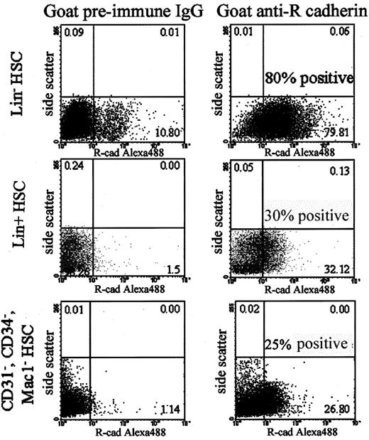
HSC expression of R-cadherin was analyzed by flow cytometry to determine if R-cadherin cell adhesion molecules were expressed at the surface of functional targeting cells. R-cadherin molecules were expressed at the surface of nearly 80% of the Lin- subpopulation, while only ∼30% of the Lin+ cells and ∼25% of CD31-/CD34-/Mac1- cells expressed R-cadherin (Figure 1). Based on the relative fluorescence intensities between the cell populations, it is likely that the Lin- HSCs also express higher concentrations of R-cadherin when compared to the smaller number of R-cadherin-positive nontargeting cell populations. Thus, most cells within the vascular targeting subpopulation express R-cadherin, while most cells from nontargeting subpopulations do not.
Expression of R-cadherin on Lin-HSCs. Flow cytometry was used to analyze cell surface expression of R-cadherin on (1) vascular targeting Lin- HSCs; (2) nontargeting Lin+ HSCs; or (3) nontargeting CD31-/CD34-/Mac1-HSCs. Ten percent of Lin- HSCs were labeled by preimmune control goat IgG, while 80% were labeled by goat anti-R-cadherin antibodies. Nontargeting subpopulations of HSCs contain significantly fewer cells positive for R-cadherin expression.
Expression of R-cadherin on Lin-HSCs. Flow cytometry was used to analyze cell surface expression of R-cadherin on (1) vascular targeting Lin- HSCs; (2) nontargeting Lin+ HSCs; or (3) nontargeting CD31-/CD34-/Mac1-HSCs. Ten percent of Lin- HSCs were labeled by preimmune control goat IgG, while 80% were labeled by goat anti-R-cadherin antibodies. Nontargeting subpopulations of HSCs contain significantly fewer cells positive for R-cadherin expression.
Blocking R-cadherin disrupts HSC targeting
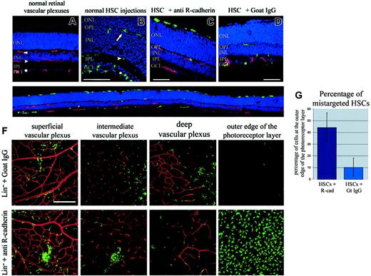
To determine if R-cadherin cell-cell adhesion functions in the targeting of HSCs to the distinct retinal vascular layers, Lin- HSCs were blocked with R-cadherin-specific blocking antibodies prior to injection. Six days after injection, normal Lin- HSCs were found within the 3 normal vascular layers: (1) the superficial vascular plexus localized within the ganglion cell layer, (2) the deep vascular plexus localized near the outer plexiform layer, and (3) the intermediate vascular plexus localized at the inner margin of the inner nuclear layer (Figure 2B). Lin- HSCs preincubated with preimmune IgG targeted identically to untreated HSCs (Figure 2D). However, many of the Lin- HSCs that were preincubated with anti-R-cadherin antibodies prior to injection no longer functionally targeted to the developing retinal vasculature (Figure 2C,E-F). Total numbers of HSCs observed in the retina were similar under both conditions, suggesting that blocking R-cadherin-mediated adhesion affects targeting but does not affect initial survival of HSCs in the retina. Targeting to the deep and intermediate vascular layers appears to be particularly affected by blocking R-cadherin adhesion, as relatively few R-cadherin-blocked HSCs were found localized within these regions. The cells localized to the superficial vascular plexus also appeared less organized and were not colocalized with the endogenous vasculature to the same extent as normal Lin- HSCs or those preincubated with preimmune IgG (Figure 2).
Blocking R-cadherin function disrupts vascular targeting of HSCs. (A) Scanning laser confocal microscopy analysis of frozen retinal cross-sections demonstrates that 3 vascular plexuses (red) are formed in the normal retina: (1) the superficial vascular plexus (star) in the GCL; (2) the intermediate vascular plexus (arrowhead) at the inner edge of the INL; and (3) the deep vascular plexus (arrow) at the outer edge of the INL. (B) Lin- HSCs from eGFP transgenic mice (green) injected intravitreally into postnatal day 6 developing mouse eyes become localized to the 3 retinal vascular plexuses. (C) Blocking R-cadherin-mediated adhesion prevented normal targeting and caused many HSCs (green) to become localized to the outer edge of the photoreceptor layer, while (D) HSCs preincubated with control preimmune IgG target normally. (E) A low-magnification image across an entire retina cross-section demonstrates the extent of HSCs mistargeting caused by blocking R-cadherin adhesion. (F) Confocal images through z-planes of the 3 normal vascular plexuses and the outer edge of the photoreceptor layer. Fewer HSCs localize to the normal vascular plexuses and many localize at the outer edge of the photoreceptors when R-cadherin adhesion is blocked. (G) The extent of mistargeting was quantified by the number of HSCs mistargeted to the outer edge of the photoreceptors relative to the total number of retinally incorporated HSCs. Error bars indicate standard deviation. GCL indicates ganglion cell layer; IPL, inner plexiform layer; INL, inner nuclear layer; ONL, outer nuclear layer (photoreceptors); blue = DAPI (nuclei); red = isolectin Griffonia simplicifolia (blood vessels); green = HSCs. Size bars = ∼50 μm. Original magnification × 20 (A, C, D, F); × 40 (B); and × 10 (E, which is a montage of 5 photos).
Blocking R-cadherin function disrupts vascular targeting of HSCs. (A) Scanning laser confocal microscopy analysis of frozen retinal cross-sections demonstrates that 3 vascular plexuses (red) are formed in the normal retina: (1) the superficial vascular plexus (star) in the GCL; (2) the intermediate vascular plexus (arrowhead) at the inner edge of the INL; and (3) the deep vascular plexus (arrow) at the outer edge of the INL. (B) Lin- HSCs from eGFP transgenic mice (green) injected intravitreally into postnatal day 6 developing mouse eyes become localized to the 3 retinal vascular plexuses. (C) Blocking R-cadherin-mediated adhesion prevented normal targeting and caused many HSCs (green) to become localized to the outer edge of the photoreceptor layer, while (D) HSCs preincubated with control preimmune IgG target normally. (E) A low-magnification image across an entire retina cross-section demonstrates the extent of HSCs mistargeting caused by blocking R-cadherin adhesion. (F) Confocal images through z-planes of the 3 normal vascular plexuses and the outer edge of the photoreceptor layer. Fewer HSCs localize to the normal vascular plexuses and many localize at the outer edge of the photoreceptors when R-cadherin adhesion is blocked. (G) The extent of mistargeting was quantified by the number of HSCs mistargeted to the outer edge of the photoreceptors relative to the total number of retinally incorporated HSCs. Error bars indicate standard deviation. GCL indicates ganglion cell layer; IPL, inner plexiform layer; INL, inner nuclear layer; ONL, outer nuclear layer (photoreceptors); blue = DAPI (nuclei); red = isolectin Griffonia simplicifolia (blood vessels); green = HSCs. Size bars = ∼50 μm. Original magnification × 20 (A, C, D, F); × 40 (B); and × 10 (E, which is a montage of 5 photos).
Many of the Lin- HSCs preincubated with R-cadherin antibody migrated completely through the retina and were observed at the outer edge of the photoreceptors near the RPE layer (Figure 2C,E-F). Almost half of the R-cadherin-blocked HSCs were found at the outer edge of the photoreceptor layer. In comparison, control retinas injected with HSCs preincubated with preimmune IgG had only about 10% of the HSCs mistargeted to this region (Figure 2G). Since almost no Lin- HSCs are normally observed within this “extra deep” layer, this small percentage of mistargeted control IgG incubated HSCs may be attributed to the fact that the preimmune IgG was able to bind to about 10% of the Lin- cells (Figure 1). One possible explanation is that the HSC-bound IgG molecules may nonspecifically prevent normal adhesion simply due to steric hindrance.
Peptide design
To address the possibility of a nonspecific steric hindrance effect of R-cadherin-blocking antibodies on HSC targeting, specific small molecule peptide antagonists were designed. Although studies have demonstrated that each of the 5 extracellular classical cadherin domains (EC1-EC5) plays an important role in mediating cadherin dimerization,20 mutational analysis has suggested that most residues that form the dimerization interface are found within the most N-terminal cadherin domain (EC1).14 This has been supported by structural information of N-cadherin molecules13,21 and E-cadherin homoassociation.22,23 Since each cadherin member prefers to form homophilic associations, it is likely that key residues mediating specificity also lie within the EC1 domain. However, relatively little is known about the mechanisms of specific homodimerization between cadherin molecules.
To determine sites within R-cadherin molecules that mediate transdimerization and confer specificity of interactions, as well as to further investigate the role of R-cadherin during developmental vascularization of the postnatal mouse retina and Lin- HSC targeting, linear and cyclic peptides were designed and tested. Tryptophan2 is already known to be critical for cadherin function,21 along with the HAV sequence at amino acid residues 79-81. In fact, linear and cyclic peptides containing the HAV sequence block N-cadherin-mediated neurite outgrowth in vitro.15 However, these sequences are absolutely conserved across all cadherin molecules and therefore cannot confer specificity of binding. Other residues also must make important contacts within the dimerization interface, with some nonconserved residues important for cadherin recognition.
Several criteria were used to identify amino acids likely to form large portions of the binding interface between adjacent cadherin molecules (Figure 3A). These criteria were (1) molecular modeling based on crystallographic studies of adhesive contacts between the N-cadherin EC1 domain13 ; (2) mutational analysis14 ; (3) preliminary studies testing the ability of small peptides to block N-cadherin-mediated neurite outgrowth15,16 ; and (4) sequence homology between cadherin family members. Colored amino acid residues (Figure 3A) were determined to have a high probability of participating in cadherin dimerization. Most contact-important residues were localized to 3 regions within EC1; amino acids 35-45, amino acids 53-59, and amino acids 79-86. Both conserved (blue) and nonconserved (red) amino acid residues were found within these regions. Amino acids 53-59 contained a majority of nonconserved residues that potentially lie within the dimerization interface. Thus, peptide mimetics based on this sequence region were designed to optimize the probability of R-cadherin specificity. Similar peptides against sequences from mouse N-cadherin, the most closely related cadherin family member, and other control peptides were designed and used for comparative analysis (Table 1).
Cadherin sequence homology and design of peptide antagonists. (A) Analysis of structural and mutational data of classic cadherin family members determined 3 regions likely to be involved in cadherin transdimerization (colored residues). Sequence homology analysis within these residues was used to identify conserved (blue) and nonconserved (red) residues between R-cadherin (green) and other classical cadherin family members above the dotted line, as well as N- and R-cadherin sequences from different species (mouse, rat, human, or chick; below the dotted line). The region corresponding to amino acids 53-59 was found to have the highest percentage of nonconserved residues likely to participate in the formation of the transdimerization interface. (B) Aggregation of R-cadherin-expressing cells was effectively inhibited by cyclic IDS peptides (IC50 ∼ 300 μM). The linear IDSMSGR peptide also inhibited R-cadherin aggregation with lower affinity. Small effects on aggregation were observed for cyclic INP and other control peptides only at high concentrations. (C) Aggregation of N-cadherin-expressing cells was effectively inhibited by cyclic INP peptides (IC50 ∼ 325 μM). Cyclic IDS was relatively ineffective at inhibiting N-cadherin-mediated aggregation, demonstrating that cyclic IDS inhibition was specific to R-cadherin-mediated aggregation. Incubation of the cells in either TC buffer (with Ca+) or TE buffer (no Ca+, + EDTA) with no inhibitors was done to establish the negative and positive baselines for cadherin-mediated aggregation. Error bars indicate standard deviation.
Cadherin sequence homology and design of peptide antagonists. (A) Analysis of structural and mutational data of classic cadherin family members determined 3 regions likely to be involved in cadherin transdimerization (colored residues). Sequence homology analysis within these residues was used to identify conserved (blue) and nonconserved (red) residues between R-cadherin (green) and other classical cadherin family members above the dotted line, as well as N- and R-cadherin sequences from different species (mouse, rat, human, or chick; below the dotted line). The region corresponding to amino acids 53-59 was found to have the highest percentage of nonconserved residues likely to participate in the formation of the transdimerization interface. (B) Aggregation of R-cadherin-expressing cells was effectively inhibited by cyclic IDS peptides (IC50 ∼ 300 μM). The linear IDSMSGR peptide also inhibited R-cadherin aggregation with lower affinity. Small effects on aggregation were observed for cyclic INP and other control peptides only at high concentrations. (C) Aggregation of N-cadherin-expressing cells was effectively inhibited by cyclic INP peptides (IC50 ∼ 325 μM). Cyclic IDS was relatively ineffective at inhibiting N-cadherin-mediated aggregation, demonstrating that cyclic IDS inhibition was specific to R-cadherin-mediated aggregation. Incubation of the cells in either TC buffer (with Ca+) or TE buffer (no Ca+, + EDTA) with no inhibitors was done to establish the negative and positive baselines for cadherin-mediated aggregation. Error bars indicate standard deviation.
Cyclic and linear peptides corresponding to residues within this region of mouse R- and N-cadherin were created along with corresponding control peptides
Peptides . | Cyclic peptides . | Linear peptides . |
|---|---|---|
| R-cad-specific peptides | Cyclic IDS (CIDSC) | IDSMSGR |
| N-cad-specific peptides | Cyclic INP (CINPC) | INPISGQ |
| Control peptides | Cyclic SDI (CSDIC) | IDSASGR |
| Cyclic RAD (CRADC) |
Peptides . | Cyclic peptides . | Linear peptides . |
|---|---|---|
| R-cad-specific peptides | Cyclic IDS (CIDSC) | IDSMSGR |
| N-cad-specific peptides | Cyclic INP (CINPC) | INPISGQ |
| Control peptides | Cyclic SDI (CSDIC) | IDSASGR |
| Cyclic RAD (CRADC) |
Peptide effects on cell aggregation
The efficacy of the peptide antagonists to disrupt cadherin-mediated adhesion was tested by an in vitro aggregation assay using mouse fibroblast cells (lineage L929), which have no endogenous cadherin expression.18 R-cadherin or N-cadherin stable transfectants were created and used to test the effects of the designed peptides on R- or N-cadherin-mediated aggregation. Successful transfections were determined by immunoblot and immunohistochemical analysis (data not shown). When tested in the aggregation assay, calcium-dependent, cadherin-specific aggregation was observed in both cell lines.
Peptides were added at varying concentrations to test their effectiveness at blocking cadherin adhesion. Cyclic IDS inhibited R-cadherin-mediated aggregation with an IC50 of approximately 300 μM (Figure 3B). The linear peptide IDSMSGR also blocked R-cadherin-mediated aggregation. However, its effectiveness (IC50 ∼ 900 μM) was 3 times lower than that of cyclic IDS. Since the cyclic peptides also proved to be much more soluble and more stable than the corresponding linear peptides, further studies were focused solely on analysis of cyclic peptides. The effects of cyclic IDS were specific for R-cadherin, as little effect on N-cadherin aggregation was observed (Figure 3C). In contrast, the corresponding N-cadherin-specific sequence, cyclic INP, inhibited N-cadherin-mediated aggregation with an IC50 just below 300 μM, similar to the effects of cyclic IDS on R-cadherin aggregation. Cyclic INP had little effect on R-cadherin-mediated aggregation. Other control peptides, cyclic RAD and cyclic SDI, had little effect on either R-cadherin- or N-cadherin-mediated aggregation. A cyclic HAV peptide, already known to be effective at blocking adhesion mediated by any classical cadherin molecules,15 was tested as a comparison. In our assay, cyclic HAV blocked R-cadherin- and N-cadherin-mediated aggregation with IC50 between 150 and 200 μM (data not shown). Thus, cyclic IDS and cyclic INP peptides selectively blocked R- or N-cadherin adhesion, respectively, with only slightly lower affinities than the nonspecific pan cadherin-blocking peptide. In comparison, the function blocking antibodies also were effective at disrupting cadherin-mediated aggregation in our in vitro assay with an IC50 of around 10 nM (data not shown).
Effects of peptides on retinal vascularization in vivo
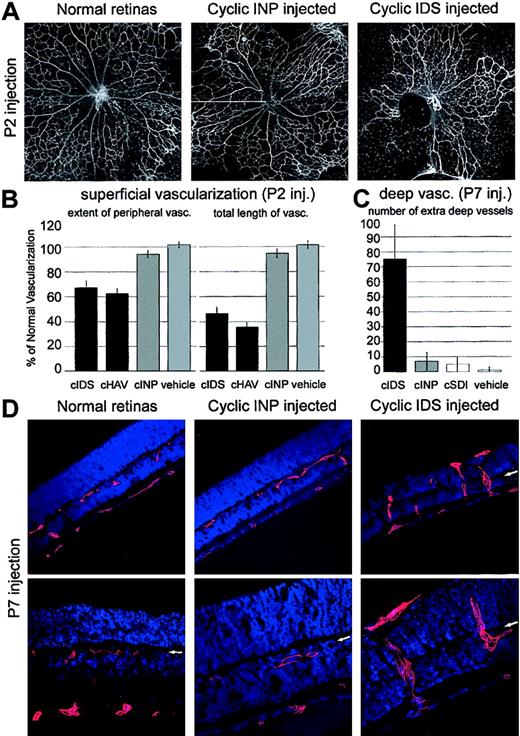
To test their effect on cadherin adhesion-mediated functions in vivo and to further confirm the specific role of R-cadherin during retinal vascular development, peptides were injected into the vitreous cavity of postnatal developing mouse eyes. When cyclic IDS or cyclic HAV peptides were injected into P2 mouse eyes and the resulting vasculature was examined 3 days later, vascular formation was disrupted with results similar to those observed after antibody injections.8 These retinas were characterized as having less extensive peripheral vascularization and fewer interconnecting vessels within the vascularized regions compared to normal, noninjected littermate controls. Overall, vascularization of the superficial layer was reduced by half with R-cadherin-blocking peptides; retinas injected with N-cadherin-specific cyclic INP peptides were relatively normal (Figure 4A,B).
R-cadherin peptide antagonists disrupt normal retina developmental vascularization. (A) When R-cadherin peptide antagonists (cyclic IDS) were injected intravitreally on postnatal day 2, formation of the superficial vasculature was greatly disrupted when compared to uninjected (normal retinas) or control (cyclic INP)—injected retinas. Each image is a montage of 5 photographs. Original magnification × 10. (B) Injection of R-cadherin blocking cyclic IDS peptides resulted in decreased vascular migration toward the retinal periphery and a reduction in the total length of retinal vessels (P < .01). These results were similar to injections of the previously characterized, nonspecific, cyclic HAV peptide. Injection of N-cadherin—specific, cyclic INP peptides resulted in relatively normal levels of retinal superficial vascularization. Error bars indicate standard deviation. (C) Injection of cyclic IDS, but not cyclic INP, peptides disrupted normal formation of the deep retinal vascular plexuses with more than a 10-fold increase in the number of extra deep vessels observed. Error bars indicate standard deviation. (D) Injection of cyclic IDS peptides at postnatal day 7, just prior to formation of the deep vascular plexus, caused vessels to migrate past the normal deep vascular plexus (arrows) and into the normally avascular photoreceptor layer. The deep vascular plexus formed normally when cyclic INP peptides were injected, demonstrating a specific role for R-cadherin during endothelial cell guidance to the 3 vascular plexuses. Original magnification × 10 (upper panels) and × 20 (lower panels).
R-cadherin peptide antagonists disrupt normal retina developmental vascularization. (A) When R-cadherin peptide antagonists (cyclic IDS) were injected intravitreally on postnatal day 2, formation of the superficial vasculature was greatly disrupted when compared to uninjected (normal retinas) or control (cyclic INP)—injected retinas. Each image is a montage of 5 photographs. Original magnification × 10. (B) Injection of R-cadherin blocking cyclic IDS peptides resulted in decreased vascular migration toward the retinal periphery and a reduction in the total length of retinal vessels (P < .01). These results were similar to injections of the previously characterized, nonspecific, cyclic HAV peptide. Injection of N-cadherin—specific, cyclic INP peptides resulted in relatively normal levels of retinal superficial vascularization. Error bars indicate standard deviation. (C) Injection of cyclic IDS, but not cyclic INP, peptides disrupted normal formation of the deep retinal vascular plexuses with more than a 10-fold increase in the number of extra deep vessels observed. Error bars indicate standard deviation. (D) Injection of cyclic IDS peptides at postnatal day 7, just prior to formation of the deep vascular plexus, caused vessels to migrate past the normal deep vascular plexus (arrows) and into the normally avascular photoreceptor layer. The deep vascular plexus formed normally when cyclic INP peptides were injected, demonstrating a specific role for R-cadherin during endothelial cell guidance to the 3 vascular plexuses. Original magnification × 10 (upper panels) and × 20 (lower panels).
R-cadherin-blocking peptides disrupted normal vascularization of the deep retinal layers as well (Figure 4C,D). When cyclic IDS peptides were injected at P7, just before vessels of the superficial vascular network dive and begin formation of the deep vascular plexus, the resultant P11 vasculature was characterized by numerous vessels that had migrated past the normal deep vascular plexus and into the normally avascular photoreceptor layer. Again, this is similar to the published effects observed when R-cadherin antibodies were injected.8 In contrast, the deep vascular plexus of eyes injected with cyclic INP peptides formed normally.
Effect of peptide antagonists on vascular targeting of bone marrow-derived HSCs
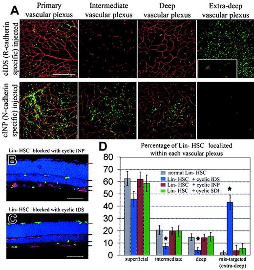
Lin- HSCs were incubated with R-cadherin peptide antagonists (cyclic IDS) or N-cadherin peptide antagonists (cyclic INP) prior to injection to further assess the requirement of R-cadherin-mediated adhesion for normal targeting of Lin- HSCs to the developing retinal vasculature. When the Lin- HSCs were preincubated with cyclic IDS peptides, between 40% and 50% of the stem cells were mistargeted to the outer edge of the photoreceptor cell layer (Figure 5), a result nearly identical to those observed when stem cells were preincubated with R-cadherin function-blocking antibodies. In some instances, a few extra deep vessels could also be observed, likely due to excessive peptide antagonists that disrupted the normal vascular development upon injection of the Lin- HSCs, cyclic IDS solution. It is unlikely that blocking R-cadherin-mediated adhesion affects HSC differentiation, as a portion of the mistargeted cells were found incorporated into the newly developed, extra deep, mistargeted vessels (Figure 5A, extra deep insert). Meanwhile, Lin- HSCs preincubated with cyclic INP peptides maintained their ability to target to and become incorporated into the normal developing vasculature at levels similar to that observed after control Lin- HSC injections. This further demonstrates the specificity of a role for R-cadherin in stem cell targeting.
R-cadherin-specific cyclic IDS peptides prevent normal EPC targeting to the developing retinal vasculature. (A) Planar images of the 3 normal retinal vascular plexuses and the outer edge of the photoreceptor layer demonstrated massive HSC mistargeting caused by R-cadherin-specific peptide antagonists (cyclic IDS), similar to the effects observed by preincubation with R-cadherin function-blocking antibodies. HSCs were mistargeted to the subretinal space rather than the superficial, intermediate, or deep vascular plexuses, compared to cyclic INP-incubated, or normal, Lin- HSCs. A small portion of mistargeted HSCs were incorporated into regions of mistargeted endogenous vasculature (insert). Scale bar represents approximately 50 μm. Original magnification of inset × 60. (B, C) Analysis of frozen retinal sections demonstrated normal targeting (B, black lines) of HSCs preincubated with cyclic INP and mistargeting (C, red lines) of HSCs to the outer edge of the photoreceptors after preincubation with cyclic IDS peptides. Scale bar represents approximately 50 μm. (D) The number of HSCs targeted to the 3 normal vascular plexuses or mistargeted to the outer edge of the photoreceptor layer was quantified as a relative percentage of the total number of retinally incorporated HSCs. Preincubation with cyclic IDS peptides greatly disrupted HSCs targeting, particularly to the intermediate and deep plexuses (P < .01, star), and greatly increased the number of mistargeted HSCs localized within the subretinal space (P < .0001, star). Error bars indicate standard deviation.
R-cadherin-specific cyclic IDS peptides prevent normal EPC targeting to the developing retinal vasculature. (A) Planar images of the 3 normal retinal vascular plexuses and the outer edge of the photoreceptor layer demonstrated massive HSC mistargeting caused by R-cadherin-specific peptide antagonists (cyclic IDS), similar to the effects observed by preincubation with R-cadherin function-blocking antibodies. HSCs were mistargeted to the subretinal space rather than the superficial, intermediate, or deep vascular plexuses, compared to cyclic INP-incubated, or normal, Lin- HSCs. A small portion of mistargeted HSCs were incorporated into regions of mistargeted endogenous vasculature (insert). Scale bar represents approximately 50 μm. Original magnification of inset × 60. (B, C) Analysis of frozen retinal sections demonstrated normal targeting (B, black lines) of HSCs preincubated with cyclic INP and mistargeting (C, red lines) of HSCs to the outer edge of the photoreceptors after preincubation with cyclic IDS peptides. Scale bar represents approximately 50 μm. (D) The number of HSCs targeted to the 3 normal vascular plexuses or mistargeted to the outer edge of the photoreceptor layer was quantified as a relative percentage of the total number of retinally incorporated HSCs. Preincubation with cyclic IDS peptides greatly disrupted HSCs targeting, particularly to the intermediate and deep plexuses (P < .01, star), and greatly increased the number of mistargeted HSCs localized within the subretinal space (P < .0001, star). Error bars indicate standard deviation.
Discussion
Tissue morphogenesis during the development of most tissues, including the neural retina, involves the selective binding of cell-cell adhesion molecules. This binding selectivity allows similarly differentiated cells to organize together and prevents the formation of incorrect tissue structures.24,25 However, despite the extensive studies on cadherin properties and function, particularly of N- and E-cadherins, a general mechanism accounting for cadherin specificity has not yet been described. In this study, peptide mimetics with selective function-blocking properties against specific cadherins were designed and synthesized. Linear and cyclic peptides were designed corresponding to amino acid residues 53-59 within cadherin domain EC1. This motif is thought to make direct contacts with the VDI sequence from an adjacent cadherin molecule at the adhesion interface.16 N-cadherin and R-cadherin are the most homologous of the cadherin family members.26 In fact, although all cadherins preferentially interact in a homophilic manner,18,27 R- and N-cadherin are the only 2 classical cadherin family members where functional heterodimers have been observed.28 Because R- and N-cadherin are so closely related, it is unlikely that these peptides would have specific antagonistic properties for these 2 cadherins, but nonspecific antagonistic properties against other cadherin family members. These studies suggest that the IDSMSGR motif (cadherin residues 53-59), particularly the IDS motif, plays an important role in mediating homoassociation of R- and N-cadherin molecules and is likely to participate in conferring cadherin specificity to other classical cadherin members as well. It is possible that only a single serine to proline substitution is required for adhesion selectivity as corresponding human sequences only differ between INS and INP. However, the selectivity of these 2 human sequences remains to be determined. This study should prove useful in the understanding of mechanisms mediating selective homophilic adhesion properties, particularly for cadherin-mediated adhesion specificity.
Endogenous retinal vascular endothelial cells use R-cadherin-mediated guidance cues to migrate along an astrocytic template during formation of the superficial retinal vascular layer and to subsequently localize and form the deep vascular plexuses within the retina.8 When R-cadherin-specific, cIDS peptide antagonists are injected during retinal vascular development, vascularization of the superficial plexus is disrupted, likely due to the interruption of R-cadherin-mediated guidance cues provided by astrocytes. Subsequently, R-cadherin molecules expressed within the regions of deep vascular plexus formation guide endothelial cells during formation of the deep and intermediate vascular plexuses. Injection of R-cadherin-specific peptide antagonists causes vessels to bypass these normal vascularized layers and migrate into the normally avascular photoreceptor layer.
We now have demonstrated that efficient targeting of Lin- HSCs to the retinal vasculature also requires R-cadherin-mediated adhesion. R-cadherin function-blocking antibodies and peptide antagonists disrupt HSC targeting to the 3 distinct vascular plexuses, causing a significant portion of the intravitreally injected stem cells to migrate through the retina and localize to the posterior margin of the photoreceptor layer. This phenotype is similar to that observed when R-cadherin functional expression is blocked during normal endogenous retinal vascularization: these vessels also migrate past the normal deep vascular plexuses to the photoreceptor/RPE cell interface. Since cyclic IDS peptides were unable to disrupt N-cadherin-mediated aggregation to a significant extent in vitro, it is unlikely that these in vivo vascular phenotypes were generated by low-affinity interactions of cyclic IDS with N-cadherin or any other cadherin type. In addition, injection of cyclic INP peptides, which were effective inhibitors of N-cadherin-mediated aggregation but not R-cadherin aggregation in vitro, did not result in significant phenotypic changes. Together these results confirm a specific role for R-cadherin during vascular guidance and targeting of bone marrow-derived cells during developmental retinal vascularization. While a number of studies have identified signals involved in mobilizing HSCs from bone marrow into the circulation,29-31 this is the first known report identifying a specific cell surface adhesion receptor that serves to target HSCs to sites of neovascularization.
The precise identity of the subpopulation of HSCs that targets and becomes incorporated into the developing vasculature remains unknown. We have demonstrated that removal of cells expressing the lineage markers CD45, CD3, Ly-6G, Mac1, or TER-119 results in a population of HSCs enriched for cells that target developing retinal vasculature.2 The precise cell surface marker signature of these cells, however, remains elusive. In addition to Lin+ cells, we have used a subpopulation of CD31-HSCs as a nontargeting control, indicating that a certain degree of endothelial differentiation may be required for vascular targeting (Figure 1; M.D. and A.O., unpublished results, 2003). However, in some cases a few CD31- were still able to functionally target the developing retinal vasculature, and HSCs selected for CD31 expression (CD31+) do not target to the same extent as Lin--selected HSCs (unpublished observations). Thus, other surface markers are likely to be important for efficient vascular targeting as well. Several recent studies have demonstrated that monocytes can differentiate into endothelial cells.1,32,33 It is possible that a fraction of the vascular targeting cells in our studies are of monocytic origin. Further studies characterizing the subpopulation of bone marrow-derived cells that target the retinal vasculature are ongoing. It is possible that a heterogeneous population of different HSCs is required for efficient vascular targeting. It is also possible that the identity of the targeting cells is environmentally dependent; the expressed identification markers may differ dramatically from the time the cells are selected and the time they become incorporated into the retinal vasculature in vivo. However, these issues do not alter the significance of our results, demonstrating the requirement for R-cadherin-mediated adhesion during targeting of a functionally defined population of cells to the developing retinal vasculature. R-cadherin adhesion may actually facilitate the development of vascular incorporating HSCs by allowing the cells to target within the environment required for appropriate differentiation and incorporation in vivo.
It is important to note that targeting of stem cells by locally delivered methods such as intravitreal injection may be different from targeting mechanisms employed by circulating bone marrow-derived HSCs. In fact, it is unclear if endogenous HSCs normally participate in formation of the developing retinal vasculature. For this study, we assessed the ability of Lin- HSCs to target to the developing retinal vasculature after systemic delivery. However, we were unable to demonstrate targeting following systemic injection, possibly due to several factors, including large sinks for HSCs in the form of other developing vascular systems in neonatal mice, as well as difficulties in injecting sufficient numbers of HSCs for targeting after nonlocal delivery methods. Using bone-marrow transplantation following lethal irradiation may help address the role of R-cadherin during targeting of circulating HSCs. However, other confounding variables should be considered when analyzing results from this type of experiment.
Ordinarily, the development of the retinal vasculature is highly regulated, forming 3 well-defined layers of vessels that never penetrate into or beyond the outer nuclear layer (ONL). Invasion of these developing vessels into the ONL and the subretinal space is only observed when R-cadherin guidance cues are blocked,8 in transgenic mice ectopically overexpressing vascular endothelial growth factor (VEGF),34 or in VLDLR knockout mice (M.D., unpublished results, 2003). These findings are all consistent with a proposed theory that vessels are induced to branch from the superficial plexus and migrate toward the deep vascular plexus in response to a stimulus gradient originating in the posterior retina.8,34 These same factors can also presumably regulate the migration of Lin- HSCs through the retinal layers. When vessels encounter R-cadherin, already expressed within the deep vascular plexuses, they are induced to stop, turn, and form the deep vascular networks. R-cadherin-mediated guidance also is likely to facilitate the localization of HSCs within these same vascular regions and their subsequent integration into the endogenous vasculature.
As suggested by others,5,35,36 circulating bone marrow-derived cells may contribute to the abnormal neovascularization observed in retinal and/or choroidal neovascularization. In the presence of R-cadherin antagonists, these cells presumably would not incorporate into the neovessels and may, in fact, accumulate in subretinal or other spaces. Potential consequences of this could include localized proliferation of these cells, leading to anatomical disruption of the retina. Our experience, however, suggests that mistargeted HSCs in the retina either leave the tissue or undergo cell death; we do not, for example, observe labeled HSCs in any extravascular sites 1 month after injection into the eye.2 HSCs first treated with R-cadherin antagonists similarly are not observed in the retina 4 weeks after injection. Long-term studies of these animals will provide more information about this potential complication of nontargeted stem cells in the eye.
R-cadherin-mediated targeting of Lin- HSCs to astrocytes and other R-cadherin-expressing cells may have application to understanding and developing treatments for a number of retinal vascular diseases. The proliferation of glial cells, or gliosis, is associated with many neovascular eye diseases,37-40 including diabetic retinopathy and age-related macular degeneration. Thus, targeting of HSCs to R-cadherin-expressing glial cells in these diseases may facilitate the stabilization of abnormal vessels associated with pathological angiogenesis or provide for targeted delivery of angiostatic molecules.2 Alternatively, blocking R-cadherin-mediated targeting may prevent recruitment of HSCs to sites of neovascularization, thus minimizing or preventing abnormal angiogenesis. If, indeed, bone marrow-derived HSCs contribute to pathological retinal and choroidal neovascularization,5,35,36 blocking the targeting of these cells with cadherin antagonists may prove to be a useful therapeutic approach in the treatment of diseases such as diabetic retinopathy or age-related macular degeneration, respectively.
Prepublished online as Blood First Edition Paper, January 15, 2004; DOI 10.1182/blood-2003-09-3012.
Supported by grants from the National Eye Institute (R01EY11254) and the Robert Mealey Program for the Study of Macular Degenerations (M.F.) and a fellowship from the San Diego chapter of the Achievement Rewards for Collegiate Scientists (ARCS) (M.I.D.).
The publication costs of this article were defrayed in part by page charge payment. Therefore, and solely to indicate this fact, this article is hereby marked “advertisement” in accordance with 18 U.S.C. section 1734.
The authors would like to thank Dr Masatoshi Takeichi of Kyoto University for generously providing the R- and N-cadherin plasmids and Dr Matthew Ritter for helpful discussions regarding stem cells.


This feature is available to Subscribers Only
Sign In or Create an Account Close Modal