Abstract
Aldolase (E.C. 4.1.2.13), a homotetrameric protein encoded by the ALDOA gene, converts fructose-1,6-bisphosphate to dihydroxyacetone phosphate and glyceraldehyde-3-phosphate. Three isozymes are encoded by distinct genes. The sole aldolase present in red blood cells and skeletal muscle is the A isozyme. We report here the case of a girl of Sicilian descent with aldolase A deficiency. Clinical manifestations included transfusion-dependent anemia until splenectomy at age 3 and increasing muscle weakness, with death at age 4 associated with rhabdomyolysis and hyperkalemia. Sequence analysis of the ALDOA coding regions revealed 2 novel heterozygous ALDOA mutations in conserved regions of the protein. The paternal allele encoded a nonsense mutation, Arg303X, in the enzyme-active site. The maternal allele encoded a missense mutation, Cys338Tyr, predicted to cause enzyme instability. This is the most severely affected patient reported to date and only the second with both rhabdomyolysis and hemolysis.
Introduction
Aldolase A is necessary for the production of adenosine triphosphate (ATP) in erythrocytes and muscle fibers, which depend on glycolysis for energy. The protein is a homotetramer, encoded by the ALDOA gene on chromosome 16q22-24. Aldolase A deficiency has been reported as a rare, autosomal recessive disorder. Clinical manifestations in 4 previously reported patients have been variable1-4 (summarized in Table 1). Hemolysis has been associated with this disorder in each patient, with myopathy in one and mental retardation in another. Two earlier reported patients were analyzed for mutations (Table 1).
Reported cases of aldolase A deficiency
. | . | Mutation . | . | Clinical description . | . | . | . | |||
|---|---|---|---|---|---|---|---|---|---|---|
| Ethnicity . | Consanguinity . | DNA . | Amino acid . | Hemolytic anemia . | Myopathy . | Mental retardation . | Reference . | |||
| Canadian Jewish | Yes | Not reported | Not reported | Yes | No | Yes | 1 | |||
| Japanese | Probable | Not reported | Not reported | Yes | No | No | 2 | |||
| Japanese | Probable | 386A>G | Asp128Gly | Yes | No | No | 2,3 | |||
| German | No | 619G>A | Glu206Lys | Yes | Yes | No | 4 | |||
| Sicilian | No | 931C>T, 1037G>A | Arg303X, Cys338Tyr | Yes | Yes | No | This report | |||
DNA sequences are numbered from A of the ATG start codon.
Study design
Patient and case report
A girl of Sicilian ancestry, born to nonconsanguineous parents, was brought for treatment as a newborn for jaundice, pyropoikilocytosis, and anemia requiring transfusion. The patient's father had normal findings on blood count and peripheral smear. Her mother had a history of newborn jaundice and elliptocytes on peripheral blood smear consistent with dominant (benign) hereditary elliptocytosis. The patient had no siblings. Initially, hereditary pyropoikilocytosis was considered. A low-expression spectrin mutation, α-LELY,5 was sought in the father but not found. From age 1 to 3, the patient had recurrent episodes of pneumonia and croup and an episode of disseminated Pseudomonas infection, without neutropenia. Neurologic examination at age 2, prompted by a seizure, revealed Gowers sign without other evidence of weakness. Cognitive function was normal. The patient required blood transfusions every 6 to 8 weeks until splenectomy at age 3, which relieved the transfusion requirement. After splenectomy (off transfusions), peripheral smear findings became more abnormal, with many elliptocytes and Howell-Jolly bodies. The patient had leg pain while climbing stairs. Elevated creatine phosphokinase (CPK) levels in the plasma were noted with febrile illnesses. On a “healthy” day at 48 months, red blood cell (RBC) and plasma enzyme levels were obtained (Table 2). An elevated serum CPK level was noted at 13 800 U/L, yet serum aldolase was normal at 6.2 U/L, suggesting relative muscle aldolase deficiency.
Erythrocyte enzyme study
. | Patient . | Normal range . |
|---|---|---|
| Enzyme levels | ||
| Erythrocyte aldolase | 0.3 U/g Hb (maternal, 1.5 ± 0.2 U/g Hb) | 1.3-2.8 U/g Hb |
| Erythrocyte LDH | 360 U/L (maternal, 260 U/L) | 90-180U/L |
| RBC enzyme levels, U/g Hb | ||
| Glucose phosphate isomerase | 69 | 48-90 |
| Hexokinase | 3.8 (↑) | 1.0-2.5 |
| Phosphoglycerate kinase | 196 (↑) | 141-179 |
| Pyruvate kinase | 40.5 (↑) | 9.0-22.0 |
| Phosphofructokinase | 5.7 | 3.0-6.0 |
| Plasma enzyme levels, U/L | ||
| Aldolase | 6.2,* 1 (↓) | 3-12 |
| Creatine phosphokinase | 13 800 (↑ ↑) | 4-150 |
Normal ranges are represented as ± 2 SD from the mean.
Enzyme activities of RBC aldolase A and LDH were measured in EDTA (ethylenediaminetetraacetic acid)–anticoagulated blood from patient and mother at 30°C. Patient's RBC aldolase level is markedly decreased. Normal enzyme ranges are derived from the Internal Committee for Standardization in Hematology.6 Patient RBC and plasma enzyme levels were obtained at age 48 months. Markedly elevated CPK levels are noted (↑ ↑). Glycolytic enzyme levels aside from aldolase A are either normal or slightly elevated (↑), as expected in hemolytic anemia. Plasma aldolase is derived primarily from muscle. The low level (↓) in the face of marked CPK elevation supports the diagnosis of muscle aldolase deficiency.
Unexpected “normal” value
Subsequent studies revealed that the patient's RBC aldolase level was low at 0.3 U/g hemoglobin (Hb), whereas levels of other glycolytic enzymes, phosphofructokinase, glucose phosphate isomerase, phosphoglycerate kinase, hexokinase, lactate dehydrogenase, and pyruvate kinase, were normal or elevated (Table 2). Episodes of rhabdomyolysis were more prominent as the patient grew older. She succumbed to severe hyperkalemia and acute rhabdomyolysis during a febrile illness with gastrointestinal hemorrhage at 54 months of age. Postmortem muscle biopsy revealed myopathic changes with small atrophic fibers and large hypertrophic fibers with increased internal nuclei, suggesting a long-standing myopathic process. Immunohistochemical staining evidence for β-sarcoglycan and spectrin was reduced, but for other sarcolemmal protein it was normal. Ragged red fibers were absent. Enzyme assays on the necropsy muscle tissue were uninformative because of problems with tissue preservation.
Erythrocyte aldolase and LDH analysis
Aldolase and lactate dehydrogenase (LDH) activities were determined from RBC lysates as described by the International Committee for Standardization in Haematology.6 Briefly, erythrocytes were isolated with Ficoll, washed, and lysed in isotonic buffer. Enzyme activities were measured by a loss in absorbance at 340 nm. Aldolase assay used a coupled assay with fructose 1,6-bisphosphate as the substrate,7 and the LDH assay substrate was pyruvate.
ALDOA gene sequencing
Genomic DNA from the patient's fibroblasts and parental whole blood samples were prepared using Puregene (Gentra Systems, Research Triangle Park, NC). Nine exons, including intron/exon junctions, were amplified with primers designed based on human aldolase A gene sequence (GenBank genomic sequence X12447). Primer sequences are available on request.
Amplified polymerase chain reaction (PCR) fragments were purified by QIAquick Extraction (Qiagen, Valencia, CA), and both strands were sequenced using the standard automated DNA sequence methodology. The sequence was confirmed for the patient and both parents. We designed an Amplification Refractory Mutation System (ARMS) assay8 to confirm the mutations in genomic DNA (ie, to rule out PCR contaminations) and to facilitate rapid screening for the mutation without sequencing.
Results and discussion
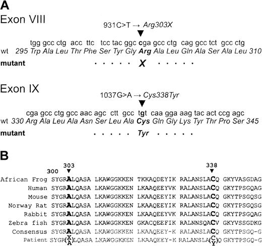
Sequence analysis of the patient's ALDOA gene (all 8 coding exons and the 5′ untranslated exon IB) revealed that the patient was heterozygous for 2 distinct novel mutations in highly conserved regions of the protein, each carried by one parent (Figure 1A). The paternal point mutation, 931C>T, introduces a premature stop codon, Arg303X. This nonsense mutation truncates the protein, producing a “null” allele. It is interesting that this nonsense allele is at Arg303, which is crucial for enzymatic activity. The guanidino group interacts with both the C1- and the C6-phosphate at different points during catalysis9-11 and acts as a “trigger” residue in conformational changes in the C-terminal region associated with catalysis.11 The maternal point mutation, 1037G>A, encodes the missense mutation Cys338Tyr. Although Cys338 is not crucial for catalysis,12 it is near a critical “hinge” region for the conformational change in the C-terminus and may be important for maintaining the structure.13 Substitution of Cys338 with tyrosine may disrupt the structure in a temperature-sensitive fashion. Both Arg303 and Cys338 are conserved in vertebrates (Figure 1B).
Genetic analysis of aldolase A gene mutations. (A) Sequence analysis of ALDOA exon VIII and IX and predicted amino acid change. A heterozygous mutation in exon VIII was found in the patient and her father. Maternal exon VIII (not shown) was wild type. In exon IX, the patient and her mother share a heterozygous mutation, which is absent in her father. (B) ALDOA sequence alignment. Partial human aldolase A polypeptide sequences from exons VIII and IX, aligned to highly conserved orthologs from other species (GenBank cDNA sequences: Xenopus, AAH46673; human, AAH16800; mouse, NP_031464; rat, NP_036627; rabbit, P00883; zebra fish, AAN04476). The patient's sequence, divergent at 2 otherwise invariant residues, is shown at the bottom.
Genetic analysis of aldolase A gene mutations. (A) Sequence analysis of ALDOA exon VIII and IX and predicted amino acid change. A heterozygous mutation in exon VIII was found in the patient and her father. Maternal exon VIII (not shown) was wild type. In exon IX, the patient and her mother share a heterozygous mutation, which is absent in her father. (B) ALDOA sequence alignment. Partial human aldolase A polypeptide sequences from exons VIII and IX, aligned to highly conserved orthologs from other species (GenBank cDNA sequences: Xenopus, AAH46673; human, AAH16800; mouse, NP_031464; rat, NP_036627; rabbit, P00883; zebra fish, AAN04476). The patient's sequence, divergent at 2 otherwise invariant residues, is shown at the bottom.
Compared with previously reported patients with aldolase A deficiency, the clinical consequences for our patient were more severe and ultimately lethal. Myopathic symptoms in the case report of Kreuder et al4 were attributed to aldolase tetramer instability. Based on the present sequence analysis, we postulate that the severity of our patient's anemia and myopathy can be attributed to one null allele and to the thermolabile nature of the remaining aldolase protein. This would explain her decompensation with fever; the instability of the protein would render it insufficient to compensate for the needs of erythrocyte and muscle energy consumption during illness. Aldolase A is reported to be the predominant or sole aldolase in leukocytes.14 It is possible that the recurrent infections in the patient, in particular disseminated Pseudomonas, resulted from defective bacterial killing. This possibility was not tested directly. Despite the severity of the enzymatic defect in this patient, her cognitive function was entirely normal. It is possible that the reported case of mental retardation was coincidental.1
Were the elliptocytosis and enzymopathy in our patient related? We believe they were coincidental. Nonspherocytic hemolytic anemia is a hallmark of glycolytic enzyme disorders of erythrocytes. In general, these disorders are not directly related to RBC membrane phenotypes. A single case report in 1986 described an infant with a partial deficiency in enolase with spherocytosis.15 In addition to hemolytic anemia and severe, progressive myopathy, our patient appears to have had a combination of dominant (mild) hereditary elliptocytosis inherited from her mother and recessive compound heterozygosity aldolase A deficiency. We cannot rule out an interaction between these genetic disorders. The glycolytic defect might have made the membrane defect more severe—for example, by intracellular adenosine triphosphate (ATP) depletion.
Aldolase is required for life. Isozymes A, B, and C have distinctive tissue distributions. Because of overlap in many tissues, deficiency of the A isozyme is predicted to be most severe in erythrocytes and muscle, where it is the sole isozyme. We conclude that hemolysis combined with evidence of weakness, muscle pain, or myopathy should prompt specific evaluation of erythrocyte aldolase along with evaluation of other glycolytic enzymes known to cause the combination of hemolytic anemia and myopathy (eg, phosphofructokinase [OMIM 232800], triose phosphate isomerase [OMIM 190450], glucose phosphate isomerase [OMIM 172400], and phosphoglycerate kinase [OMIM 311800]).
Prepublished online as Blood First Edition Paper, November 13, 2003; DOI 10.1182/blood-2003-09-3160.
Supported by National Institutes of Health grants HL04184 (E.J.N.) and DK43521 (D.R.T.).
The publication costs of this article were defrayed in part by page charge payment. Therefore, and solely to indicate this fact, this article is hereby marked “advertisement” in accordance with 18 U.S.C. section 1734.
We thank Dr Patrick Gallagher (Yale University, New Haven, CT) for analysis of α-spectrin mutations. We also thank the patient's family for their participation in these studies.


This feature is available to Subscribers Only
Sign In or Create an Account Close Modal