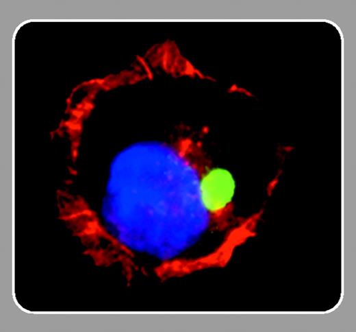
Much interest has centered around the recent recognition that not only neurons, but also lymphocytes, can form synapses. Some of the proteins involved in regulating signal transduction at the neuronal and immunologic synapses contain an EVH1 domain, named after a region of homology initially identified in the proteins Ena and VASP. EVH1 domain proteins include the Homer subfamily (Homer-1a, -1b, -1c, -2a, -2b, and -3) and have also been implicated in cellular processes that involve actin dynamics.1 The approximately 110–amino acid EVH1 domain has little homology but a high 3-dimensional structural similarity to the pleckstrin homology domain found in many signal transduction molecules, and it specifically binds proline-rich motifs: PPxxF in the case of Homer family members, and FPPPP in the case of Ena and VASP.2 Homer proteins (except Homer-1a) also contain a coiled coil (CC) domain that mediates specific self-interactions and promotes the formation of larger molecular complexes or scaffolds. This property represents an essential part of their function as adaptors in synaptic signaling complexes. Because Homer-1a, an immediate early gene product, lacks the CC region, it will fail to promote formation of large complexes but will bind to target proteins and therefore may participate in dissociating signaling complexes. Homer proteins have been studied mostly in neurons, where they have have been found to play a role in synaptogenesis and spatial localization of metabotropic glutamate receptors, with which they directly interact, and also in the intracellular trafficking of receptors. In the hematopoietic system, however, only little is known so far about the role of these proteins.
In this issue of Blood, Ishiguro and Xavier (page 2248) present their remarkable findings about Homer-3, the only isoform that is expressed in the thymus. They observed that in T cells (Jurkat or primary CD4+ cells) stimulated with anti-CD3– or anti-CD28–coated beads, Homer-3 is very rapidly recruited to the area of contact with the beads and this takes place prior to the redistribution of actin characteristic of the synapse formation. This recruitment is mediated by the CC domain. Shortly thereafter, Homer-3 translocates to the nucleus in an EVH1 domain–dependent process, while other components of the immunologic synapse stay behind. So if Homer-3 goes to the nucleus, then what does it do there? Ishiguro and Xavier reasoned that since the cellular distribution of Homer-3 is regulated dynamically during the early phase of T-cell activation, with a kinetic similar to that of immediate early gene induction, there might be a functional relationship between the two. They followed this intuition and examined the c-fos gene as an example of an immediate early gene that is induced upon T-cell activation. Using a reporter gene driven by the serum response element (SRE) of the c-fos gene, Ishiguro and Xavier showed that Homer-3 expression reduces activation through the SRE in Jurkat cells. This effect is mediated by the EVH1 domain of Homer-3; the CC domain appears to be dispensible. In addition, down-regulation of endogenous Homer-3 by siRNA led to increased activation of the SRE, consistent with the idea that nuclear Homer-3 is a negative regulator of SRE activity. The authors then showed that Homer-3 interacts with one of the transcription factors binding to the SRE, CCAAT/enhancer binding protein β (C/EBPβ) which has in its N-terminal transcription activation domain a consensus motif for interaction with the EVH1 domain of Homer. Interaction of Homer-3 with C/EBPβ does not impair binding of C/EBPβ to its cognate DNA site but masks the activation domain and presumably alters its capacity to interact with the general transcription machinery. Thus, Homer-3 is reminiscent of other proteins, such as β-catenin and CASK/LIN-2, that also have a dual life in the cytosol and in the nucleus. A number of interesting questions remain to be elucidated; for example, what triggers the departure of Homer-3 from its synaptic location? Is some posttranslational protein modification involved? How is the nuclear targeting achieved—or are there other nuclear targets? One can anticipate that the further study of Homer proteins in the hematopoietic system will reveal important insights about T-cell activation and function.


This feature is available to Subscribers Only
Sign In or Create an Account Close Modal