Abstract
Elevated levels of interleukin 7 (IL-7) have been correlated with various T-cell depletion conditions, including HIV infection, and suggested as an indicator of HIV disease progression (AIDS and death). Here, the assessment of pathogenic simian immunodeficiency virus (SIVmac239) infection in rhesus macaques demonstrated a clear association between a significant elevation in IL-7 levels and disease progression. In 5 macaques that progressed to simian AIDS and death, elevated IL-7 levels were unable to restore T-cell homeostasis. In contrast, increased IL-7 levels were followed by relatively high and stable T-cell numbers in the SIV-infected macaques with a slow-progressing phenotype. Further, studies in sooty mangabeys that do not progress to simian AIDS and that maintain stable T-cell numbers despite high levels of viral replication support the importance of IL-7 and T-cell homeostasis in disease progression. These data suggest that during pathogenic SIV infection with high viral replication, elevated IL-7 levels are unable to recover T-cell homeostasis, thereby leading to disease progression. The utility of IL-7 as a potential immunotherapeutic agent to improve HIV/SIV-related T-cell depletion may therefore depend on controlling the pathogenic effects of viral replication prior to the administration of IL-7. (Blood. 2004;103:973-979)
Introduction
Relatively stable numbers of T cells are maintained in the periphery throughout an individual's life as a result of homeostatic control of T-cell production and elimination.1,2 T-cell generation can be achieved through de novo production of naive T cells in the thymus as well as postthymic expansion of mature T cells in the periphery.3 T cells produced in the thymus are unique because they contain a diverse T-cell receptor (TCR) repertoire.4-6 In contrast, peripheral expansion involves both the antigen-driven and homeostatic proliferation of T cells resulting in an increase in the number of T cells with a limited number of potential TCRs.6,7 During T-cell-depleting conditions such as in HIV infection, both the expansion of antigen-specific T cells (peripheral expansion) and de novo generation of new T cells (thymic output) are important for effectively maintaining T-cell homeostasis and preventing opportunistic infections.
The cytokine interleukin 7 (IL-7) is unique in its ability to homeostatically increase T-cell generation from both peripheral and thymic origins.8-10 IL-7-induced proliferation occurs without altering the naive or memory phenotype of the T cells undergoing proliferation.10,11 In addition to its role in proliferation, IL-7 also increases the survival of T cells by increasing the expression of antiapoptotic factor Bcl-2.12 Interestingly, levels of IL-7 are often elevated in T-cell-depleting conditions, including chemotherapy and HIV infection.8,13 These studies suggest that increased production of IL-7 by stromal and dendritic cells present in immune tissues throughout the body may be a homeostatic mechanism of regulating T-cell proliferation, and possibly thymic output.8,13 In patients with HIV, elevated levels of IL-7 have been correlated with progression to AIDS.13,14 Furthermore, in vitro studies in peripheral blood mononuclear cells (PBMCs) and thymic organ cultures have demonstrated that IL-7 can increase HIV replication.15,16
Recently, there is considerable interest in the use of IL-7 as an immunotherapeutic agent in patients with HIV because of the limited immune recovery seen in some patients undergoing highly active antiretroviral therapy (HAART).17,18 Experiments undertaken in murine models have shown the usefulness of IL-7 as a potential therapeutic modality for immune reconstitution.19 In addition to potential immunologic benefits, IL-7 therapy in the SCID-hu mouse model has been shown to be an effective means of derepressing the latently infected resting T cells.20 Nonhuman primates remain one of the best models to study human AIDS.21-23 Through the assessment of endogenous IL-7 levels in monkeys infected with simian immunodeficiency virus (SIV) in this study, we describe the role of IL-7 in disease progression and provide a framework for undertaking future IL-7 therapy interventions in this model. Assessment of the immunologic and virologic events that occur following SIV infection indicate that elevated plasma IL-7 levels represent a homeostatic regulation mechanism to offset the decline in CD4+ T cells. The inability of IL-7 to maintain T-cell numbers in macaques infected with SIVmac239 results in a new elevated IL-7 “set point” that may be a contributing factor to the progression to simian AIDS and death in the SIV-infected macaques.
Materials and methods
Animals and viral infection
Uninfected control macaques (n = 12) and SIV-infected macaques (6 SIVsmE660+ and 4 SIVmac239+) obtained from the Yerkes, California, and New England primate research centers were used in the cross-sectional analysis. All the macaques used in the cross-sectional study were between 2 and 7 years of age. An additional 6 macaques were exclusively used in the longitudinal study (RM1-RM6) and were between 3 and 5 years of age when infected with SIVmac239. RM1, RM2, and RM3 had undergone a sham thymectomy in which a small thymus biopsy was removed 3 months prior to SIV infection. RM4, RM5, and RM6 had their thymus tissue removed (thymectomy) 3 months prior to SIV infection. The surgery was performed carefully to eliminate all thymic tissue that could be observed. Completeness of thymectomy was further verified in each animal by visual inspection at the time of death. RM1 to RM6 were given intravenous inoculations of 2.5 × 10⁵ 50% tissue culture infectious doses (determined in CEMx174 cells) with a SIVmac239 viral stock. Plasma from 24 sooty mangabeys (11 natural SIVsmm infected, 13 uninfected) was obtained from the Yerkes Regional Primate Research Center. Local animal care and use committee and National Institutes of Health protocols were strictly followed in the maintenance of animals used in these studies.
Monoclonal antibodies used for flow cytometry
Antibodies used for immunophenotyping of PBMCs were anti-CD4 (clone SK3), anti-CD8 (clone SK1), anti-CD45RA (clone L48), anti-CD62L (clone SK11; BD Immunocytometry Systems, San Jose, CA), anti-CD3e (clone SP34; BD Pharmingen, San Diego, CA), and anti-Ki67 (clone R0840; Dako, Carpinteria, CA). The antibodies were conjugated to the following fluorphores: fluorescein isothiocyanate (FITC; CD45RA, CD3), phycoerythrin (PE; CD62L, CD4, Ki67), peridinin chlorophyll protein (PerCP; CD8), or allophycocyanin (APC; CD4).
Immunophenotyping of PBMCs
PBMCs were isolated with Ficoll-Hypaque gradient and assessed with 4 fluorometric markers to determine the absolute number of specific cell subsets. Percentage of naive T cells was determined using the antibodies CD62L and CD45RA, and the percentage of proliferating T cells was determined by Ki67 staining.24 After the antibody incubations were completed, the cells were washed in flow cytometry buffer (1 × phosphate-buffered saline, 1% bovine serum albumin, and 0.1% NaN3) and fixed in 1% paraformaldehyde.
Assessment of TREC levels
PBMCs isolated via Ficoll-Hypaque gradient were sorted into CD8+ and CD8-depleted (CD4+) T-cell populations using the MACS columns (Miltenyi Biotec, Auburn, CA). T-cell receptor excision circle (TREC) levels were determined in these magnetically sorted populations using TaqMan real-time polymerase chain reaction (PCR) as previously described.24 Real-time PCR was performed on the ABI Prism 7700 sequence detector (Applied Biosystems, Foster City, CA).
Viral load analysis
SIV plasma viral load was determined as previously described.25
Quantification of cytokines
Blood for IL-7 estimation was collected in EDTA (ethylenediaminetetraacetic acid)-containing tubes and allowed to sit on ice overnight before centrifugation for the isolation of plasma. IL-7 levels in the plasma of SIV-infected macaques and mangabeys were determined using the commercial sandwich enzyme immunoassay kits available for humans (R&D Systems, Minneapolis, MN). The specificity of the IL-7 enzyme-linked immunosorbent assay (ELISA) for rhesus macaques and sooty mangabeys was ascertained by blocking experiments with anti-IL-7 antisera (R&D Systems). The minimum detectable level of IL-7 was 0.10 pg/mL. The reproducibility of the IL-7 ELISA was assessed through the incorporation of a control plasma sample in each assay, variation was between 0.1% and 5%.
Statistical analysis
The nonparameteric Spearman rank correlation coefficient was used to assess the degree of positive and negative association between IL-7 and other host factors as well as viral load. For each pairing of observations the hypothesis that the correlation coefficient was zero was tested versus the 2-sided alternative that it was not zero. Additional statistical analysis was done by nonparametric Mann-Whitney U test (Prism version 3.0 for Mac; GraphPad Software, San Diego, CA). For all statistical analyses, results were considered significant if the probability was less than .05 (P < .05).
Results
Plasma IL-7 levels are elevated in SIV-infected macaques
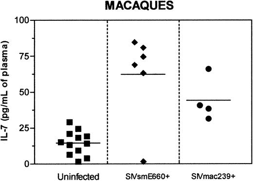
Analysis of IL-7 in mice and humans established a role for this cytokine in T-cell homeostasis and determined that IL-7 is elevated during T cell-depleting conditions, including chemotherapy and HIV infection.8,13 Here analysis of plasma IL-7 levels was undertaken during the SIV infection of monkeys to assess its importance toward the homeostatic regulation of T cells in nonhuman primates. First, a cross-sectional analysis was carried out by comparing plasma IL-7 levels in uninfected and SIV-infected macaques. Macaques infected with 2 distinct viral strains, SIVsmE660 and SIVmac239, were assessed. Infection of macaques with SIVmac239 resulted in simian AIDS in 4 to 11 months at which time the levels of plasma IL-7 were significantly elevated when compared to the uninfected controls (P < .004; [Figure 1]). A similar increase in plasma IL-7 levels (P < .02) was observed in SIVsmE660+ macaques infected for 12 to 19 months without any clinical manifestations of simian AIDS (Figure 1). In summary, both SIVmac239 and SIVsmE660 infections result in increased IL-7 levels in macaques.
Cross-sectional analysis of plasma IL-7 levels in rhesus macaques. Plasma IL-7 levels are depicted for 12 uninfected (▪), 6 SIVsmE660-infected asymptomatic at the time of sampling (♦), and 4 SIVmac239-infected rhesus macaques which progress to simian AIDS and death (•). The minimum detectable level of IL-7 was 0.10 pg/mL.
Cross-sectional analysis of plasma IL-7 levels in rhesus macaques. Plasma IL-7 levels are depicted for 12 uninfected (▪), 6 SIVsmE660-infected asymptomatic at the time of sampling (♦), and 4 SIVmac239-infected rhesus macaques which progress to simian AIDS and death (•). The minimum detectable level of IL-7 was 0.10 pg/mL.
IL-7 levels are elevated and maintained at high levels at times prior to simian AIDS and death
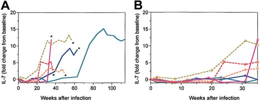
To expand on the observations made from the cross-sectional analysis, a longitudinal study was undertaken. IL-7 levels were assessed in both SIV-infected normal (nonthymectomized) macaques (RM1, RM2, and RM3) as well as macaques that had been thymectomized (RM4, RM5, and RM6) prior to SIVmac239 infection. Our goal was to use the thymectomized macaques to determine the influence of the thymus and thymic-derived T cells on the regulation of IL-7 and disease progression. A longitudinal assessment of the plasma IL-7 levels throughout the disease course identified 2 distinct phases that followed the SIVmac239 infection (Figure 2A). Initially, all 6 SIVmac239-infected macaques exhibited low levels of IL-7 in the plasma similar to their preinfection levels (first 24-62 weeks after infection). Following a relatively short transition period a new, elevated plasma IL-7 level (increased 2- to 15-fold) was established and maintained in each of the 6 macaques (Figure 2A). The absence of a thymus (RM4, RM5, and RM6) did not dramatically affect plasma IL-7 levels in the SIVmac239-infected macaques. However, a trend was observed in which the elevation of IL-7 levels occurred at slightly earlier time points in the thymectomized macaques (Figure 2B) implying that limiting T-cell renewal may affect plasma IL-7 levels. Following SIVmac239 infection in these macaques a range of disease outcomes was observed, although no specific differences in disease progression were associated with thymectomy. SIVmac239-infected macaques died of simian AIDS at 35 weeks (RM1), 37 weeks (RM4), 49 weeks (RM5), 57 weeks (RM6), and 62 weeks (RM2) after infection (Figure 2A). Interestingly, in the macaques that progressed to simian AIDS (RM1, RM2, RM4, RM5, and RM6) IL-7 levels were significantly elevated (P < .05) and maintained at high levels until death. This indicates an association between elevated IL-7 levels and disease progression.
Longitudinal analysis of plasma IL-7 levels in SIVmac239-infected macaques. Six juvenile macaques—3 nonthymectomized macaques (continuous lines; RM1, magenta; RM2, dark blue; RM3, blue/gray) and 3 thymectomized macaques (broken lines; RM4, red; RM5, orange; RM6, yellow/green)—were included in the longitudinal study. (A) Plasma IL-7 levels as fold change from baseline are depicted through the disease course. Macaques that died of simian AIDS are indicated by an asterisk. (B) Plasma IL-7 levels as fold change from baseline depicted for first 35 weeks after infection to elucidate the changes occurring during the early time points.
Longitudinal analysis of plasma IL-7 levels in SIVmac239-infected macaques. Six juvenile macaques—3 nonthymectomized macaques (continuous lines; RM1, magenta; RM2, dark blue; RM3, blue/gray) and 3 thymectomized macaques (broken lines; RM4, red; RM5, orange; RM6, yellow/green)—were included in the longitudinal study. (A) Plasma IL-7 levels as fold change from baseline are depicted through the disease course. Macaques that died of simian AIDS are indicated by an asterisk. (B) Plasma IL-7 levels as fold change from baseline depicted for first 35 weeks after infection to elucidate the changes occurring during the early time points.
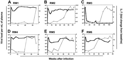
One of the mechanisms by which IL-7 could influence disease progression is through an increase in viral replication. Indeed, recent studies in the SCID-hu mouse model20 and human thymocytes16 have shown the direct role of IL-7 in HIV viral replication. However, in this study the elevated IL-7 levels did not correlate with any change in plasma viral loads (Figure 3). The levels of plasma viremia were high and remained relatively stable within the 5 macaques that progressed to simian AIDS (RM1, RM2, RM4, RM5, and RM6). In addition, the undetectable viral load in RM3 remained below the level of detection following the IL-7 increase (Figure 3C). Therefore, assessment of plasma viremia did not exhibit any evidence that elevated plasma IL-7 levels resulted in an increase in SIV replication.
Elevated IL-7 levels do not alter viral load in SIVmac239-infected macaques. Plasma viral load (black lines, +; viral RNA copies/mL) and plasma IL-7 levels (gray lines, ▦) presented as fold change from the baseline are depicted for macaques RM1 through RM6 (A-F, respectively).
Elevated IL-7 levels do not alter viral load in SIVmac239-infected macaques. Plasma viral load (black lines, +; viral RNA copies/mL) and plasma IL-7 levels (gray lines, ▦) presented as fold change from the baseline are depicted for macaques RM1 through RM6 (A-F, respectively).
Effect of elevated IL-7 levels on T-cell proliferation and maintenance of T-cell levels in SIVmac239-infected macaques
Failure to restore proper T-cell homeostasis by the elevated IL-7 levels may be one of the reasons for disease progression and simian AIDS seen in 5 SIV-infected macaques. Recovery of T-cell homeostasis would be expected to require an increase in peripheral T-cell proliferation. To determine the influence of IL-7 on T-cell proliferation in macaques, the percentage of T cells expressing the protein Ki67 was quantified throughout the disease course. In the majority of macaques an early peak in Ki67 activity occurred in both CD4+ and CD8+ T cells during the acute phase of the infection (weeks 2-8) immediately following a peak in plasma viremia, whereas IL-7 levels remained low (Figure 4A,C,E,G,I,K). Following the acute phase peak, Ki67 levels were observed to be elevated at numerous time points throughout the infection (Figure 4A,C,E,G,I,K). In the 5 macaques that progressed to simian AIDS (RM1, RM2, RM4, RM5, and RM6), no specific increase in CD4 or CD8 T-cell proliferation was observed following the increase in plasma IL-7 levels. In contrast, the long-term asymptomatic/nonprogressor RM3 did exhibit an increase in proliferation within both the CD4+ and CD8+ T-cell subsets (Figure 4E) that occurred just following the IL-7 increase.
Longitudinal analysis of T-cell proliferation and T-cell numbers in SIVmac239-infected macaques. The percentage of proliferating (Ki67+) CD4+ (dark green, ○) and CD8+ T cells (light green, ♦) in peripheral blood (A,C,E,G,I,K) and plasma IL-7 levels (red, ▪) as fold change from the baseline are plotted for macaques RM1, RM2, RM3, RM4, RM5, and RM6. Absolute numbers of CD4+ (dark blue, ▿) and CD8+ T cells (light blue, ▴; B,D,F,H,J,L) are given as markers of T-cell homeostasis. Dotted horizontal lines indicate preinfection levels of CD4+ (dark blue) and CD8+ (light blue) T cells that represent the homeostatic T-cell levels in the absence of SIV infection. The nonthymectomized macaques are shown in panels A-F and the thymectomized macaques in panels G through L.
Longitudinal analysis of T-cell proliferation and T-cell numbers in SIVmac239-infected macaques. The percentage of proliferating (Ki67+) CD4+ (dark green, ○) and CD8+ T cells (light green, ♦) in peripheral blood (A,C,E,G,I,K) and plasma IL-7 levels (red, ▪) as fold change from the baseline are plotted for macaques RM1, RM2, RM3, RM4, RM5, and RM6. Absolute numbers of CD4+ (dark blue, ▿) and CD8+ T cells (light blue, ▴; B,D,F,H,J,L) are given as markers of T-cell homeostasis. Dotted horizontal lines indicate preinfection levels of CD4+ (dark blue) and CD8+ (light blue) T cells that represent the homeostatic T-cell levels in the absence of SIV infection. The nonthymectomized macaques are shown in panels A-F and the thymectomized macaques in panels G through L.
Mackall and colleagues have demonstrated that IL-7 can increase both thymic and postthymic T-cell proliferation to maintain T-cell homeostasis following murine bone marrow transplantation.19 Within the SIVmac239-infected macaques the absolute numbers of CD4+ T cells as well as CD8+ T cells were assessed throughout the course of SIV infection to ascertain the potential role of IL-7 in proper maintenance of T-cell numbers (Figure 4B,D,F,H,J,L). Our goal was to assess whether the increased IL-7 levels corresponded to increased T-cell numbers; and if so, would the T-cell level recover to the preinfection level. Within the 5 macaques that progressed to simian AIDS (Figure 4B,D,H,J,L), the increased IL-7 levels were generally associated with low CD4 levels that remained stable (RM2, RM5, and RM6) or continued to decline (RM1 and RM4). In fact, there was a drastic decline in CD4+ T cells from a mean average of 1352 counts to 233 counts in these macaques. In none of these 5 macaques did increased IL-7 levels appear to impart any benefits to CD4+ T-cell recovery. In contrast, macaque RM3 (long-term asymptomatic, still alive) exhibited a relatively stable and high CD4+ T-cell level that approached preinfection levels (Figure 4F). We hypothesize that the ability of CD4+ T cells in macaque RM3 to proliferate at times of high IL-7 and achieve relatively high and stable CD4+ T-cell levels indicates that the CD4+ T cells retain function with regard to their ability to respond to homeostatic signals. Further evidence to support this hypothesis was obtained from 3 macaques infected with SIVsmE660 that lacked evidence of disease progression even after 12 to 19 months. In these macaques the elevated IL-7 levels (Figure 1) corresponded with high CD4 levels (1626-1767 CD4 cells/μL blood) and low viral loads (2-3 × 10² viral RNA molecules/mL plasma). Assessment of CD8+ T-cell levels identified many similarities to the changes described for the CD4+ T-cell subsets with some interesting distinctions (Figures 4). In 2 macaques, RM5 and RM6, the increased IL-7 levels were followed by a recovery of the CD8+ T-cell numbers to the preinfection levels (Figure 4J,L). The CD8+ T-cell levels in these macaques then remained near or above the preinfection level until the macaques died of simian AIDS and death. The increase in CD8+ T cells following the IL-7 increase establishes a potential role for IL-7 in the homeostatic maintenance of T-cell levels in the SIV-infected macaques replicating virus at high levels. In summary, during SIV-induced disease progression with high viral replication, elevated IL-7 levels were unable to recover proper CD4+ T-cell levels. Whereas when the viral replication is controlled, elevated IL-7 levels are able to enhance CD4+ T-cell proliferation as well as maintain relatively stable and high T-cell numbers.
Increased IL-7 levels correlate with depletion of T-cell subsets
A number of studies have shown a clear correlation between T-cell depletion and an elevation in IL-7 levels in various clinical conditions including HIV infection and cancer chemotherapy.8,13 To determine which of the immunologic and virologic parameters correlated with the increased IL-7 levels during SIV infection in macaques, the Spearman rank correlation test was used (8-15 longitudinal time points from each macaque). The P value representing all 6 macaques as a composite value along with the median Rho values are depicted in Table 1. We observed that the plasma IL-7 levels correlating significantly (negative correlation; .05 or less as statistically significant) with a number of the immunologic parameters including the levels of B cells, CD3+ T cells, CD4+ T cells, naive (CD62L+/CD45RA+) CD4+ T cells, naive (CD62L+/CD45RA+) CD8+ T cells, CD4/TREC+ T cells, and CD8/TREC+ T cells (Table 1). TRECs are a marker for recent thymic emigrants and represent an indirect measure of thymic output.26 Decreasing CD4/TREC+ and CD8/TREC+ levels correlated with the elevation in the plasma IL-7 levels (Table 1). However, the correlation of CD8/TREC+ with IL-7 was particularly compelling (highly significant, P < .0001). Declining CD4+ T- cell levels, one of the markers of disease progression, also had a highly significant negative correlation (P < .0001) with elevated IL-7 levels. However, no correlation was observed with the CD8+ T cells, Ki67+ T cells, or the level of plasma viremia (Table 1). These results indicate that declining CD4+ T cell levels and CD8/TREC+ cells correlate best with the increasing IL-7 levels present in the SIV-infected macaques.
Spearman rank correlation of plasma IL-7 levels with immunologic and viral parameters in SIV-infected macaques
Variable . | P . | Rho . |
|---|---|---|
| TREC+/CD8+ | < .0001 | −0.62 |
| TREC+/CD4+ | .01 | −0.55 |
| CD4 cell count | < .0001 | −0.62 |
| CD8 cell count | .08 | −0.35 |
| CD3 cell count | .002 | −0.52 |
| Percent naive CD4+ | .04 | −0.35 |
| Percent naive CD8+ | .01 | −0.70 |
| Percent Ki67+/CD4+ | .37 | 0.27 |
| Percent Ki67+/CD8+ | .78 | 0.30 |
| B-cell count | .03 | −0.56 |
| Total lymphocytes | .11 | −0.35 |
| Viral load | .54 | 0.27 |
Between 8 and 15 time points throughout the disease course were considered for the correlation of each immune parameter with IL-7 levels in each SIV-infected macaque. The P depicted represents a compilation of the P values from all 6 macaques. P < .05 was considered significant and is given in the table along with median of the 6 Rho values.
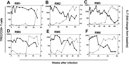
To determine whether the IL-7 increase occurred prior to or following the decline in T-cell subsets, 2 of the best correlates of IL-7 increase, CD4+ T cells and CD8/TREC+ cells, were assessed graphically along with IL-7 levels. With regard to CD4+ T-cell levels, the decline in CD4+ cells generally occurred prior to the increase in IL-7 levels (RM2, RM3, RM5, and RM6; Figures 4D,F,J,L). In fact, CD4+ T-cell decline occurred as early as 25 weeks before the elevation of IL-7 levels (RM2; Figure 4D). In the remaining 2 macaques that progressed rapidly to simian AIDS (RM1 and RM4), the CD4+ T-cell decline occurred concurrently or following the increase in plasma IL-7 levels (Figure 4B,H). Analysis of the TREC data are problematic because elevated IL-7 levels are predicted to increase proliferation of naive T cells,27,28 thereby causing a dilution of the TRECs. We observed CD8/TREC+ levels declining concurrently with (RM1, RM3, RM4, RM5, and RM6; Figure 5A,C-F) or slightly earlier (RM2; Figure 5B) than the increase in plasma IL-7 levels. Based on these results it is difficult to discern whether the declining CD8/TREC+ levels are influencing the IL-7 levels or if the increasing IL-7 levels are inducing naive T-cell proliferation and thereby reducing the CD8/TREC+ levels.
Temporal assessment of the depletion in T-cell receptor excision circles (TREC/CD8+) and IL-7 increase in SIVmac239-infected macaques. The longitudinal assessment of TRECs within the CD8+ T-cell population (black lines, ▵) with plasma IL-7 levels (gray lines, ▦) are presented for SIVmac239-infected macaques RM1 (A), RM2 (B), RM3 (C), RM4 (D), RM5 (E), and RM6 (F).
Temporal assessment of the depletion in T-cell receptor excision circles (TREC/CD8+) and IL-7 increase in SIVmac239-infected macaques. The longitudinal assessment of TRECs within the CD8+ T-cell population (black lines, ▵) with plasma IL-7 levels (gray lines, ▦) are presented for SIVmac239-infected macaques RM1 (A), RM2 (B), RM3 (C), RM4 (D), RM5 (E), and RM6 (F).
In summary, the declining absolute CD4+ T-cell levels were observed to correlate significantly with the increasing plasma IL-7 levels. The observation that the CD4 levels decline prior to the IL-7 increase suggests that during pathogenic SIV infection, IL-7 levels are increased as a homeostatic response to declining T-cell levels in the periphery.
Maintenance of T-cell homeostasis in mangabeys despite high viral loads
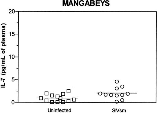
The SIV/macaque animal model is generally used to assess AIDS pathogenesis due to the high viral replication and disease progression observed in the majority of infected animals. Our studies, as described, indicate a role for IL-7 in the maintenance of T-cell homeostasis in SIV-infected macaques and that the loss of T-cell homeostasis may have an impact on disease progression. Another monkey species, the sooty mangabeys, are naturally infected with a related SIV strain that results in a very different disease phenotype. SIVsmm infection in sooty mangabeys is a nonpathogenic infection that does not result in disease progression or simian AIDS.29-31 SIV replicates in the mangabeys to high levels as in macaques29-31 ; still, mangabeys are able to maintain proper T-cell counts.32 Therefore, nonpathogenic SIV infection of mangabeys permits assessment of IL-7 levels in the presence of high viral load but in a functionally intact immune environment with normal CD4+ T-cell levels.29-32 SIVsm infection of mangabeys resulted in only a slight increase in plasma IL-7 levels (not statistically significant; Figure 6) when compared to the several-fold increase observed in the SIV-infected macaques (Figure 1), further indicating the correlation between IL-7 and disease progression. Recently we have shown that SIV-infected mangabeys are able to maintain proper T-cell numbers through the maintenance of high regenerative capacity and attenuated immune activation.32 These studies using this nonpathogenic HIV animal model further indicate the correlation between IL-7 levels and disease progression.
Cross-sectional analysis of plasma IL-7 levels in sooty mangabeys. Plasma IL-7 levels are depicted for 13 uninfected (□) and 11 SIVsm-infected (○) sooty mangabeys, a species that does not progress to simian AIDS. Mean of each group is shown by horizontal line. The minimum detectable level of IL-7 was 0.10 pg/mL.
Cross-sectional analysis of plasma IL-7 levels in sooty mangabeys. Plasma IL-7 levels are depicted for 13 uninfected (□) and 11 SIVsm-infected (○) sooty mangabeys, a species that does not progress to simian AIDS. Mean of each group is shown by horizontal line. The minimum detectable level of IL-7 was 0.10 pg/mL.
Discussion
Relatively stable numbers of T cells are maintained in the periphery through the regulation of both thymus-derived (de novo) T-cell production and postthymic proliferation of mature T cells.27,33,34 IL-7 is one of the key cytokines involved in the regulation of T-cell homeostasis.27,28,35 Here we report that plasma IL-7 levels are dramatically elevated during pathogenic infection in macaques, whereas during the nonpathogenic infection of mangabeys, IL-7 levels are only slightly increased. Using these 2 monkey species with distinct disease outcomes, we demonstrate a correlation between a significant elevation of IL-7 levels and disease progression and provide further evidence for the importance of T-cell homeostasis.
Studies in various clinical conditions, including HIV infection in humans, have shown an elevation in IL-7 levels as a homeostatic response to the decline in T-cell numbers.8,13,14 In this study, analysis of immunologic events in SIVmac239-infected macaques identified a similar increase in plasma IL-7 levels following a decline in CD4+ T cells. Although the mechanisms behind the IL-7 increase are not yet clear, the time delay between CD4+ T-cell decline and IL-7 increase indicate that the regulation may be functioning at the level of increased IL-7 production.8,13 The production of IL-7 by the stromal/dendritic cells may be due to the monitoring of specific T-cell subsets. In addition to total CD4 levels, recent thymic emigrant levels may also be a factor because thymectomy led to a marginally earlier increase in plasma IL-7 levels. IL-7 levels may also be elevated in part from a reduction in the number of IL-7 receptors (due to both declines in T-cell numbers and down-regulation of IL-7 receptor expression) as has been shown by others.36,37
In the 5 SIVmac239-infected macaques that progressed to simian AIDS and death, elevated IL-7 levels were followed by insufficient CD4+ T-cell recovery. This suggests that failure to restore proper T-cell homeostasis by elevated IL-7 levels may be contributing to disease progression in these macaques. Normally, in the absence of a SIV/HIV infection an increase in plasma IL-7 levels may be a transient event, triggered by declining T-cell levels, to stimulate both de novo and peripheral T-cell proliferation.27,34,38 However, infection by SIV in macaques or HIV in humans represents a unique challenge to homeostatic regulation because large numbers of T cells are being continuously killed by the direct and indirect effects of the high viral replication.38 In each of these macaques the elevated IL-7 levels remained stably high until death. Hence, this new, elevated IL-7 “set point” may be indicative of an inherent SIV-induced change to the immune system of the macaque that cannot be reversed by elevated IL-7 levels. Significant phenotypic changes on the T cells including down-regulation of IL-7 receptors induced by SIV infection would further complicate the ability of the immune system to maintain homeostasis because fewer cells are capable of responding to the increased IL-7 levels.36,37 These data suggest that the normal IL-7 function to homeostatically regulate T cells is impaired in the SIV-infected macaques during disease progression.
Recently, in vitro studies as well as studies in the SCID-hu mouse models have demonstrated the ability of IL-7 to increase HIV replication.15,16,20,39,40 In addition, IL-7 has been shown to induce the replication of “latent” virus by the activation of resting T cells.20,41 These studies suggest that IL-7 could have a direct role in disease progression by increasing viral replication. In this study, it was very difficult to discern any effect of IL-7 on viral replication in 5 of the macaques that progressed to simian AIDS, possibly due to the high viral loads present prior to IL-7 increase (Figure 3). However, it is possible that IL-7 could be increasing SIV replication in lymphoid organs without influencing plasma viral loads; therefore, the assessment of lymphoid compartments requires further study. Overall, these data indicate that having ongoing active SIV replication in macaques at times when IL-7 levels are elevated could accelerate disease progression.13,27
Evidence for the role of IL-7 and loss of T-cell homeostasis in SIV disease progression also comes from our studies of the nonpathogenic SIV infection in sooty mangabeys. Sooty mangabeys are the natural host for the SIVsmm virus. Despite high levels of viral replication similar to those observed during pathogenic infection in humans and macaques, mangabeys have stable CD4 counts and do not progress to simian AIDS.29,31 In earlier studies we have shown that mangabeys maintain their T-cell regenerative capacity.32 Further studies in mangabeys have demonstrated a significant correlation between plasma IL-7 levels and T-cell proliferation during SIV T-cell depletion, suggesting that effective homeostatic mechanisms are in place to maintain T-cell numbers despite high viral replication rates32 (M.P. et al, manuscript submitted, October 2003). These studies provide additional evidence that proper T-cell homeostasis is important for protection from developing AIDS and the direct consequences of viral replication alone is not sufficient to account for disease progression.32
Although the majority of the SIVmac239-infected macaques proceed to simian AIDS and death after the IL-7 levels increase, one macaque remained disease free. This long-term asymptomatic/nonprogressor phenotype is observed on occasion and has been associated with specific major histocompatibility complex (MHC) haplotypes,42 although the exact mechanism by which some macaques control their SIV infection is not known. Careful analysis of this macaque in which viral replication is suppressed determined that elevated IL-7 levels are followed by increased T-cell proliferation and maintenance of reasonably high CD4 counts compared to other macaques (Figure 4). Cross-sectional analysis of 3 SIVsmE660-infected macaques (Figure 1) that remain alive after 12 to 19 months of infection with low viral loads (2-3 × 10² viremia) showed similar results of elevated IL-7 levels and reasonably high T-cell numbers (absolute CD4 counts range from 1626 to 1767cells/μL blood). These studies indicate that during conditions in which viral replication is controlled IL-7 increase is able to maintain T-cell homeostasis. Further, the finding that increased IL-7 levels did not result in increased plasma viral load in the slow-progressor macaque RM3 is encouraging for the use of IL-7 as an immunotherapeutic agent. Indeed, these findings are in agreement with a recent study of SIV-infected macaques treated with antiretroviral therapy and given recombinant human (rh) IL-7 for 9 continuous days.43 Administration of exogenous IL-7 resulted in an increase in peripheral T-cell proliferation and no increased viremia following rhIL-7 administration.43 Here the increase in endogenous IL-7 resulted in a transient increase in T-cell proliferation (24 weeks), a finding that may have implications for designing future experiments using IL-7 therapy. In addition to its role in T-cell proliferation, recent studies have demonstrated that IL-7 can maintain T-cell numbers through increased survival of T cells.12,44 These results indicate that IL-7 could be useful for immune recovery when viral replication is suppressed.
HAART suppresses HIV replication, thereby preventing further immune destruction. However, many patients have limited immune recovery due to immune degradation already imparted from the direct and indirect effects of the HIV infection.17,18 This indicates that an adjunct therapy given along with HAART to improve immune reconstitution might be advantageous for HIV patients.45,46 The experiments described here, as well as by other groups,43 indicate the utility of the SIV/macaque animal model for assessing various immune therapeutics that might be administered to assist immune recovery during HAART. Importantly, the influence of any therapy, such as exogenous IL-7 administration, could potentially accelerate the rate of disease progression making it important that the initial experiments be conducted in the SIV animal model. The studies described here depicting endogenous IL-7 levels during T-cell depletion provide the necessary framework from which any future exogenous IL-7 or similar therapies could be compared and evaluated in nonhuman primate models.
Prepublished online as Blood First Edition Paper, October 2, 2003; DOI 10.1182/blood-2003-03-0874.
Supported by National Institutes of Health grants AI35522 (D.L.S.) and DE12926 (D.L.S.), and the Elizabeth Glaser Pediatric AIDS Foundation (D.L.S.).
The publication costs of this article were defrayed in part by page charge payment. Therefore, and solely to indicate this fact, this article is hereby marked “advertisement” in accordance with 18 U.S.C. section 1734.
The authors thank Stephanie Ehnert, David Maggio, and Chris Ibegbu for assistance with processing blood samples at the Yerkes Regional Primate Center for sample collection; Richard Koup and Yukari Okamoto for helpful discussions and guidance; Ashley Barry at the Emory Vaccine Center for assisting in the acquisition and preparation of plasma samples from SIV+ macaques; William-Frawley for statistical analysis; Mike Piatek and Jeff Lifson for undertaking the quantification of plasma viral load; Felecia Ware and Courtney Matthews for excellent technical assistance; and Victor Garcia, Louis Picker, Celia LaBranche, Jeffrey Milush, Dejiang Zhou, and Jason Mendoza for helpful comments regarding the preparation of the manuscript.


This feature is available to Subscribers Only
Sign In or Create an Account Close Modal