HEMOSTASIS, THROMBOSIS, AND VASCULAR BIOLOGY
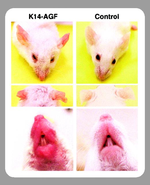
The complex network of positive and negative regulators of vascular homeostasis welcomes a newcomer with apparently unique properties. After finding that the angiopoietin-related protein designated angiopoietin-related growth factor (AGF) acts as a growth factor for epidermal keratinocytes,1 in this issue of Blood Oike and colleagues (page 3760) describe a number of properties that identify AGF as a possibly relevant player in the angiogenesis field. Transgenic mice expressing AGF in epidermal keratinocytes (named K14-AGF mice) show a peculiar phenotype: they are grossly red, particularly in the ears, snout, and foot sole. When electron microscopy was used to investigate the skin of these blush mice, authors found an increased frequency of capillary-sized vessels with apparently normal pericytes. As epidermal keratinocytes are known for releasing a relevant amount of crucial angiogenic factors such as vascular endothelial growth factor (VEGF) and basic fibroblast growth factor (bFGF), the authors investigated by quantitative reverse-transcription polymerase chain reaction (RT-PCR) the expression levels of these and other angiogenic growth factors in the skin of K14-AGF mice. The finding that VEGF, bFGF, angiopoietin 1 (Ang1), and other angiogenic factors were not increased (or even decreased) in the skin of K14-AGF mice was a clear hint of the angiogenic properties of AGF. Other classical tools for the measurement of angiogenic activity, such as matrigel plug assays and mouse corneal assays, confirmed the positive effect of AGF on in vivo vascular formation. Furthermore, a vascular permeability assay indicated that AGF induces vascular leakiness, albeit with lower efficacy when compared with VEGF, also known as vascular permeability factor. Conversely to VEGF, however, AGF does not increase endothelial cell growth nor does it have synergistic activity on VEGF-induced endothelial cell proliferation.
What now seems urgent to understand about AGF? First of all, we lack a cognate receptor. Although AGF contains the fibrinogen-like domain that is the angiopoietins' binding site to the TIE2 receptor, AGF (like other angiopoietin-related proteins) does not bind to TIE1 or TIE2. In addition to tyrosine kinases (the family of VEGF and angiopoietins receptors), the list of suspects should include integrins such as αvβ3, which has been recently identified as a possible receptor for the angiopoietin-related protein angiopoietin-like protein 3 (ANGPTL3).2 A second relevant question is whether AGF, in addition to angiogenesis, plays a role in vasculogenesis (ie, in the progenitor cell–derived generation of new blood vessels). The finding that AGF directly promotes the endothelial cell chemotactic activity deserves further investigation, because it might be possible that AGF (as VEGF) promotes the mobilization of endothelial progenitors from the marrow to the peripheral blood. Finally, in spite of the evident blush phenotype, there are normal AGF levels in the circulation of K14-AGF mice compared with controls. Considering the recent evidence that microenvironmental concentrations (rather than total dose) of angiogenic growth factors such as VEGF may dictate the threshold between pathologic and physiologic angiogenesis,3 it would be of interest to assess microenvironmental AGF levels in diseases where angiogenesis (or vasculogenesis) plays a distinct role.


This feature is available to Subscribers Only
Sign In or Create an Account Close Modal