Abstract
Cytochrome P450 1B1 (CYP1B1), a drug-metabolizing extrahepatic enzyme, was recently shown to be overexpressed in multiple types of cancer. Such tumor-associated genes may be useful targets for anticancer therapy, particularly cancer immunotherapeutics. We identified HLA-A*0201–binding peptides and a naturally processed and presented T-cell epitope capable of inducing CYP1B1-specific cytotoxic T lymphocytes (CTLs) in HLA-A2 transgenic mice. Furthermore, the induction of CYP1B1-specific T cells was demonstrated in healthy donors and cancer patients. These T cells efficiently lysed target cells pulsed with the cognate peptide. More important, HLA-A2–matched tumor cell lines and primary malignant cells were also recognized by CYP1B1-specific CTLs. These findings form the basis of a phase 1 clinical trial exploring a DNA-based vector encoding CYP1B1 for widely applicable cancer immunotherapy conducted at the Dana-Farber Cancer Institute.
Introduction
Cytochrome P450 1B1 (CYP1B1) is an extrahepatic cytochrome P450 enzyme that has been associated with the activation of environmental carcinogens.1,2 Expression of CYP1B1 is upregulated early during malignant transformation.3 Furthermore, CYP1B1 was reported to be overexpressed in most human malignancies with minimal expression on critical healthy tissues.4 Such shared tumor-associated antigens may be useful targets for the development of widely applicable cancer immunotherapeutics.5 The expression of the antigen in some rare healthy tissues does not necessarily exclude such genes from serving as immunological targets, as has been shown for other tumor antigens such as MUC-1,6 survivin,7-9 telomerase,10,11 ras,12 or p53.13 We were therefore interested in determining whether a gene such as CYP1B1, expressed early during the carcinogenic process, might serve as a target for cytotoxic T lymphocytes (CTLs).
In the present report, we characterize the CTL response to HLA-A*0201–restricted epitopes derived from CYP1B1. One CYP1B1-derived epitope was isolated and identified from HLA-A*0201 expressed in several tissue-specific tumor cells. Additional epitopes were predicted. The immunogenicity of CYP1B1 was demonstrated in HLA-A2 transgenic mice. Functional CYP1B1-specific T cells capable of lysing tumor cells were established from healthy donors and cancer patients. Preliminary results from a clinical trial targeting CYP1B1 as a tumor antigen suggest that this antigen might be an attractive candidate to be integrated in widely applicable cancer immunotherapeutics.
Patients, materials, and methods
Healthy volunteer and patient samples
Following informed consent and approval by the Dana-Farber Cancer Institute's Review Board, peripheral blood from healthy donors and cancer patients (multiple myeloma, n = 6; follicular lymphoma, n = 1; prostate cancer, n = 1) was obtained by leukapheresis or phlebotomy. Primary follicular lymphoma (FL), acute lymphoblastic leukemia (ALL), and acute myeloid leukemia (AML) samples and malignant tissue blocks were obtained from discarded specimens. Healthy tissue specimens were obtained from the tissue library at IMPATH Biopharmaceutical Services (New York, NY).
Cell lines
The cell lines K029 (melanoma) and 36M (ovarian carcinoma) were kind gifts of Drs Dranoff and Cannistra (Harvard Medical School, Boston, MA). T2, COS, U266, HS-Sultan, IM-9, SK-MEL-2, SK-OV-3, JY, KATO III, and EL-4 cell lines were obtained from ATCC (Manassas, VA). EL-4 cells were transfected with the HLA-A2/Kb cDNA inserted into the pSV2neo vector.
Immunohistochemistry of CYP1B1
Sections of paraffin-embedded tumor specimens were prepared for immunostaining following standard procedures. Slides were then incubated for 1 hour with 150 to 200 μL polyclonal rabbit anti-CYP1B1 (generous gift of Dr Marcus, University of New Mexico, Albuquerque)4 or nonspecific polyclonal rabbit immunoglobulin. Slides were rinsed and treated with Link, Label, and Substrate (Biogenix StrAviGen Multilink kit; Biogenex, San Ramon, CA) and counterstained with hematoxylin. Analysis of healthy tissue sections was performed by IMPATH Biopharmaceutical Services on frozen tissue sections. In brief, OCT compound (Miles Laboratories, Naperville, IL) embedded tissues were cut and fixed in acetone. After blocking by hydrogen peroxide, slides were incubated with the monoclonal mouse–antihuman CYP1B1 5D3 antibody14 (10 μg/mL) or a murine immunoglobulin G1k (IgG1k) control antibody (DAKO, Carpinteria, CA) for 30 minutes. Tissues were counterstained with hematoxylin (AMTS, Lodi, CA). Breast cancer specimens were used as positive controls, and healthy liver was included as negative control. Samples were evaluated by pathologists, and the staining intensity of test slides was judged relative to the intensity of a control slide containing an adjacent section stained with an irrelevant negative control. In keeping with standard pathology practice, staining was reported at the highest level of intensity observed in all tissue elements.
Western blot analysis
CYP1B1 protein was isolated following standard protocols for preparation of the microsomal protein fraction by differential speed centrifugation. Recombinant human (rh) CYP1B1 (0.015 pmol; Gentest, Woburn, MA) was used as a control. Membranes were probed with purified monoclonal mouse–antihuman CYP1B1 clone 5D3,14 and secondary goat–antimouse horseradish peroxidase (HRP; SC Biotechnologies, Santa Cruz, CA). Bands were visualized by enhanced chemiluminescence detection (Amersham Pharmacia, Piscataway, NJ).
Peptides and peptide prediction
Peptides were purchased from Sigma Genosys Biotechnologies (The Woodlands, TX), Harvard Medical School Biopolymers Laboratory (Boston, MA), New England Peptides (Fitchburg, MA), and Multiple Peptide Systems (San Diego, CA): CYP77 (LARRYGDV), CYP190 (FLDPRPLTV), CYP239 (SLVDVMPWL), HTLV-TAX11 (LLFGYPVYV), EBV-BMLF1 280 (GLCTLVAML), the idiotype derived peptide (AHTKDGFNF), MAGE-3 F271 (FLWGPRALV), and hepatitis B virus (HBV) core F18 (FLPSDYFPSV). Binding of peptides to HLA-A*0201 was predicted using 3 algorithms: BIMAS,15 LPpep (kindly provided by Z. Weng, Boston University), and SYFPEITHI.16 The peptides were ranked for each algorithm and sorted by a cumulative score.
HLA-A*0201 peptide binding and complex stability assay
Following previously described methods, peptide binding was assayed using T2 cells.10 For complex stability T2 cells were washed 3 times in serum-free Iscove modified Dulbecco medium (IMDM) after peptide incubation, and aliquots of cells were replated and incubated at 37°C in the absence of exogenous peptide. HLA-A*0201 expression was measured by flow cytometry using fluorescein isothiocyanate (FITC)–conjugated monoclonal antibody (mAb) BB7.2 (ATCC) at 0, 2, 4, 6, and 24 hours after peptide withdrawal. The increase of HLA-A*0201 expression on T2 cells reflects the stabilization of major histocompatibility complexes (MHCs) by the addition of exogenous peptides and was quantified using the fluorescence index (FI = (MFIpeptide-pulsed T2/MFIunpulsed T2) – 1). The half-life of HLA-A*0201 complexes on the surface was calculated using linear regression analysis (y = yo + a × e exp(– b × x)) (SigmaPlot). Peptides were also tested for their capacity to bind recombinant HLA-A*0201 molecules in vitro, as previously described.17 The HBV core 18-27 peptide was used as the radiolabeled probe.
HLA-A*0201 isolation and peptide repertoire analysis
An automated high-performance liquid chromatography (HPLC)–based immunoaffinity chromatography system was used to rapidly purify the HLA molecules.18 The intact peptide repertoire was isolated by acid extraction and separated by reverse-phase HPLC. Peptide sequencing was accomplished by automated microcapillary LC/MS/MS analysis using ion trap technology as previously described.19 Briefly, aliquots (0.5-5 μL) of each peptide-containing fraction were concentrated using a microtrap (peptide captrap; Michrom BioResources, Auburn, CA) in place of the sample loop in the autoinjector and were analyzed by either of 2 ion trap systems (LCQ classic or LCQ Deca; Thermo-Finnigan, San Jose, CA) equipped with automated data-dependent selection of precursor ions for subsequent MS/MS analysis. After acquisition of an MS/MS spectrum, the precursor ion was dynamically written to an exclusion list where it resided for 30 seconds before an MS/MS spectrum for this precursor ion could be collected for a second time. The complete data set of MS/MS spectra was searched against a protein database that contained homologues of the CYP1B1 protein. A synthetic homologue of CYP190 was analyzed by LC/MS/MS to confirm the detection of naturally processed CYP1B1 peptides.
Generation of CTLs
Cytotoxicity assay
CTL lines were used after at least 4 antigenic stimulations in standard chromium Cr 51 release assays, as previously described.10 For testing endogenous processing of CYP1B1-derived peptides, COS cells stably expressing HLA-A*0201 were transfected with mini-gene constructs encoding either enhanced green fluorescent protein (EGFP) linked to huCYP1B1 aa170-213, EGFP linked to huCYP1B1 aa205-352, or EGFP alone. COS cells were sorted for EGFP expression before use. Alternatively, a recombinant vaccinia virus containing full-length human CYP1B1 cDNA21 was generated22 and used to infect HLA-A*0201+ monocyte-derived and matured dendritic cells (DCs)23 for 16 to 18 hours (multiplicity of infection [MOI], 10).
Human IFN-γ ELISPOT assay
ELISPOT analysis for interferon-γ (IFN-γ) secretion using human peripheral blood mononuclear cells (PBMCs) was carried out as previously described.24 Peptides from human T-cell leukemia virus-1 (HTLV-1) and Epstein-Barr virus (EBV) BMLF-1 were used as negative and positive controls, respectively.
CTL induction in HLA-A2/Kb transgenic mice
Female HLA-A2/Kb transgenic mice were obtained from The Scripps Research Institute (La Jolla, CA).25 v/huCYP1B1d3 consists of a 1.6-kb full-length human CYP1B1 cDNA coding sequence with introduced nucleotide changes that produce 3 single amino acid substitutions (Trp57Cys, Gly61Glu, and Gly365Trp) in the huCYP1B1 coding sequence, to inhibit the enzymatic activity of the CYP1B1 protein (data not shown) cloned into cytomegalovirus (CMV) promoter-based mammalian expression vector. Plasmid DNA was made with plasmid purification kits (Qiagen, Chatsworth, CA) and encapsulated in plasminogen (PLG) microparticles26 and then was injected intramuscularly (total dose, 100 μg DNA) at 2-week intervals and assayed 9 to 12 days after the last immunization.
Mouse IFN-γ ELISPOT assay
Murine CD8+ T-cell responses to CYP1B1 were analyzed by IFN-γ ELISPOT (R&D Systems, Minneapolis, MN). Pooled spleen cells from 2 mice per treatment group were enriched for CD8+ T cells and were plated at 1 × 10⁵ cells/well. T cells were stimulated with 1 × 10⁵ EL4-A2Kb cells/well pulsed with 10 μg/mL peptide or infected with recombinant vaccinia virus before plating (MOI, 10). Plates were incubated for 24 hours, developed, and analyzed by automated image analysis (Zellnet Consulting, New York, NY).
Results
CYP1B1 protein is highly expressed in malignant but not healthy cells
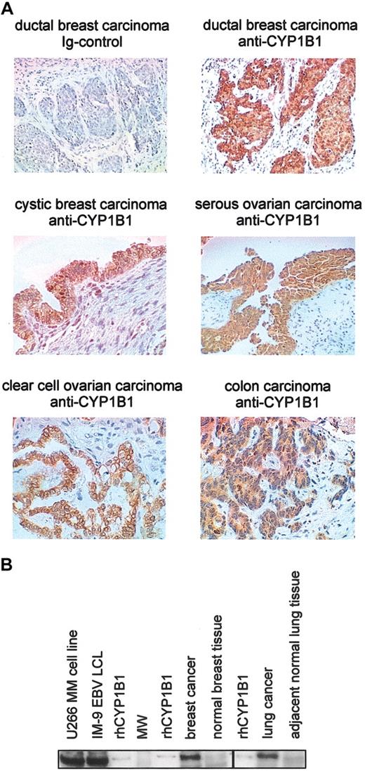
Among the proteins associated with early events of malignant transformation induced by chemical carcinogens, cytochrome P450 1B1 protein has been reported to be overexpressed in most cancers tested.4,14,27 Expression of mRNA for CYP1B1 has been reported for some healthy tissues,28-31 and a single report has also suggested protein expression in several human tissues.32 We extended these findings and demonstrate homogenous and significant protein overexpression in multiple randomly selected tumor specimens (Figure 1; Table 1). A comprehensive screen of 32 healthy tissues (3 specimens for each) derived from autopsy material from otherwise healthy patients who died of trauma (Table 1) was also conducted. Breast, ovarian, and colon carcinoma (Figure 1A) demonstrated high to very high CYP1B1 staining in neoplastic cells, whereas stromal compartments and surrounding healthy tissue were negative. Significant CYP1B1 protein overexpression was observed in 9 of 9 cancer cell lines of various histologies and in primary tumor specimens compared with healthy adjacent tissue (Figure 1B; Table 1; and data not shown). Among all healthy tissues tested, fallopian tube showed the highest level with an apical cellular distribution. Breast, uterine, and ureter specimens showed intermediate to high levels of staining (++) with less than 20% of tissue-specific cells displaying high intensity. Weak to intermediate staining (+) was detected in 2 of 3 skin samples with less than 20% of cells displaying intermediate intensity. In addition, variable and weak staining (+/–) was detected in 10% to 25% of cells of prostate (2 of 3 samples), 20% of pancreas (2 of 3), 50% of pituitary (1 of 3), 40% of colon (1 of 3), 10% of bladder (1 of 3), 20% of small intestine (1 of 3), and 10% of thymus (1 of 3) tissue-specific cells. Taken together, CYP1B1 is expressed in some healthy tissues but is strongly overexpressed in all malignancies tested to date.
Expression of CYP1B1 protein in healthy and malignant tissue. (A) Expression of CYP1B1 in human tumors detected by immunohistochemistry using a polyclonal antibody. All tissues were also stained with an immunoglobulin control (upper left panel and data not shown). (B) Western blot analysis of microsomal fractions (30 μg per sample) from healthy and malignant tissue using the monoclonal CYP1B1 antibody (similar results were obtained using the polyclonal antibody). Healthy and malignant lung tissue samples were from the same patient; healthy and malignant breast tissue samples were from 2 persons. As a positive control, 0.015 pmol rhCYP1B1 was loaded.
Expression of CYP1B1 protein in healthy and malignant tissue. (A) Expression of CYP1B1 in human tumors detected by immunohistochemistry using a polyclonal antibody. All tissues were also stained with an immunoglobulin control (upper left panel and data not shown). (B) Western blot analysis of microsomal fractions (30 μg per sample) from healthy and malignant tissue using the monoclonal CYP1B1 antibody (similar results were obtained using the polyclonal antibody). Healthy and malignant lung tissue samples were from the same patient; healthy and malignant breast tissue samples were from 2 persons. As a positive control, 0.015 pmol rhCYP1B1 was loaded.
Analysis of CYP1B1 expression
Tissue . | Expression . |
|---|---|
| Cancer | |
| Acute lymphocytic leukemia* | +++ |
| Acute myeloid leukemia* | +++ |
| Breast cancer*† | +++ |
| Colon carcinoma* | +++ |
| Esophagus carcinoma† | +++ |
| Lung cancer* | +++ |
| Lymphoma* | +++ |
| Multiple myeloma*‡ | +++ |
| Melanoma*‡ | +++ |
| Ovarian carcinoma*† | +++ |
| Rhabdomyosarcoma† | +++ |
| Normal | |
| Fallopian tube* | +++ |
| Breast* | ++ |
| Uterus (cervix, endometrium)* | ++ |
| Ureter* | ++ |
| Skin* | + |
| Prostate* | +/- |
| Pancreas* | +/- |
| Pituitary gland* | +/- |
| Colon* | +/- |
| Bladder* | +/- |
| Small intestine* | +/- |
| Thymus* | +/- |
| Adrenal gland* | - |
| Blood cells* | - |
| Bone marrow* | - |
| Cerebellum* | - |
| Cerebral cortex* | - |
| Eye (retinal)* | - |
| Heart* | - |
| Kidney (glomerulus, tubule)* | - |
| Liver* | - |
| Lung* | - |
| Lymph node* | - |
| Ovary* | - |
| Parathyroid* | - |
| Placenta* | - |
| Skeletal muscle* | - |
| Spinal cord* | - |
| Spleen* | - |
| Stomach* | - |
| Testis* | - |
| Thyroid* | - |
For normal tissues, tissue sections from 3 subjects were evaluated for CYP1B1 expression. Breast carcinoma was used as a positive control, always showing high expression. +++ indicates more than 20% of cells with high expression in all 3 normal samples; ++, intermediate expression, or less than 20% of cells with high expression in at least one sample; +, low expression, or less than 20% of cells with intermediate expression in at least one sample; +/-, weak expression in at least one sample; and -, no expression in any sample tested.
Tissues analyzed by immunohistochemistry (IHC).
Tissues analyzed by Western blot.
Analysis not performed on primary tumor tissue.
Elution of CYP1B1-derived epitopes from tumor cells
A biochemical approach was undertaken to identify epitopes presented by HLA-A*0201 from several tumor samples, including multiple myeloma, gastric carcinoma, colorectal adenocarcinoma, and EBV-transformed B cells. Automated HPLC-based immunoaffinity chromatography was followed by HPLC-based peptide repertoire fractionation and mass spectrometry. Peptide sequencing was accomplished by automated LC/MS/MS analysis using ion trap technology.33
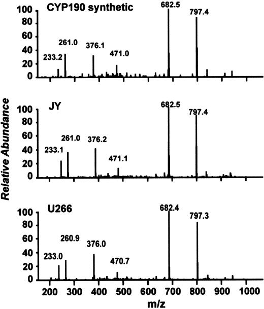
A search of MS/MS spectra against a protein database containing only homologues of the CYP1B1 protein revealed one epitope derived from CPY1B1 (referred to as CYP190; FLDPRPLTV; Figure 2). To confirm the nature of CPY190, a synthetic homologue was characterized with respect to HPLC elution profile and the precursor ion and MS/MS fragmentation pattern. CYP190 was confirmed using the ion trap by specifically targeting and fragmenting all m/z values in the appropriate peptide-containing fractions isolated from tumor cells (Figure 2).
LC/MS/MS spectra. Comparison of LC/MS/MS spectra for the HLA-A*0201–associated, naturally processed and presented CYP190 peptide isolated from the EBV-transformed B-cell line JY (middle panel) and the myeloma cell line U266 (lower panel) compared with the synthetic homologue (upper panel). The primary sequence of CYP190 is depicted in the upper left corner. Retention times were identical (data not shown), and the fragmentation patterns confirm the CYP190 sequence identity. Similar spectra were obtained from gastric carcinoma, several colorectal adenocarcinomas, and EBV-transformed B-cell lines derived from multiple subjects (data not shown).
LC/MS/MS spectra. Comparison of LC/MS/MS spectra for the HLA-A*0201–associated, naturally processed and presented CYP190 peptide isolated from the EBV-transformed B-cell line JY (middle panel) and the myeloma cell line U266 (lower panel) compared with the synthetic homologue (upper panel). The primary sequence of CYP190 is depicted in the upper left corner. Retention times were identical (data not shown), and the fragmentation patterns confirm the CYP190 sequence identity. Similar spectra were obtained from gastric carcinoma, several colorectal adenocarcinomas, and EBV-transformed B-cell lines derived from multiple subjects (data not shown).
Prediction of additional CYP1B1-derived epitopes
Additional epitopes for characterization of CTL responses against CYP1B1 were predicted using 3 computational algorithms (BIMAS, SYFPEITHI, and LPpep). Among the 10 most likely candidates, the peptide/HLA-A*0201 complex stability (t1/2) was the highest for CYP190, whereas the predicted CYP239 epitope (SLVDVMPWL) consistently showed the highest binding affinity (FI) in a cellular binding assay (Table 2). These results were confirmed by affinity measurements to recombinant HLA-A*0201 (IC50) using an inhibition affinity assay17 (Table 2). Based on these findings the epitopes CYP190 and CYP239 were chosen for immunologic analysis.
Binding affinity and HLA/peptide complex stability of CYP1B1-derived and control peptides to human HLA-A*0201
. | . | . | . | T2 assay . | . | . | |
|---|---|---|---|---|---|---|---|
| Peptide . | Protein . | Position . | Sequence . | Maximum, FI* . | t1/2, h† . | IC50, nmol‡ . | |
| CYP190 | CYP1B1 | 190 | FLDPRPLTV | 3.7 | 10.1 | 67 | |
| CYP239 | CYP1B1 | 239 | SLVDVMPWL | 3.9 | 3.3 | 63 | |
| Controls | |||||||
| Positive | MAGE-3 | 271 | FLWGPRALV | 3.2 | 3.4 | ND | |
| Negative | Idiotype | 98 | AHTKDGFNF | 0 | ND | ND | |
FI indicates mean fluorescence with peptide/mean fluorescence without peptide; ND, not determined.
The known HLA-A*0201-binding peptide from MAGE-3 and a nonbinding peptide from an idiotype sequence were used as positive and negative controls. Results from 1 of 5 representative experiments are shown.
Time to half-maximal FI after withdrawal of peptide was calculated using linear regression analysis.
Peptide concentration necessary to inhibit binding of a labeled reference peptide HBV core 18-27 to HLA-A*0201 by 50%.
Immunity against CYP1B1 in HLA-A2 transgenic mice
In vivo immunogenicity of CYP1B1 was assessed using HLA-A2/Kb transgenic mice.25 Mice were vaccinated with a plasmid encoding full-length mutated human CYP1B1 (v/huCYP1B1d3) or vector control (v). To target DNA for uptake by antigen-presenting cells (APCs), the plasmid DNA was encapsulated in biodegradable microparticles composed of PLG. After 3 intramuscular vaccinations, CD8+-enriched splenocytes showed specific IFN-γ reactivity against EL4-A2/Kb cells expressing full-length huCYP1B1 (Figure 3A). Nonimmunized animals or animals immunized with a control vector showed no IFN-γ response. We further investigated whether mice immunized with full-length huCYP1B1d3 would show reactivity against the HLA-A*0201 binding epitopes defined above. All animals immunized with v/huCYP1B1d3 regularly showed strong IFN-γ production when EL4-A2/Kb cells were pulsed with CYP190 (3 of 3 experiments; Figure 3B). Weak reactivity against CYP239 was seen only in one experiment, suggesting that CYP190 is the immunodominant epitope when stimulating with the whole huCYP1B1 cDNA.
Ex vivo IFN-γ ELISPOT analysis of T-cell reactivity in HLA-A2/Kb mice vaccinated with a huCYP1B1-encoding DNA construct. (A) HLA-A2/Kb mice were primed and boosted twice with PLG-microparticle–encapsulated v/huCYP1B1d3 or control vector or were not vaccinated. Twelve days after the second boost, CD8+-enriched spleen cells were tested for reactivity against EL4-A2/Kb cells infected with vaccinia encoding huCYP1B1 (vac-huCYP1B1), vaccinia wild-type (vac-wt), or noninfected control. Results are shown as IFN-γ spot-forming cells (SFCs)/106 CD8+ T cells. (B) The same spleen cells from vaccinated and control mice were used to measure IFN-γ release in response to EL4-A2/Kb cells untreated or pulsed with the CYP190 peptide. (C) IFN-γ ELISPOT analysis of spleen cells from mice primed and boosted twice with v/huCYP1B1d3 in PLG microparticles. EL4-A2/Kb cells pulsed with the HLA-A*0201–binding human epitope CYP190 or the Kb-binding shared mouse/human epitope CYP77 were used as stimulators. In all assays, PHA was used as a positive control (data not shown), and IFN-γ background secretion was determined without stimulation.
Ex vivo IFN-γ ELISPOT analysis of T-cell reactivity in HLA-A2/Kb mice vaccinated with a huCYP1B1-encoding DNA construct. (A) HLA-A2/Kb mice were primed and boosted twice with PLG-microparticle–encapsulated v/huCYP1B1d3 or control vector or were not vaccinated. Twelve days after the second boost, CD8+-enriched spleen cells were tested for reactivity against EL4-A2/Kb cells infected with vaccinia encoding huCYP1B1 (vac-huCYP1B1), vaccinia wild-type (vac-wt), or noninfected control. Results are shown as IFN-γ spot-forming cells (SFCs)/106 CD8+ T cells. (B) The same spleen cells from vaccinated and control mice were used to measure IFN-γ release in response to EL4-A2/Kb cells untreated or pulsed with the CYP190 peptide. (C) IFN-γ ELISPOT analysis of spleen cells from mice primed and boosted twice with v/huCYP1B1d3 in PLG microparticles. EL4-A2/Kb cells pulsed with the HLA-A*0201–binding human epitope CYP190 or the Kb-binding shared mouse/human epitope CYP77 were used as stimulators. In all assays, PHA was used as a positive control (data not shown), and IFN-γ background secretion was determined without stimulation.
Human and mouse CYP1B1 show 75% sequence identity and 81% homology. Nevertheless, CYP190 differs in 4 and CYP239 in 2 amino acids between mice and humans. Therefore, we elected to identify an additional human epitope that was 100% identical to the murine sequences. Using MHC binding prediction algorithms (SYFPEITHII, BIMAS) and IFN-γ ELISPOT screening, we identified a Kb-restricted peptide, CYP77, that is identical in human and mouse. HLA-A2/Kb transgenic mice were immunized using the same vaccination strategy with the full-length v/huCYP1B1d3 construct. Immunity was detected against CYP77 in 2 of 2 vaccination experiments (Figure 3C). The frequency of CTLs reacting against the self-epitope CYP77 in these experiments reached 50% of the frequency of CYP190-specific CTLs. Complete histologic examination of all major organs, including fallopian tube, mammary gland, and uterus, did not reveal any pathologic changes associated with autoimmune phenomena in empty vector- or v/huCYP1B1d3-vaccinated mice (data not shown). Taken together, immunity to CYP1B1 was induced efficiently in HLA-A2/Kb transgenic mice. Despite the efficient induction of immunity to the shared self-peptide CYP77, there was no evidence of autoimmune phenomena.
CYP190- and CYP239-reactive T cells in healthy volunteers and cancer patients
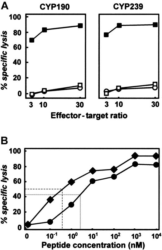
After demonstrating immunogenicity of CYP1B1-derived epitopes in HLA-A2 transgenic mice, we investigated whether these epitopes would also trigger specific and functional CTL responses in HLA-A*0201–positive healthy donors and cancer patients. Peptide-specific T cells were expanded by weekly stimulations with either CYP190 or CYP239 peptide presented on autologous APCs. In more than 70% of healthy HLA-A*0201–positive donors tested, CYP190- and CYP239-specific CD8+ T cells were generated that specifically lysed peptide-pulsed T2 cells (Figure 4A; Table 3). Moreover, specific T cells were also successfully expanded in vitro from 2 of 2 cancer patients against CYP190 and from 5 of 5 patients against CYP239. T-cell lines were peptide specific because target cells loaded with irrelevant peptides were not lysed (Figure 4A). HLA restriction was demonstrated by the lack of lysis of HLA-A*0201–mismatched target cells (data not shown). Avidity of CTL lines was estimated in peptide titration studies (Figure 4B) indicating that CYP190- and CYP239-specific CTLs were of intermediate to high avidity.34 Epitope-specific CTLs were enumerated by IFN-γ ELISPOT assay. For both peptides the frequency of epitope-specific T cells ranged between 0.5% and 3% in expanded CTL lines (data not shown). These results are comparable to previously published data for human telomerase reverse transcriptase (hTERT),24 gp100,35 or proteinase-3.36 To assess whether these CYP1B1-derived epitopes are part of a preexisting antitumor immune response, we analyzed T cells derived from peripheral blood of 5 HLA-A*0201–positive healthy donors and 8 HLA-A*0201–positive cancer patients using IFN-γ ELISPOT assay (Table 4). We were unable to detect CYP190- or CYP239-specific CTLs above background level in either group, suggesting that these CYP1B1-derived epitopes are not targeted by the endogenous antitumor immune response.
CTL recognizing the CYP190 or CYP239 peptide can be generated from cancer patients and healthy donors. (A) After 4 ex vivo antigenic stimulations, CTLs raised from healthy HLA-A*0201 donors against CYP190 or CYP239 peptide specifically lysed T2 cells pulsed with 20 μg/mL cognate peptide (▪) but not unpulsed T2 cells (□) or T2 cells pulsed with an irrelevant peptide (○, F271 from MAGE-3). (B) Cytotoxicity of CYP190-specific (•) and CYP239-specific (♦) CTLs against T2 cells pulsed with increasing concentrations of cognate peptide (effector-target ratio, 10:1). Dashed lines reflect the peptide concentration at which half-maximal lysis was achieved. Results are representative of 2 independent experiments.
CTL recognizing the CYP190 or CYP239 peptide can be generated from cancer patients and healthy donors. (A) After 4 ex vivo antigenic stimulations, CTLs raised from healthy HLA-A*0201 donors against CYP190 or CYP239 peptide specifically lysed T2 cells pulsed with 20 μg/mL cognate peptide (▪) but not unpulsed T2 cells (□) or T2 cells pulsed with an irrelevant peptide (○, F271 from MAGE-3). (B) Cytotoxicity of CYP190-specific (•) and CYP239-specific (♦) CTLs against T2 cells pulsed with increasing concentrations of cognate peptide (effector-target ratio, 10:1). Dashed lines reflect the peptide concentration at which half-maximal lysis was achieved. Results are representative of 2 independent experiments.
Efficiency for the induction of peptide-specific CTLs from healthy donors and cancer patients
. | Healthy donors . | Cancer patients . |
|---|---|---|
| CYP190 | 6 of 8 | 2 of 2 |
| CYP239 | 15 of 21 | 5 of 5 |
. | Healthy donors . | Cancer patients . |
|---|---|---|
| CYP190 | 6 of 8 | 2 of 2 |
| CYP239 | 15 of 21 | 5 of 5 |
Data shown are donors or patients for whom CTL could be generated ex vivo.
Detection of IFN-γ–secreting cells by ELISPOT analysis in the peripheral blood of cancer patients and healthy donors
. | Diagnosis . | CYP190* . | CYP239 . | TAX† . | EBV . | OKT-3 . |
|---|---|---|---|---|---|---|
| Cancer patients | MM | 15 | 5 | 10 | 116 | 1335 |
| MM | 0 | 5 | 5 | 152 | 1289 | |
| FL | 0 | ND | 2 | 60 | 756 | |
| CaP | 16 | ND | 5 | 37 | 654 | |
| MM | ND | 0 | 0 | 46 | 922 | |
| MM | ND | 5 | 0 | 10 | 670 | |
| MM | ND | 0 | 2 | 18 | 1489 | |
| MM | 0 | ND | 0 | 216 | 1205 | |
| Healthy donors | HD | 0 | 0 | 0 | 168 | 792 |
| HD | 0 | 0 | 7 | 109 | 252 | |
| HD | 0 | 0 | 0 | 120 | 1894 | |
| HD | 0 | 0 | 0 | 335 | 756 | |
| HD | 7 | 0 | 7 | 65 | 562 |
MM indicates multiple myeloma; FL, follicular lymphoma; ND, not determined; CaP, cancer of the prostate; and HD, healthy donor.
All values given in spot-forming cells/100 000 CD8+ T cells.
HTLV-TAX, negative control; EBV-BMLF-1, positive control.
Recognition of endogenously processed CYP190 and CYP239 epitopes
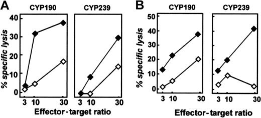
Recognition of endogenously processed CYP1B1-derived peptides by human CTLs was evaluated using (1) COS cells transfected with HLA-A*0201 and minigene constructs containing the CYP190 (aa173-205) and CYP239 (aa213-352) epitopes, (2) DCs infected with a vaccinia construct encoding full-length CYP1B1 cDNA, and (3) HLA-matched tumor cell lines and primary tumors. CYP190- and CYP239-specific CTLs showed significant lysis of CYP1B1 minigene-transfected COS cells compared with vector-control–transfected COS cells (Figure 5A). Similarly, DCs infected with the vaccinia construct containing CYP1B1, but not the wild-type vaccinia virus, were lysed by CYP190- and CYP239-specific CTLs (data not shown). As exemplified in Table 5, a variety of tumor cells, including multiple myeloma (U266), ovarian carcinoma (36M), melanoma (K029), and EBV-transformed lymphoid cell lines (IM-9), were lysed by CYP1B1-specific CTLs. HLA-A*0201–positive healthy monocytes used as controls were not lysed. Lysis of tumor cell lines was equally demonstrated for CTLs derived from healthy donors or cancer patients. HLA-A*0201–negative tumor cell lines were not killed (data not shown). As exemplified in Figure 5B using follicular lymphoma (FL) cells, CYP190- and CYP239-specific CTLs demonstrated comparable lysis of HLA-A*0201–matched primary tumor cells. The particular HLA-A*0201–mismatched FL sample used as the control consistently showed a higher background for CYP190-specific CTLs. In addition, CYP190- and CYP239-specific CTLs displayed specific lysis of HLA-A*0201–positive acute leukemia blasts (data not shown).
CYP1B1-derived peptides are endogenously processed and presented by tumor cells. (A) Specific lysis of COS/A*0201 cells transfected with a construct encoding EGFP + CYP1B1 amino acids (aa) 170-213 by CYP190-specific CTLs (left panel, ♦) or aa 205-352 by CYP239-specific CTLs (right panel, ♦) but not control EGFP-transfected COS cells (⋄). (B) Lysis of primary follicular lymphoma cells by CYP190-specific and CYP239-specific CTLs (♦, HLA-A*0201–positive; ⋄, HLA-A*0201–negative FL cells). All graphs are representative of at least 2 independent experiments. Expression of CYP1B1 by tumor cells was confirmed by Western blotting.
CYP1B1-derived peptides are endogenously processed and presented by tumor cells. (A) Specific lysis of COS/A*0201 cells transfected with a construct encoding EGFP + CYP1B1 amino acids (aa) 170-213 by CYP190-specific CTLs (left panel, ♦) or aa 205-352 by CYP239-specific CTLs (right panel, ♦) but not control EGFP-transfected COS cells (⋄). (B) Lysis of primary follicular lymphoma cells by CYP190-specific and CYP239-specific CTLs (♦, HLA-A*0201–positive; ⋄, HLA-A*0201–negative FL cells). All graphs are representative of at least 2 independent experiments. Expression of CYP1B1 by tumor cells was confirmed by Western blotting.
Lysis of HLA-A*0201–positive tumor cell lines
. | Effector-target ratio . | . | . | . | . | . | |||||
|---|---|---|---|---|---|---|---|---|---|---|---|
| . | CYP190 . | . | . | CYP239 . | . | . | |||||
| Cell line . | 30 . | 10 . | 3 . | 30 . | 10 . | 3 . | |||||
| U266 | 31 | 14 | 6 | 38 | 23 | 10 | |||||
| 36M | 14 | 4 | 0 | 35 | 20 | 10 | |||||
| K029 | 30 | 14 | 7 | 45 | 18 | 4 | |||||
| IM-9 | 44 | 29 | 12 | 36 | 16 | 4 | |||||
| monocytes A2+ | 2 | 0 | 0 | 0 | 0 | 0 | |||||
Lysis of HLA-A*0201—negative control cell lines was consistently less than 10% at the 30:1 effector-target ratio (data not shown). The NK cell target K562 was not lysed (data not shown). Results are representative of at least 2 and as many as 15 experiments. Expression of CYP1B1 in target cells was confirmed by Western blot analysis (Figure 1 and data not shown).
Discussion
Here, we propose CYP1B1 as a shared tumor-associated antigen expressed in almost all human malignancies tested so far. Biochemical analyses revealed expression of at least one CYP1B1-derived epitope (CYP190) on HLA-A*0201 molecules derived from tumor cells. Further epitopes are most likely to be presented as demonstrated for the CYP239 epitope. However, at least in HLA-A*0201 transgenic animals, CYP190 seems to be the immunodominant epitope. Importantly, immunity to epitopes from murine CYP1B1 could also be induced in vivo. Although not directly detectable in the peripheral blood of healthy donors and cancer patients, fully functional CYP1B1-specific T cells were generated from most healthy volunteers and all cancer patients tested, demonstrating an intact and expandable T-cell repertoire for CYP1B1. Considering the above findings and the role of CYP1B1 as a carcinogen-activating enzyme during the early events of malignant transformation and progression, we have initiated clinical trials targeting CYP1B1.
CYP1B1 displays certain unique properties. Longitudinal studies in animal models have established stable overexpression of CYP1B1 throughout the malignant transformation.3 CYP1B1 has been implicated in carcinogenesis by environmental carcinogens such as dioxins21 and polycyclic aromatic hydrocarbons (PAHs).37 PAHs are metabolized by CYP1B1 to highly active epoxides, thereby causing DNA adduct formation,1,2 an early step in tumor development. CYP1B1 has also been linked to endogenous estrogen-related carcinogenesis in humans in breast, uterine, and other tumors.38,39 CYP1B1 catalyzes the 4-hydroxylation of 17β-estradiol, and the product (4-hydroxyestradiol) and its metabolites have been implicated in direct and indirect free radical–mediated DNA damage.40 Further support for the role of CYP1B1 in carcinogenesis is derived from studies of CYP1B1–/– mice.41 Challenge of these mice with the prototypic PAH 7,12-dimethylbenz[a]anthracene (DMBA) leads to a significantly reduced incidence of lymphoma and skin tumors compared with wild-type mice. Likewise, the expression of CYP1B1 on healthy tissues appears unique. The pattern of expression might be related to the linkage of CYP1B1 to endogenous estrogen-related carcinogenesis and to the finding that estrogen metabolites may be involved in direct and indirect free radical–mediated DNA damage in these tissues. The expression of CYP1B1+ cells in the fallopian tube, breast, and uterus is of some concern when targeting this self-antigen in immunotherapy strategies. The detection of whole protein by immunohistochemistry or Western blotting in healthy tissue is an important aspect during the characterization of novel tumor antigens. However, it must be taken into account that the use of different antibodies and techniques32 will lead to somewhat different results. For tumor antigen discovery, immunohistochemistry therefore can only function as a screening tool followed by detailed immunologic analysis as demonstrated here. Moreover, protein expression does not necessarily reflect antigen processing of the immunologically relevant epitopes in tissues that tested positive for protein expression. There might be significant differences between various cell types in processing and presenting immunogenic epitopes, and it is possible that some healthy tissues only poorly present these epitopes. This is best addressed in vivo in animal models or early clinical trials. Indeed, preliminary results from a phase 1 clinical trial conducted at Dana-Farber Cancer Institute targeting CYP1B1 with the same DNA construct described in this study did not reveal any autoimmunity against CYP1B1 despite the fact that anti-CYP1B1–specific immunity was induced in basically all patients vaccinated42 (John Gribben, Dana-Farber Cancer Institute, oral personal communication, April 2003).
Success of cancer immunotherapeutics might require a combination of tumor antigens administered to cancer patients capable of responding to antigen challenge. Optimally, each patient would have a healthy T-cell repertoire and minimal tumor burden. CYP1B1 might therefore be integrated into cancer immunotherapy with other widely expressed tumor antigens including, but not limited to, NY-ESO1,43 hTERT,10 MDM-2,44 cyclin B1,45 and survivin.7 The clinical benefits of combining these widely expressed tumor-associated antigens are now under consideration.
Prepublished online as Blood First Edition Paper, July 17, 2003; DOI 10.1182/blood-2003-05-1374.
Supported by Deutsche Forschungsgemeinschaft (B.M.) and Multiple Myeloma Research Foundation (B.M.); Deutsche Krebshilfe und Dr Mildred Scheel Stiftung (M.S.v.B.-B.); National Institutes of Health grants P01-CA-66996 and P01-CA-78378 (L.M.N.), R01-06-086 (D.H.S.), K08-CA-88444-01 (K.S.A.), and K08-CA-87720-01 (M.O.B.); the Sankyo Foundation of Life Science (N.H.); the Cancer Research Fund of the Damon Runyan-Walter Winchell Foundation (R.H.V.); a Special Fellowship of the Leukemia and Lymphoma Society (J.L.S); and a Translational Research Award by the Leukemia and Lymphoma Society (J.L.S.).
The publication costs of this article were defrayed in part by page charge payment. Therefore, and solely to indicate this fact, this article is hereby marked “advertisement” in accordance with 18 U.S.C. section 1734.
We thank our patients for their commitment to this project; Dr G. Dranoff for scientific discussion; Drs K.C. Anderson, S.M. Domchek, D.C. Fisher, J.W. Friedberg, D.J. George, W.N. Haining, P.G.G. Richardson, R.L. Schlossman, and D.S. Doss, RN and K.F. Stephans, RN for referral of patients; Drs M.F. Loda and D.J. Sugarbaker for providing tissue specimens; Dr M.D. Fleming (all Dana-Farber Cancer Institute [DFCI]) for expert pathology opinion; K. Beul, J. Daley, M. Bedor, K. Hoar, D. Schnipper, I. Menezes (all DFCI), and L. Baker (Zycos Inc) for technical assistance; and Drs J.A. Mollick (DFCI) and S. Calaman (Zycos Inc) for advice with microsomal protein preparations.


This feature is available to Subscribers Only
Sign In or Create an Account Close Modal