Abstract
Whereas bleeding is the most frequent adverse event encountered in patients receiving glycoprotein (GP) IIb/IIIa inhibitors, there are currently no recommendations for how to treat such patients. The present study tested the hypothesis that infusion of desmopressin (DDAVP) reverses the in vitro platelet dysfunction induced by GPIIb/IIIa inhibitors (+l-aspirin). Study group 1 (10 healthy volunteers) received a DDAVP infusion to establish dose-response curves for the in vitro inhibition of platelet function by eptifibatide, abciximab, and tirofiban together with l-aspirin before and after DDAVP. In a randomized, double-blind, placebo-controlled, crossover study (group 2) volunteers received l-aspirin and a standard eptifibatide infusion. Thereafter, DDAVP or a physiologic saline infusion was given over 30 minutes. In group 1, all GPIIb/IIIa inhibitors prolonged collagen-epinephrine (CEPI) and collagen-adenosine diphosphate (CADP) closure times (CTs), measured with the platelet function analyzer 100 (PFA-100). DDAVP caused a shift in the concentration response curves to the right of all 3 GPIIb/IIIa inhibitors. In group 2, DDAVP accelerated the normalization of CADP-CT and CEPI-CT after the stop of eptifibatide infusion with a maximum effect at 1.5 hours to 2 hours. In contrast, CEPI-CT remained above normal in the placebo group for more than 4 hours. In conclusion, DDAVP accelerates normalization of the in vitro platelet dysfunction induced by GPIIb/IIIa inhibitors (+l-aspirin). (Blood. 2003;102:4594-4599)
Introduction
Platelet aggregation plays a central role in the formation of hemostatic plugs and arterial thrombi. Endothelial damage and plaque rupture result in the exposure of substances like collagen that promote platelet adhesion, activation, aggregation, and subsequent thrombus formation. GPIIb/IIIa is a platelet membrane receptor for fibrinogen, von Willebrand factor (VWF), vitronectin, and fibronectin, but the binding of fibrinogen and VWF is most critical for platelet aggregation. These polyvalent adhesive proteins crosslink GPIIb/IIIa (αIIb/βIII integrin) on the surface of activated platelets to cause platelet aggregation.1
Since several large randomized trials have shown the therapeutic efficacy of GPIIb/IIIa blockers in patients with acute coronary syndromes, the blockers have become a mainstay for the treatment of acute coronary syndromes.2 As expected from their mechanism of action, bleeding is the principle adverse effect from GPIIb/IIIa inhibitors. Yet, there are no antidotes to counteract GPIIb/IIIa blockers when treated patients are bleeding, although case reports or retrospective studies describe successful management of patients with platelet transfusions who received the GPIIb/IIIa inhibitor abciximab and underwent emergency bypass grafting.3-6
It has been demonstrated that the vasopressin agonist desmopressin (DDAVP) improves primary hemostasis and is a first-line therapy for mild-to-moderate VWF deficiency or hemophilia.7 In addition, DDAVP has been shown to improve congenital or acquired platelet dysfunction such as found in uremia and liver cirrhosis, and drug-induced bleeding related to heparin, hirudin, dextran, aspirin, or ticlopidine.8 DDAVP is known to increase plasma levels of VWF and factor VIII.9 An increased presence of high multimeric forms of VWF after DDAVP has been related to its haemostatic effectiveness9,10 and to its release from endothelial cells. It has recently been demonstrated that the release of VWF from endothelial storage pools improves primary hemostasis measured with the platelet function analyzer (PFA-100).11,12 Thus, DDAVP infusion increases von Willebrand factor ristocetin cofactor (VWF:RCo) activity in patients with type 1 von Willebrand disease, and prolonged collagen-epinephrine (CEPI) and collagen-adenosine diphosphate (CADP) closure times (CTs) were normalized in all of these patients after DDAVP infusion.11 The in vivo VWF release effects the normalization of CT values in vitro.
We tested the hypothesis that DDAVP infusion would reverse the in vitro platelet dysfunction induced by GPIIb/IIIa inhibitors due to its well-known pro-haemostatic properties.9
Patients, materials, and methods
Study design
The study was approved by the Ethics Committee of the University of Vienna. Written informed consent was obtained from all subjects. Part 1 of this study was an open, prospective trial in 10 healthy volunteers (group 1), who received DDAVP (Octostim; Ferring AG, Vienna, Austria). Dose-response curves were established for the in vitro platelet inhibition by eptifibatide (Integrilin; AESCA, Traiskirchen, Austria), tirofiban (Aggrastat; Merck Sharp & Dome, Vienna, Austria), or abciximab (Reopro; Eli Lilly, Vienna, Austria) together with a fixed concentration of acetylsalicylic acid (Aspisol L-ASA; Bayer, Vienna, Austria) before and after the increase in VWF activity induced by DDAVP. Part 2 was a randomized, placebo-controlled crossover study trial in 10 other healthy volunteers (group 2), who had not been part of group 1, receiving DDAVP and placebo. The randomization was performed by randomization lists in blocks of 2 and subjects crossed over to the alternative treatment after a wash-out period of 8 days.
Subjects
Twenty healthy, nonsmoking, male volunteers (19 to 40 years, body mass index [BMI] 24 ± 3 kg/m2) were included into the study. Physical health was defined as the absence of disease detectable by medical history, physical examination, and routine laboratory and virologic parameters as described elsewhere.13 Exclusion criteria were history of drug allergy or recent (within 3 weeks) intake of any drugs.
Treatment
In group 1, DDAVP (0.3 μg/kg in 50 mL physiologic saline) was infused over 30 minutes after the baseline blood sample had been drawn. Further venous samples were taken with minimal stasis (whenever possible no tourniquet was applied for blood sampling) at 15 and 30 minutes, and 1, 2, 4, 6, and 8 hours after dosing from an antecubital vein contralateral to the infusion.
To mimic a clinical situation in group 2, all subjects received acetylsalicylic acid (250 mg intravenous bolus) as well as a standard eptifibatide infusion (180-μg/kg bolus followed by a continuous infusion of 2 μg/kg per minute for 2 hours) on both study days, after the baseline blood sample (-2 hours) had been drawn. For this purpose, we chose eptifibatide because abciximab more frequently induces severe thrombocytopenia in patients and even in healthy subjects.14,15 As would be done in case of bleeding, the eptifibatide infusion was then stopped. Thereafter, a 50-mL DDAVP (0.4 μg/kg) or a physiologic saline (placebo) infusion was given over 30 minutes. A second cannula on the arm (contralateral to the infusion) permitted further blood samplings at 0.5, 1, 1.5, 2, 4, 6, and 8 hours after dosing. Before and after blood sampling, the venous line was rinsed with 5 mL physiologic saline to avoid plugs in the cannula without the need for anticoagulants, discarding 10 mL of blood before any blood sampling.
All volunteers (groups 1 and 2) reported 24 hours after the drug administration for a final blood sample (venipuncture with 21-gauge butterfly needle).
Blood collection
Blood was drawn into 4-mL siliconized glass tubes (Vacutainer Greiner bio-one, Kremsmünster, Austria) containing 3.8% sodium citrate for determination of the in vitro aspirin effect on collagen-epinephrine (CEPI) closure times (CTs)12,16 in group 1. However, in citrated blood samples the inhibitory effect of eptifibatide is overestimated17 and the CEPI-CT is prolonged by aspirin.16 Thus, for determination of the effects of GPIIb/IIIa inhibitors in the members of study group 2, collagen-adenosine diphosphate (CADP)-CT, CEPI-CT, and platelet aggregation units, specimens were drawn into 4-mL native glass tubes (Vacutainer Greiner bio-one) containing 400 μL lepirudin (final concentration: 25 μg/mL lepirudin whole blood) (Refludan; a kind gift from Hoechst, Vienna, Austria).
Blood counts
Platelet counts were performed with a cell counter (Sysmex Counter, Milton Keynes, United Kingdom).
VWF:RCo levels
VWF:RCo was assayed by turbidometry using a commercial kit from Behring (Marburg, Germany) which consists of lyophilized platelets and ristocetin.
Platelet function analyzer 100 (PFA-100)
In this study we used the PFA-100 (Dade Behring, Vienna, Austria), which measures in vitro platelet plug formation under high shear stress.18 The instrument records the time necessary for the occlusion of the aperture, defined as CT.19 Aspirin prolongs the CEPI-CT of activated blood, whereas the CADP-CT is minimally affected.16 All measurements were done within 1 hour of blood sampling.
Aspirin and GPIIb/IIIa inhibitors: in vitro incubation experiments
Whole blood (980 μL) was incubated with 20 μL acetylsalicylic acid (final concentration: 50 mg/L) for 3 minutes at room temperature to compare the influence of acetylsalicylic acid in citrated and lepirudinised anticoagulated whole blood on the CADP-CT and CEPI-CT. The selected concentrations of GPIIb/IIIa inhibitors we used were based on previous reports20-25 and on our prestudy experiments (data not shown). For the determination of the CADP-CT, lepirudin anticoagulated whole blood (980 μL) was incubated with acetylsalicylic acid (10 μL; final concentration: 50 mg/L) and either eptifibatide (10 μL; final concentration: 200, 400, 800, and 1600 ng/mL), abciximab (10 μL; final concentration: 1000, 1500, and 3000 ng/mL), or tirofiban (10 μL; final concentration: 20, 40, and 80 ng/mL) for 3 minutes at room temperature. The time period for the in vitro incubation experiments was selected on the basis of a previous report16 and on our prestudy experiments (data not shown).
Rapid platelet function assay
In group 2, the aggregation of fibrinogen-coated beads was measured with the rapid platelet function assay (Accumetrics; Genozyme Virotech, Rüsselsheim, Germany).26 The rapid platelet function assay provides information on platelet function that mirrors platelet aggregation and reflects GPIIb/IIIa receptor blockade.27 Thrombin receptor activating peptide in the cartridge activates platelets, which bind and agglutinate fibrinogen-coated beads with a consequent increase in light transmittance. The light absorbance of the sample is measured as a function of time, and the rate of agglutination is quantified as platelet aggregation units. To calculate the percent inhibition for the subjects who received the GPIIb/IIIa inhibitor eptifibatide, a baseline platelet aggregation unit value was obtained prior to administration of the GPIIb/IIIa inhibitor drug. Lepirudin anticoagulated whole blood was analyzed according to the manufacturer's instructions at room temperature within 1 hour of sample collection.
Bleeding time
The bleeding time was determined before any treatment (-2 hours) and immediately after the DDAVP infusion using a commercially available template method (Simplate II R; Eppelheim, Baden-Württemburg, Germany). A blood pressure cuff was put on the upper arm (pressure: 40 mm Hg) and a standardized horizontal incision (5 mm long and 1 mm deep) was created on the volar part of the forearm. Thereafter, blood was wicked from the cut with filter paper until the bleeding stopped (maximal observation period: 30 minutes), at which point the time was recorded (normal range, 3 to 10 minutes).
Statistical methods
For descriptive statistics, all data are expressed as means plus or minus the standard deviation (SD) for description of results in text or ranges unless otherwise stated. We used a 2-way analysis of covariance (ANCOVA) for analysis of treatment effects first (treatment = independent factor, subject = covariant, outcome variable = dependent factor in a repeated-measures design). All subsequent statistical comparisons were done by the Friedmann ANOVA and the Wilcoxon signed ranks test for post hoc comparisons. A 2-tailed P value of less than .05 was considered significant.
Results
Basal closure times
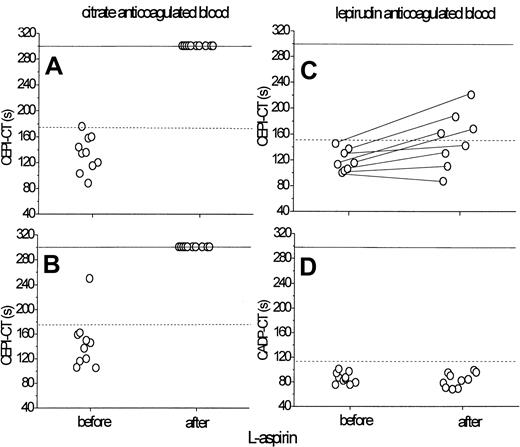
In good agreement with previous reports,18 basal CEPI-CT of citrated blood averaged 133 seconds (range, 88-176 seconds), excluding 1 outlier shown in Figure 1, and mean basal CADP-CT was 109 seconds (range, 78-133 seconds). In lepirudinised blood CTs were reduced as compared with citrate: the basal CEPI-CT averaged 120 seconds (range, 73-155 seconds) and the basal CADP-CT was 87 seconds (range, 57-117 seconds).
Effect of l-aspirin on in vitro platelet plug formation in citrated or lepirudin anticoagulated blood.l-aspirin was added in vitro to whole blood (50 mg/L; A) or infused into volunteers (250 mg; B) for determination of collagen-epinephrine (CEPI) closure time (CT) in citrated blood. For the determination of CEPI and collagen-adenosine diphosphate (CADP) CTs in lepirudinised blood, l-aspirin was infused into volunteers (250 mg; C, D). l-aspirin prolonged the CEPI-CT (n = 10) in citrated blood (A, B) whereas in lepirudinised blood the CEPI-CT (n = 8) was less influenced (C) and the CADP-CT (n = 10) was not prolonged in vitro (D). Dotted lines refer to upper normal limits.
Effect of l-aspirin on in vitro platelet plug formation in citrated or lepirudin anticoagulated blood.l-aspirin was added in vitro to whole blood (50 mg/L; A) or infused into volunteers (250 mg; B) for determination of collagen-epinephrine (CEPI) closure time (CT) in citrated blood. For the determination of CEPI and collagen-adenosine diphosphate (CADP) CTs in lepirudinised blood, l-aspirin was infused into volunteers (250 mg; C, D). l-aspirin prolonged the CEPI-CT (n = 10) in citrated blood (A, B) whereas in lepirudinised blood the CEPI-CT (n = 8) was less influenced (C) and the CADP-CT (n = 10) was not prolonged in vitro (D). Dotted lines refer to upper normal limits.
Aspirin effect in vitro: comparison between citrated and lepirudinised blood
l-aspirin, added in vitro to whole blood (50 mg/L) (Figure 1A) or infused into volunteers (250 mg) (Figure 1B) prolonged the CEPI-CT to more than 300 seconds in vitro in citrated blood (P = .005). In contrast, in lepirudinised blood, the CEPI-CT was much less affected (from 120 ± 21 seconds to 151 ± 43 seconds, P < .05; Figure 1C) and the CADP-CT was not prolonged in vitro (87 ± 13 seconds before aspirin, 83 ± 12 seconds after aspirin, P > .05; Figure 1D) after infusion of 250 mg l-aspirin.
VWF:RCo
In group 1, DDAVP increased plasma VWF:RCo levels 2.8-fold from 132 ± 60 U/mL to 377 ± 65 U/mL at 1 hour (P = .005 vs baseline; Figure 2). Likewise, in group 2, VWF:RCo increased 2.8-fold from 108 ± 18 U/dL to 302 ± 26 U/dL 30 minutes after infusion of DDAVP and reached a maximum at 1.5 hours (397 ± 50 U/dL; P = .005 vs baseline and between treatment). Placebo did not affect VWF:RCo levels at any time (range, 110 to 140 U/dL).
Desmopressin infusion improves the in vitro platelet dysfunction induced by l-aspirin. Ten healthy volunteers received DDAVP (0.3 μg/kg) over 30 minutes. l-aspirin (50 mg/L) (▴) was added in vitro to blood samples before and after DDAVP infusion. Horizontal dashed lines show the upper and lower limits of normal CEPI-CT and CADP-CT values. Data are presented as mean ± SEM. *P < .05, **P < .01, and ***P = .005 versus baseline. DDAVP increased von Willebrand ristocetin cofactor activity (VWF:RCo) levels (top) and thereby shortened both CEPI-CT (citrated blood; middle) and CADP-CT (lepirudinised blood; bottom). DDAVP decreased the aspirin-induced inhibition of platelet function (middle; ▴) with a normalization at 30 minutes and a persistent response for 8 hours.
Desmopressin infusion improves the in vitro platelet dysfunction induced by l-aspirin. Ten healthy volunteers received DDAVP (0.3 μg/kg) over 30 minutes. l-aspirin (50 mg/L) (▴) was added in vitro to blood samples before and after DDAVP infusion. Horizontal dashed lines show the upper and lower limits of normal CEPI-CT and CADP-CT values. Data are presented as mean ± SEM. *P < .05, **P < .01, and ***P = .005 versus baseline. DDAVP increased von Willebrand ristocetin cofactor activity (VWF:RCo) levels (top) and thereby shortened both CEPI-CT (citrated blood; middle) and CADP-CT (lepirudinised blood; bottom). DDAVP decreased the aspirin-induced inhibition of platelet function (middle; ▴) with a normalization at 30 minutes and a persistent response for 8 hours.
Platelet counts
Platelet counts were not affected by treatment with DDAVP in group 1. The maximum change in any individual was 11%. In group 2, platelet counts were not affected by any drug; compared with baseline, the maximum change in platelet counts in any individual was 4% after eptifibatide infusion. The maximum change in the DDAVP period was 7% and in the placebo period it was 10%.
DDAVP, aspirin, and GPIIb/IIIa inhibitors: relationship and concentration effect
DDAVP infusion shortened both CEPI-CT and CADP-CT with a maximum effect at 30 minutes; CEPI-CT decreased from 133 ± 27 seconds to 71 ± 12 seconds and CADP-CT decreased from 86 ± 18 seconds to 55 ± 7 seconds (P < .01; Figure 2, group 1). More important, after adding l-aspirin in vitro to the blood samples, DDAVP decreased the l-aspirin-induced inhibition of platelet function in a biphasic manner: (1) complete normalization was seen immediately after DDAVP infusion: CEPI-CT was reduced from 292 ± 28 seconds to 145 ± 61 seconds (P < .01) at 30 minutes (Figure 2); and (2) the aspirin effect was significantly mitigated up to 2 hours after the start of DDAVP infusion in all subjects.
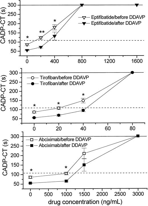
Further, adding eptifibatide, tirofiban, or abciximab in vitro to whole blood, all 3 GPIIb/IIIa inhibitors concentration-dependently prolonged both CEPI-CT and CADP-CT in vitro (P < .01, n = 10; Figure 3, group 1). After DDAVP infusion, higher concentrations of all 3 antagonists were necessary to inhibit platelet plug formation (Figure 3). Hence, DDAVP inhibited the effect of all 3 GPIIb/IIIa inhibitors at submaximal concentrations and caused a shift in their concentration response curves to the right.
Desmopressin shifts concentration response curves of GPIIb/IIIa antagonists to the right. GPIIb/IIIa inhibitors were added to whole blood in vitro before and after DDAVP infusion. Eptifibatide (top), tirofiban (middle), and abciximab (bottom) concentration-dependently inhibited platelet function, as measured by the PFA-100. Horizontal dashed lines show the upper limit of normal CT values. The horizontal solid lines show the maximal measurable CT. Data are presented as mean ± SEM. *P < .05, **P < .01 (before DDAVP vs after DDAVP). DDAVP infusion caused a shift in the concentration response curves of all 3 GP IIb/IIIa inhibitors to the right in lepirudinised blood (n = 10).
Desmopressin shifts concentration response curves of GPIIb/IIIa antagonists to the right. GPIIb/IIIa inhibitors were added to whole blood in vitro before and after DDAVP infusion. Eptifibatide (top), tirofiban (middle), and abciximab (bottom) concentration-dependently inhibited platelet function, as measured by the PFA-100. Horizontal dashed lines show the upper limit of normal CT values. The horizontal solid lines show the maximal measurable CT. Data are presented as mean ± SEM. *P < .05, **P < .01 (before DDAVP vs after DDAVP). DDAVP infusion caused a shift in the concentration response curves of all 3 GP IIb/IIIa inhibitors to the right in lepirudinised blood (n = 10).
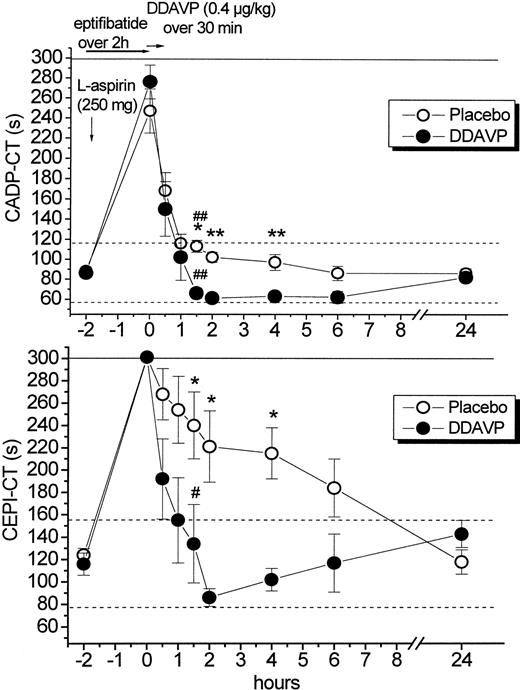
Infusion of eptifibatide prolonged both CEPI-CT and CADP-CT in lepirudinised blood: CEPI-CT increased from 120 ± 21 seconds to more than 300 seconds (P = .005) and CADP-CT increased from 87 ± 13 seconds to 262 ± 62 seconds (P < .001) 2 hours after start of eptifibatide infusion (Figure 4, group 2). DDAVP shortened both CEPI-CT and CADP-CT after discontinuation of eptifibatide infusion with a maximum effect at 1.5 to 2 hours (P < .05 and P < .01, between treatments; Figure 4). Compared with the values immediately after eptifibatide infusion, CADP-CT decreased from 276 ± 17 seconds (0 hours) to 66 ± 5 seconds 1.5 hours after start of DDAVP infusion (P < .01), and from 246 ± 22 seconds to 113 ± 6 seconds in the placebo period at 1.5 hours (P < .01). In parallel, DDAVP decreased CEPI-CT from more than 300 seconds to 134 ± 35 seconds at 1.5 hours (P < .05), whereas CEPI-CT decreased from more than 300 seconds to 240 ± 30 seconds in the placebo period at 1.5 hours (P > .05; Figure 4).
Desmopressin accelerates reversal of in vitro platelet dysfunction after discontinuation of eptifibatide infusion as compared with placebo. On both study days, 10 healthy volunteers received l-aspirin (250 mg intravenous bolus) and a standard eptifibatide infusion for 2 hours. After the stop of eptifibatide infusion, DDAVP or placebo were infused. Eptifibatide prolonged both CEPI-CT and CADP-CT in lepirudinised blood. Data are presented as mean ± SEM. *P < .05, **P < .01 between groups; #P < .05, ##P < .01 compared with the values after eptifibatide infusion. DDAVP significantly accelerated normalization of both CEPI-CT and CADP-CT after stop of eptifibatide infusion with a maximum effect at 1.5 to 2 hours whereas the initial CADP-CT values (0-1 hour) were not affected. In contrast, CEPI-CT remained above normal in the placebo group for more than 4 hours, possibly reflecting an aspirin-like defect.
Desmopressin accelerates reversal of in vitro platelet dysfunction after discontinuation of eptifibatide infusion as compared with placebo. On both study days, 10 healthy volunteers received l-aspirin (250 mg intravenous bolus) and a standard eptifibatide infusion for 2 hours. After the stop of eptifibatide infusion, DDAVP or placebo were infused. Eptifibatide prolonged both CEPI-CT and CADP-CT in lepirudinised blood. Data are presented as mean ± SEM. *P < .05, **P < .01 between groups; #P < .05, ##P < .01 compared with the values after eptifibatide infusion. DDAVP significantly accelerated normalization of both CEPI-CT and CADP-CT after stop of eptifibatide infusion with a maximum effect at 1.5 to 2 hours whereas the initial CADP-CT values (0-1 hour) were not affected. In contrast, CEPI-CT remained above normal in the placebo group for more than 4 hours, possibly reflecting an aspirin-like defect.
Rapid platelet function assay
The mean level of platelet inhibition was more than 92% in all subjects immediately after the stop of the eptifibatide infusion (Table 1). A satisfactory level of inhibition (> 80%)23 was measured until 1.5 hours after the end of the eptifibatide infusion. A low level of inhibition of GPIIb/IIIa receptors (< 80%) was obtained 2 hours after the stop of eptifibatide infusion regardless of concomitant treatment with placebo or DDAVP (Table 1).
Effect of eptifibatide on platelet glycoprotein GPIIb/IIIa receptor blockade measured with the rapid platelet function assay
Hours after eptifibatide infusion . | Placebo period, %* . | Desmopressin period, %* . |
|---|---|---|
| 0 | 95 ± 1 (93-100) | 94 ± 1 (92-97) |
| 0.5 | 90 ± 2 (80-97) | 90 ± 2 (83-95) |
| 1.0 | 81 ± 2 (73-90) | 87 ± 1 (83-91) |
| 1.5 | 82 ± 1 (80-83) | 85 ± 1 (82-90) |
| 2.0 | 77 ± 2 (73-80) | 78 ± 3 (72-88) |
Values are expressed as mean ± SEM, with the range in parentheses.
Bleeding time
Eptifibatide increased the bleeding time from 7 minutes (range, 4-11 minutes) to 22 minutes (range, 13-30 minutes) in the placebo period and from 6 minutes (range, 3-8 minutes) to 19 minutes (range, 8-30 minutes) in the DDAVP period (P = .005 vs baseline). No significant shortening of bleeding time was observed immediately after the end of DDAVP infusion (P > .05).
Correlation between methods
Under basal conditions CADP-CT correlated with CEPI-CT (r = 0.85, P = .014) and both with the bleeding time (r = 0.66-0.68, P < .05). CADP-CT showed a good correlation with the bleeding time (r = 0.83, P = .003) 30 minutes after eptifibatide infusion. In contrast, the rapid platelet function assay showed only a trendwise correlation with the bleeding time (r = 0.63, P > .05) and did not correlate with CEPI-CT or CADP-CT. Interestingly, 1.5 hour after start of DDAVP infusion, both CEPI-CT or CADP-CT were essentially corrected to normal, whereas rapid platelet function assay-blockade was still 85%.
Safety aspects
We believe this is the first study where healthy volunteers received currently accepted standard doses of both eptifibatide and l-aspirin. All clinical adverse effects observed were attributed to concomitant infusion of DDAVP (Table 2). None of the 20 volunteers (groups 1
Adverse events after drug infusion
. | Treatment . | . | . | ||
|---|---|---|---|---|---|
| . | Group 1 . | Group 2 . | . | ||
| Symptoms . | DDAVP; n . | Eptifibatide and l-aspirin; n . | Eptifibatide, l-aspirin, and DDAVP;n . | ||
| Headache | 4 | 2 | 4 | ||
| Hypotension* | 3 (systemic) | 3 (systemic) | 5 (systemic) | ||
| 8 (diastolic) | 5 (diastolic) | 7 (diastolic) | |||
| Facial flush | 1 | 0 | 2 | ||
| Nausea | 1 | 0 | 0 | ||
| Thrombocytopenia† | 0 | 0 | 0 | ||
Hypotension was defined as a greater than 10% drop in blood pressure.
Thrombocytopenia was defined as less than 150 × 109/L.
Discussion
This is the first study to demonstrate that DDAVP accelerates normalization of the in vitro platelet dysfunction induced by GPIIb/IIIa inhibitors (+l-aspirin) in healthy subjects. For repeated assessment of primary hemostasis, we used the PFA-100.18 As the inhibitory effect of eptifibatide is overestimated in citrated blood samples,17 lepirudin-anticoagulated blood was used for determination of all measurements.
First, group 1 showed the expected 3-fold increase in VWF: RCo levels after DDAVP infusion; DDAVP thereby shortened both CEPI-CT and CADP-CT (Figure 2). Second, DDAVP normalized the aspirin-induced in vitro platelet dysfunction immediately at the end of infusion and significantly ameliorated the aspirin defect of platelet function up to 2 hours (Figure 2). Third, DDAVP shifted concentration response curves for all 3 GPIIb/IIIa inhibitors to the right in vitro (Figure 3). However, DDAVP did not antagonize the effect at maximal concentrations of GPIIb/IIIa inhibitors. Thus, DDAVP antagonizes the effect induced by GPIIb/IIIa inhibitors, particularly at submaximal concentrations in vitro.
The results of group 1 were further confirmed in group 2. First, DDAVP infusion did not shorten the 3-fold prolongation in bleeding time compared with placebo 0.5 hours after stop of eptifibatide infusion. This is in good agreement with the lack of DDAVP effect on closure times (PFA-100) immediately after the end of DDAVP infusion (Figure 4). At the time when the bleeding time was measured (0.5 hours) CADP-CT was not significantly different between groups, although CEPI-CT was trendwise shorter in the DDAVP period (possible due to normalization of the aspirin component of platelet defect). CADP-CT averaged 160 seconds, which is similar to patients, after coronary artery bypass graft, who excessively bleed, but who are responsive to platelet transfusions.28
Second, DDAVP accelerated the normalization of CADP-CT and CEPI-CT after stop of eptifibatide infusion with a maximum effect at 1.5 to 2 hours. In contrast, CEPI-CT remained above normal in the placebo group for more than 4 hours, possibly reflecting an aspirin-like defect (Figure 4). Hence, DDAVP appears to have no measurable effect at maximal concentrations of GpIIb/IIIa inhibitors in vivo confirming the ex vivo experiments (Figure 3). Compared with placebo, DDAVP accelerates normalization of platelet dysfunction induced by eptifibatide with a maximum effect at 1.5 to 2 hours, when plasma eptifibatide levels are reduced to submaximal levels (Figure 4). The receptor occupancy of 80% to 85% (measured by the rapid platelet function assay; Table 1) was still in the therapeutic range29 at that time. Summarizing, DDAVP can help to normalize the in vitro platelet dysfunction quickly even at therapeutic levels of GPIIb/IIIa blockade and is expected to shorten bleeding episodes.
The CADP-CT correlated with the bleeding time under basal conditions and after infusion of eptifibatide. After eptifibatide infusion this correlation reached r2 = 0.69. There was a relatively good agreement (r = 0.83) between the in vivo bleeding time and ex vivo assessment of platelet function by PFA-100. The value of the bleeding time in the prediction of bleeding disorders, however, is controversially discussed. In contrast, the rapid platelet function assay showed only a trendwise correlation with CADP-CT and the bleeding time. The lack of a perfect correlation between these methods is likely attributable to differences between in vivo and in vitro systems with or without high shear rates.
The current study has some limitations. First, bleeding complications during administration of GPIIb/IIIa inhibitors are not very common and for this reason, a randomized study in bleeding patients is not easily conducted. The relative contribution of GPIIb/IIIa inhibitors to bleeding may be difficult to differentiate from heparin-induced bleeding in the individual case. Hence, we have to assume that GPIIb/IIIa inhibitors could enhance bleeding in patients who receive these agents along with aspirin and heparin. However, DDAVP responsiveness in patients with acute coronary syndromes has not been formerly studied; whether such patients will respond to DDAVP in the same manner as healthy individuals is not clear. Second, eptifibatide was given as a single bolus in this trial, as our study was designed before the ESPRIT-study (enhanced suppression of the platelet receptor glycoprotein IIb/IIIa using integrilin therapy [in patients undergoing percutaneous coronary intervention (PCI)]) was published, in which eptifibatide bolus was given twice. However, the double bolus primarily affects plasma concentrations early after start of infusion, and we have nonetheless achieved clinically relevant receptor occupancy rates of 90% to 95%. Third, and importantly, the PFA-100 and rapid platelet function assay test are artificial measures of primary hemostasis and do not necessarily reflect in vivo hemostasis. Finally, there may be concerns about using DDAVP in patients after PCI because of potential adverse effects, such as fluid retention or thrombotic events due to DDAVP, as indicated by case reports. We assume that the former problem can be managed by close observation of patients and fluid restriction, and that restoration of normal flow and normal shear rates after PCI will decrease the likelihood of acute thrombosis which might be facilitated by VWF release.
Despite these limitations, the following clinical recommendations may be given: DDAVP should be used whenever bleeding is suspected to stem from GPIIb/IIIa inhibitors. As long as there are no clinical trials in bleeding patients, we recommend the following course of action based on our “proof of concept study”: (1) stop the GPIIb/IIIa infusion; (2) obtain a platelet count and activated partial thromboplastin time (aPTT); (3) if possible, measure the degree of platelet inhibition with a rapid bedside test; (4) administer a DDAVP infusion; and/or (5) transfuse platelet concentrates in case of major or life-threatening bleeding or urgent need for normalization of platelet function in case of surgery.
In conclusion, DDAVP accelerates normalization of in vitro platelet dysfunction induced by GPIIb/IIIa inhibitors as soon as levels of GPIIb/IIIa inhibitors are reduced to levels of borderline effectiveness.
Prepublished online as Blood First Edition Paper, August 14, 2003; DOI 10.1182/blood-2002-11-3566.
Supported by Jubiläumsfonds der Österreichischen Nationalbank (Nr 9345).
The publication costs of this article were defrayed in part by page charge payment. Therefore, and solely to indicate this fact, this article is hereby marked “advertisement” in accordance with 18 U.S.C. section 1734.


This feature is available to Subscribers Only
Sign In or Create an Account Close Modal