Abstract
The gene encoding ribosomal protein S19 (RPS19) has been shown to be mutated in 25% of the patients affected by Diamond-Blackfan anemia (DBA), a congenital erythroblastopenia. As the role of RPS19 in erythropoiesis is still to be defined, we performed studies on RPS19 expression during terminal erythroid differentiation. Comparative analysis of the genomic sequences of human and mouse RPS19genes enabled the identification of 4 conserved sequence elements in the 5′ region. Characterization of transcriptional elements allowed the identification of the promoter in the human RPS19 gene and the localization of a strong regulatory element in the third conserved sequence element. By Northern blot and Western blot analyses of murine splenic erythroblasts infected with the anemia-inducing strain Friend virus (FAV cells), RPS19 mRNA and protein expression were shown to decrease during terminal erythroid differentiation. We anticipate that these findings will contribute to further development of our understanding of the contribution of RPS19 to erythropoiesis.
Introduction
The mammalian ribosome is composed of 4 RNA species and 80 different ribosomal proteins.1-5 One of these proteins, the ribosomal protein S19 (RPS19) is localized at the beak of the small ribosomal subunit 40S6 and mutations in the gene encoding RPS19 have been identified in 25% of the patients affected by Diamond-Blackfan anemia (DBA), a rare congenital erythroblastopenia.7 8 However, the mechanistic understanding of the relation between RPS19 and erythropoiesis and, specifically, the impact of RPS19 mutations in DBA remains to be defined.
At the transcriptional level, all ribosomal protein genes must be coordinated to allow for efficient and balanced protein synthesis. Although mammalian ribosomal protein genes are not clustered9 but rather dispersed throughout the genome,2 they are transcribed at very similar rates due to the equivalent strength of their promoters.10 Such coordinated activity of the promoters of these numerous ribosomal protein genes is regulated transcriptionally through the binding of transcription factors to specific promoter sequence elements.11 The cis-acting transcriptional regulatory elements and the trans-acting factors, which bind to these elements, have been characterized in the region located upstream of the translation initiation codon, in a few human12,13 and mouse ribosomal protein genes.10,11 14-25 However, transcriptional control of most of these genes, including the RPS19 gene, remains to be defined.
In the present study, we provide information on the transcriptional regulatory elements in the 5′ region upstream of the translation initiation codon in the human RPS19 gene. Alignment of the human and mouse genomic sequences from this region allowed us to delineate 4 consensus regions present in both sequences. The Neural Network Promoter Prediction software (LBNL, Berkeley, CA)14-16 identified a human RPS19 gene promoter, which was confirmed to have promoter activity. Furthermore, we found a strong transcriptional regulatory element in the third consensus region, upstream of the translation initiation site but downstream of the promoter element. A putative transcription factor, which is likely a member of the nuclear factor–κB (NF-κB)/Rel family such as c-Rel or Rel-A subunits of NF-κB, repressed promoter activity. Northern blot and Western blot analyses of terminally differentiating murine splenic erythroblasts infected with the anemia-inducing strain Friend virus (FAV cells)17 were performed to assess RPS19 mRNA and protein expression during erythropoiesis. RPS19 mRNA and protein expression were shown to decrease during terminal erythroid differentiation. We anticipate that these findings will contribute to further development of our understanding of the contribution RPS19 to erythropoiesis.
Materials and methods
Mouse RPS19 gene characterization
The murine RPS19 gene (GenBank accession no. AF16207) was characterized by analysis of bacterial artificial chromosome (BAC) clones obtained by polymerase chain reaction (PCR) screening of mouse BAC library CitbCJ7 derived from embryonic stem (ES) cell lines of the 129/SvJ strain (Research Genetics, Huntsville, AL). Briefly, the putative organization of the murine gene was deduced by aligning the sequence of the human gene with the sequences of various murine expressed sequence tags (EST) clones. Based on the sequence of adjacent exons, 2 pairs of primers were designed in order to span intron 2 and intron 4. This strategy was used since the existence of several pseudoprocessed genes made use of other approaches untenable.18 There were 4 BAC clones, which were positive using both primer sets, that were found to contain the entireRPS19 gene, as determined by direct BAC DNA sequencing, and by sequencing of PCR products (Applied Biosystems 373 DNA sequencer and ABI Big Dye Terminator sequencing kits; Perkin Elmer, Foster City, CA).19 The mouse RPS19 gene was mapped using The Jackson Laboratory BSS interspecific backcross [(C57BL/6JEi X SPRET/Ei) F1 X SPRET/Ei] panel.20 A sequence containing a polymorphism within the 3′ untranslated region (UTR) (C57BL/6Jei, 240-bp fragment; SPRET/Ei, 380 bp) was amplified with 2 RPS19-specific primers and used to follow the segregation of alleles in the 94 backcross progeny from the BSS panel on ethidium bromide–stained 2% agarose gels. The mouseRPS19 gene was also mapped by fluorescence in situ hybridization (FISH) analysis, using the BAC DNA labeled by random priming with digoxigenin and stained with Texas red on mouse metaphase chromosomes isolated from C57 black mice.21Chromosome-specific probes were randomly labeled with biotin and stained with avidin–fluorescein isothiocyanate (FITC).22
Alignment of human and mouse genomic sequences
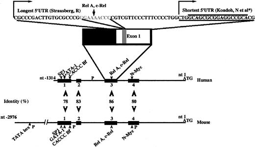
Comparison of the genomic organization of RPS19 genes was performed using the previously published human genomic sequence8 (GenBank accession no. AF092906) and the mouse genomic sequence, which we characterized in the present study (Figure1). The 5′ region upstream of the translation initiation start was examined using various promoter and binding site recognition tools: TESS (University of Pennsylvania), TSSG (Baylor College of Medicine), TSSW (Baylor College of Medicine), NNPP/Eukaryotic and Prokaryotic (LBNL),14-16 and Matinspector/TRANSFAC (Baylor College of Medicine).
Consensus regions in human and mouse genomic organization of the 5′ end region upstream of initiation start ATG.
Black boxes indicate regions of homology in the genomic sequence upstream of the translation start site, within both species, human and mouse. Numbers indicate the percentage of identity. Arrowheads show the consensus position of potential transcription factors binding sequences identified in both mouse and human sequences. “P” indicates potential promoter sites. In the third consensus region, the sequence of the 2 5′ UTRs described by Strausberg (GenBank accession no. BC000023) and Kondoh et al (GenBank accession no. NM_001022)25 is indicated. Arrows indicate the 2 transcriptional starts. The gray characters in exon 1 indicate the binding site for a potential κB element, Rel-A, c-Rel.
DNA cloning
Constructs designed to analyze regulatory elements in the humanRPS19 5′ region upstream of the translation initiation site were cloned into pGL3 Luciferase Reporter Vector (Promega, Madison, WI). This strategy allows quantitative analysis of sequences and factors that potentially regulate mammalian gene expression by cloning putative eukaryotic transcriptional regulatory sequences upstream of the luciferase reporter gene. In cells transfected with pGL3 plasmid, the luciferase reporter gene (luc+) expression is driven by and directly proportional to the promoter activity of the inserted sequences upstream of luc+, since the empty vector lacks eukaryotic promoter and enhancer sequences. Either a full-length wild-type human sequence upstream of the translation initiation start ATG or 2 mutated constructs recapitulating 2 mutations found in patients with DBA in this region (missense mutation G>T at −460 and a 4-bp insertion at −629 and −625 [Figure2A]) were amplified by PCR with a forward primer creating a Mlu1 restriction site (5′-AAT ACG ACG CGT CGG AAG GCA AAT GGA CTG CCT AAG CTA C-3′) and a reverse primer creating a BlgII restriction site (5′-ACG GGA AGA TCT TCC GAG GGA GAA AGT CAA GCA TGT GAA C-3′). Digestion of the full-length wild-type construct with ApaI restriction enzyme allowed us to generate a mutated construct by releasing anApaI 107-bp fragment (−405 nucleotide [nt] to −299 nt), containing a putative N-Myc transcription factor binding site. The other mutated constructs (Figure 2A) in which we deleted either (1) each of the 4 consensus regions in the human and mouse 5′ sequence upstream of the translation initiation start codon, (2) the human sequence predicted to be a promoter sequence by computer software, NNPP (LBNL),14-16 or (3) the specific binding sites for potential transcription factors, N-Myc, c-Rel, Rel-A, were generated by splice overlap extension method.23 Fidelity of sequence of all pGL3 constructs was confirmed by sequencing with ABI Big Dye Terminator sequencing kit using an Applied Biosystems 373 DNA sequencer (Perkin Elmer).19
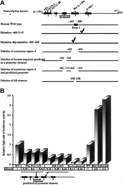
Deletions made in the 5′ end upstream of initiation start ATG in the human RPS19 gene and measured promoter activities.
(A) We identified 4 consensus regions (▪) between the mouse and human 5′ sequence upstream of the initiation start ATG. The human genomic sequence upstream of the translational initiation start site is represented as the black line at the top of the panel. The predicted promoter found in the human sequence between the second and third consensus regions is also indicated. The consensus binding motifs for putative transcription factors in the consensus regions 1, 3, and 4 are noted with arrowheads. We cloned the full-length human 5′ end (wild-type RPS19 gene at the top of the panel and mutants below) in a promoterless pGL3 vector, which contained a luciferase reporter gene. The 2 mutations found in patients affected by DBA in the 5′ sequence upstream of the translational initiation start site were also cloned into pGL3 vector. Their positions are indicated. We deleted each of the consensus regions, the predicted promoter, both in a same construct the predicted promoter and the consensus region 3, and finally the putative κB element in the consensus region 3. The number of the flanking nucleotide indicates the location of each construct. (B) We measured the activity of the luciferase reporter gene (y-axis) driven by RPS19 5′ end wild-type (WT) and mutated constructs: deletion of the human predicted promoter (Pre Pro), deletion of the consensus region 3 (Del 3), deletion of both consensus region 3 and predicted promoter (Pre Pro+Del 3), deletion of binding motif for potential transcription factors from the NF-κB/Rel family (Del κB element). When we deleted the RPS19 human gene predicted promoter (Pre Pro, 3 clones each tested in 3 independent experiments), we observed a 40% decrease in luciferase reporter gene activity compared with wild-type RPS19 gene. After deletion of the consensus region 3 (Del 3, 3 clones each tested in 3 independent experiments), we obtained a greater decrease in luciferase reporter gene activity (decrease of 66%). More importantly, when we deleted both the predicted promoter and consensus region 3 (Pre Pro+Del 3, 2 clones in 2 independent experiments), we noted an even larger decrease in the luciferase reporter gene (decrease of 86%). The positions of Pre Pro and Del 3 deletion in the 5′ flanking human region upstream of initiation start are indicated at the bottom of the panel. To investigate transcription factors, which may regulate theRPS19 gene promoter activity in the third consensus region, we measured the luciferase reporter gene (y-axis) driven by humanRPS19 in which we deleted the binding motif for potential transcription factors c-Rel/Rel-A (Del κB element, clones 1 and 2). Unexpectedly, the luciferase reporter gene activity was not decreased but in fact increased 3-fold compared with the wild-typeRPS19 gene. Data from individual experiments are shown.
Cell culture
Immortalized human embryonic fibroblast cells (293T cells) were cultured in Dulbecco modified Eagle medium (DMEM) (Life Technologies, Gaithersburg, MD), supplemented with 1% glutamine, 10% fetal bovine serum (FBS), and 1% penicillin-streptomycin. Murine splenic erythroblasts infected with the anemia-inducing strain Friend virus (FAV cells) were generously provided by Mark J. Koury (Vanderbilt University, Nashville, TN).17,24 Briefly, CD2F1 mice were infected with the anemia-inducing strain Friend virus, and 2 weeks later, after velocity sedimentation at unit gravity, immature splenic erythroblasts were isolated and cultured with 0.2 U EPO/mL (human recombinant EPO, 130 000 U/mg protein; AMGEN, Thousand Oaks, CA).17 24Cells at t = 0 hour of culture were mostly proerythroblasts, which underwent terminal erythroid differentiation during culture. At 44 hours, the cultures contained predominantly enucleating erythroblasts and reticulocytes.
Cell transfection
293T cells were transfected with 3 μg DNA per 300 000 cells with Lipofectamine 2000 (Life Technologies). At 24 hours after transfection, cells were assayed for luciferase activity using a luminometer (Dynex, Chantilly, VA). Transfection efficiency was normalized by cotransfection with a second plasmid encoding renilla luciferase.
Production and purification of chicken antibody against mouse RPS19 protein
We produced an antibody raised in chicken-against-mouse His-tagged RPS19 protein with the assistance of Washington Biotechnology (Baltimore, MD). Mouse RPS19 cDNA was cloned,NsiI-XhoI, into pET31b(+) vector (Novagen, Madison, WI) and transformed in BL21 bacteria (Stratagene, La Jolla, CA). Of particular note, this cloning strategy results in removal of the insoluble protein originally encoded by the empty pET31b(+) vector. This vector was chosen because it allows C-terminal expression of the protein of interest with only 8 additional amino acids including the 6xHis tag. Mouse RPS19 protein was expressed as a His-tagged recombinant RPS19 protein after induction of BL21 bacteria with 1 mM isopropyl β-D thiogalactopyranoside (ITPG) (Sigma, St Louis, MO). After induction, bacteria were spun down and lysed by freeze-thaw. Lysed bacteria were resuspended in binding buffer (Novagen) supplemented with 0.1 mM diisopropyl fluorophosphate (DFP) as a protease inhibitor (Sigma) and 6 M urea in order to solubilize the RPS19 protein. Bacteria suspension was then sonicated for 60 seconds at 4°C and centrifuged at 17 000 rpm for 10 minutes at 4°C. Mouse recombinant His-tagged RPS19 protein was purified on a nickel column according to the manufacturer's instructions (Histidine Binding Buffer Kit; Novagen) with some modifications. RPS19 protein solubilized in binding buffer containing 6 M urea was bound to the column. The column was subsequently washed with wash buffer containing decreasing concentrations of urea in order to renature the protein as much as possible. Recombinant RPS19 protein was then eluted with elution buffer and dialyzed in phosphate-buffered saline (PBS). Two hens were immunized with purified mouse His-tagged recombinant RPS19. Prior to immunization, nonspecific IgYs and after immunization, specific IgYs (corresponding to chicken IgGs) contained in egg yolk were subject to delipidation. Then, nonspecific IgYs were just precipitated using Eggcellent IgY precipitation reagent according to the manufacturer's instructions (Pierce, Rockford, IL) whereas the specific IgYs were affinity-purified using an Affi-Gel 10 column (Bio-rad, Hercules, CA) previously coupled with mouse recombinant RPS19 His-tagged protein. Specific IgYs bound to mouse-affigel column RPS19 were eluted with 0.2 M glycine-HCl, pH 2.2, neutralized with 1 M Tris, pH 8.5, extensively dialyzed against PBS, and quantified by absorbance at 280 nm. This affinity-purified antibody was used in immunoblotting and immunofluorescence experiments.
Northern blotting
Total RNA (10 μg) extracted from FAV cells was separated on 1% agarose genetic technology grade (GTG) gel and transferred by capillarity to a nylon membrane (Amersham, Piscataway, NJ) overnight at room temperature using NorthernMax one-hour transfer buffer (Ambion, Austin, TX). The membrane was prehybridized for 6 hours at 65°C, then hybridized overnight at 65°C with a 361-bp α-[32P] deoxycytosine triphosphate (dCTP)–labeled cDNA probe amplified from RPS19 cDNA and with a 2-kb α-[32P] dCTP-labeled cDNA probe amplified from human β-actin, as a control. The membrane was then washed and autoradiographed for 8 hours at −80°C.
Immunoblotting
Proteins were subject to 15% sodium dodecyl sulfate–polyacrylamide gel electrophoresis (SDS-PAGE), then transferred onto a polyvinylidenefluoride (PVDF) membrane (Millipore, Portsmouth, NH) for one hour at a constant current using a semidry electroblotter according to the manufacturer's instructions (Integrated separation system; Enprotech, Natick, MA). Protein transfer was assessed after staining PVDF membrane with red Ponceau. The membrane was blocked for one hour at room temperature in Blotto solution (10 mM Tris, pH 7.5, 140 mM NaCl, 1% bovine serum albumin [BSA], 4% nonfat milk, 1% donkey serum [anti-RPS19 blotting] or 1% goat serum [antiactin and anti-4.1R blotting], 1% Tween-20, 0.02% sodium azide). The membrane was then incubated overnight at 4°C with various affinity-purified primary antibodies, either an antibody against His-tagged recombinant mouse RPS19 raised in chicken, or an IgG1 kappa light chain antibody against chicken actin raised in mouse (clone C4) (ICN Biomedicals, Aurora, OH), or an antibody against a 16-kDa His-tagged recombinant protein encoding mouse 4.1R exon 13, raised in rabbit. The 4.1R antibody was a kind gift from Drs Loren Walensky and Solomon Snyder (The Johns Hopkins University School of Medicine, Baltimore, MD). These antibodies were diluted in Blotto solution at 0.05 μg/mL to 0.3 μg/mL for the anti-RPS19 antibody, at the dilution 1:20 000 for the antiactin antibody, and at 0.1 μg/mL for the anti-4.1R antibody. After 2 rinses, 3 washes of 10 minutes each, and a short blocking in Blotto solution without serum for 15 minutes, the membrane was incubated for 1 hour at room temperature with secondary antibodies coupled to the horseradish peroxidase, donkey anti–chicken (RDI, Flanders, NJ) for RPS19 detection, goat anti–mouse (Jackson ImmnoResearch Laboratories, West Grove, PA) for actin detection, and goat anti–rabbit (Sigma) for 4.1R detection, diluted 1:5000, 1:20 000, and 1:200 000, respectively, in Blotto solution without serum. After extensive washing as described above, the membranes were probed with enhanced chemiluminescence reagent R (ECL; Nen Life Science Products, Boston, MA).
Results
Mouse RPS19 gene
Subcloning of an 18-kb fragment from BAC clones into a bluescript vector enabled us to sequence the entire mouse RPS19 gene (5.1 kb), a 2.5 kb region upstream of the 5′ UTR, and a 5.8 kb region downstream of the 3′ UTR (GenBank accession no. AF216207). It is composed of 5 coding exons and 4 introns. The structure of the murine gene is very similar to that of the human gene. RPS19 was nonrecombinant with Lipe, D7Mit20, andD7Ertd462e, placing RPS19 5.5 cM distal to the centromere on mouse chromosome 7, a region that shows conserved synteny with human 19q13.1-q13.2, where the human RPS19 gene segregates.1 Our data have been added to Mouse Genome Database under accession no. J: 58250 and can be accessed through the World Wide Web (http://www.jax.org). No obvious potential candidate mouse mutations map to the region containing RPS19 in chromosome 7 (1997 Chromosome Committee data). Linkage data were confirmed by FISH analysis of BAC DNA containing the RPS19 gene localized to the proximal end of chromosome 7 (data not shown).
Genomic sequence analysis
Alignment of 5′ sequences upstream of the translation initiation site in mouse and human RPS19 genes identified 4 distinct regions with significant identity. These 4 regions in human and mouse sequences respectively were: (1) region 1 from position −1219 to −1054, and −1160 to −998 (77% identity); (2) region 2 from position −992 to −934, and −933 to −874 (83% identity); (3) region 3 from position −631 to −474, and −609 to −455 (86% identity); (4) region 4 from position −398 to −263, and −381 to −249 (80% identity) (Figure 1). Interestingly, consensus region 3 spanned the exon 1 of the RPS19 gene, which corresponded to the 2 previously identified 5′ UTRs of the gene, from −557 to −488 for the longer 5′ UTR described by Strausberg et al50(GenBank entry accession no. BC000023) and from position −509 to −488 for the shorter one described by Kondoh et al25(GenBank accession no. NM_001022) (Figure 1).
The computer software, NNPP,14-16 identified only one potential promoter region in the 1314-bp long region upstream of the translation initiation start codon from −761 to −712 (Figure 1). The human RPS19 gene had almost all the features of other mammalian ribosomal protein promoters: absence of a canonical TATA-box, transcription start site at a C residue embedded in a polypyrimidine stretch (13 nt), and a short (22 nt) and CG rich (82%) 5′ UTR. A search for transcription factor binding motifs present in both mouse and human RPS19 genes revealed the existence of a weak consensus for a putative GATA-1 element (position −1090 to −1078 and −1148 to −1138, in mouse and human RPS19genes, respectively), an optimal motif for a putative CACCC binding factor (position −1011 to −1004 and −1068 to −1061, in mouse and human RPS19 genes, respectively) in consensus region 1. The same complete consensus for putative c-Rel and Rel-A transcription factors (position −517 to −508 and −538 to −530 in mouse and human RPS19 genes, respectively) and a partial consensus for a binding region for putative NF-κB (position: −516 to −507 and −538 to −530 in mouse and humanRPS19 genes, respectively) were found in consensus region 3. A complete consensus sequence for a putative binding region for the N-Myc transcription factor (position −345 to −334 and position −363 to −352 in mouse and human RPS19genes, respectively) was identified in consensus region 4 (Figure 1).
Promoter analysis of human RPS19 gene
The presence of a functional promoter in the region upstream of the translation initiation start codon in the human RPS19gene was confirmed by the fact that this promoter was able to drive transcription of the luciferase reporter gene (Figure 2B). In the promoter assay system used, the luciferase activity generated by the human wild-type RPS19 construct was at least 40-fold higher than that measured by transfection with empty vector.
No alteration in luciferase activity was observed when we introduced into the region upstream of the translational initiation start codon the 2 mutations found in patients with DBA (Figure 2A): the missense mutation G>T at −460 and 4-bp insertion GCCA (−629 nt to −625 nt; data not shown). Furthermore, neither the deletion of consensus regions 1, 2, or 4, nor that of the transcription factor N-Myc binding motif had any effect on luciferase reporter gene activity (data not shown). In marked contrast, deletion of the predicted promoter site in the human sequence resulted in decreased luciferase reporter gene activity (Figure 2B). This enabled us to locate a transcriptional regulatory element of the human RPS19 gene promoter, spanning from −761 nt to −712 nt upstream of translational initiation start site, and only −204 nt and −252 nt upstream of the 2 transcription start points described in human by Strausberg et al50 (GenBank accession entry no. BC000023) and Kondoh et al25 (GenBank accession no. NM_001022), respectively. It should be noted, however, that the deletion of this promoter element resulted in only a partial decrease in promoter activity (40%) compared with the wild-type promoter. This strongly suggested the existence of another important regulatory region in the 5′ flanking region of the RPS19 gene. Indeed, we found that deletion of consensus region 3 resulted in a dramatic decrease inRPS19 promoter activity (66%) compared with the wild-type promoter (Figure 2B). These data were reproducible in 3 independent experiments (Figure 2B). Strikingly, when both consensus region 3 and the promoter region were deleted, we observed an even larger decrease in luciferase reporter gene activity, implying an additive effect of these 2 regions in controlling humanRPS19 promoter activity (Figure 2B). In order to identify putative transcription factors, which may regulate humanRPS19 promoter function in consensus region 3, we mutated the binding region of a putative κB element conserved in both mouse and human sequences. Unexpectedly, this mutation resulted in a dramatic increase in the RPS19 promoter luciferase activity (Figure2B). Bioinformatic analysis suggests that this region contains putative binding sites for transcription factors c-Rel and Rel-A.
RPS19 gene and protein expression
We investigated by Northern blot analysis the level of RPS19 mRNA in FAV cells, kindly provided by Mark Koury and colleagues.17 24 Over the time course of 0 to 44 hours in suspension culture, the proerythroblasts at 0 hours matured into enucleating erythroblasts by 44 hours. RPS19 mRNA expression decreased markedly compared with actin mRNA expression during terminal erythroid differentiation (Figure 3).
RPS19 mRNA decreased during terminal erythroid differentiation.
We studied RPS19 gene expression by Northern blot analysis of FAV cells. As shown in the panel on the left, the expression of RPS19 mRNA decreased over the time course 0 to 44 hours of terminal erythroid differentiation. The expression of β-actin mRNA is shown in the panel on the right. Molecular weight standard (MWs) was loaded and the position of the markers is indicated.
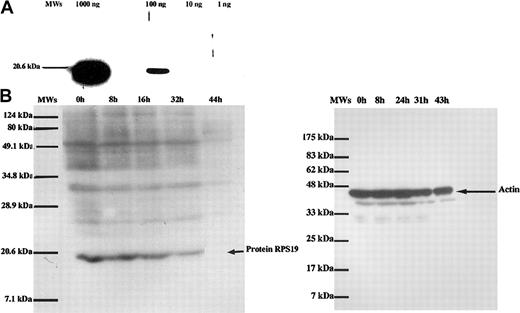
To monitor the level of RPS19 protein expression during erythroid differentiation, we produced an antibody raised in chicken against mouse recombinant His-tagged RPS19. After affinity purification, anti-RPS19 antibody used in a range of concentrations of 0.02 μg/mL to 0.1 μg/mL was able to detect 100 ng of human recombinant RPS19 protein (Figure 4A). Over the time course of 0 to 44 hours of the terminal differentiation of FAV cells, there was a progressive decrease in RPS19 protein expression with no detectable level of protein at 44 hours in enucleating erythroblasts (Figure 4B). This result was reproducible in 3 independent experiments. Importantly, specificity of the decrease in RPS19 expression during terminal erythroid differentiation was further supported by the finding that the expression of cytoskeletal protein 4.1R, a protein known to accumulate late in erythroid differentiation, showed increased expression (data not shown), whereas the expression of actin did not change during erythroid differentiation over the same time course (Figure 4B).
RPS19 protein expression during terminal erythroid differentiation.
(A) We tested the sensitivity of our antibody raised in chicken-against-mouse recombinant His-tagged RPS19 protein to detect the recombinant protein. The antimouse RPS19 was able to readily detect 100 ng of recombinant RPS19 protein. A molecular weight standard (MWs) was loaded in the first lane. (B) RPS19 protein expression in FAV cells during terminal erythroid differentiation. As with RPS19 mRNA expression, RPS19 protein expression also decreased during terminal erythroid differentiation. The arrow indicates the location of the RPS19 protein in the gel. The expression of actin during the same time course of terminal erythroid differentiation is shown in the right panel. In contrast to the marked reduction in RPS19 protein expression during the late stages of erythropoiesis, the level of actin expression showed little change. MWs was loaded in the first lane, and the positions of the molecular weights are indicated above lanes.
Discussion
The present study highlights several interesting features of theRPS19 gene expression. The comparison of human and mouse sequences upstream of the translation initiation start site enabled us to identify 4 consensus regions containing putative binding motifs for 5 transcription factors. These include SP1, GATA1, CACCCBf in the first consensus region, c-Rel and Rel-A in the third consensus region, and N-Myc transcription factor in the fourth consensus region. The strong homology between human and mouseRPS19 genes in the promoter region already reported for theRPL13A and RPS1113 genes suggested to us that some conserved regions and transcription binding motifs could also be functionally involved in the regulation ofRPS19 promoter activity. The location of the humanRPS19 gene promoter predicted by the computer program NNPP14-16 was confirmed at position −761 nt to −712 nt from the initiation start ATG, only −204 nt and −252 nt from the 2 transcription start points described in the human gene. Furthermore, we identified a strong regulatory element in the third consensus region. Importantly, when both the promoter element and the third consensus region were simultaneously deleted, the RPS19 gene promoter activity was dramatically reduced, suggesting an additive effect of these 2 regions. If we define the minimal promoter to be the 5′ sequence upstream of translational start site ATG without both the putative promoter sequence and the third consensus region, the putative promoter activity is approximately 7-fold higher than the minimal promoter.
In order to further investigate the role of consensus region 3 in promoter function, we deleted binding sites for putative transcription factors within that region. Surprisingly, we found that deletion of the putative binding site for c-Rel and Rel-A subunits of NF-κB resulted in an unexpected 3-fold increase in promoter activity, suggesting that a yet-to-be-defined transcription factor binds this NF-κB/Rel site, and represses promoter activity.
These results were in accordance with the general features of other mammalian ribosomal protein gene promoters, as previously described for the mouse RPL30,10,11,26-28RPL32,10,27-35 and RPS1610,34,36-38 genes. These features included (1) location of regulatory transcription elements in a short region of 200 nt immediately upstream of the transcriptional start site; (2) existence of regulatory elements located downstream of the transcription cap; and (3) regulatory elements close to the cap site which play a major role in the promoter function. In human RPS6,12 and in mouse RPS16 and RPL32, the farther-upstream elements contributed to but were not essential for promoter activity.11,31,32,36 The only exception described to date is the mouse RPL30 gene promoter in which the major determinant elements of transcriptional activity were located farther from the cap site.11,26,27 The RPS19 gene, like other housekeeping genes and ribosomal protein genes,12,13,36,37 contains several SP1 binding sites, often located very far upstream of the transcriptional start site. At least 6 potential SP1 binding motifs are present in the humanRPS19 gene (all located in GC-rich regions), but they do not appear essential for promoter activity. Furthermore, in accordance with the features of mammalian ribosomal protein gene promoters,10,12,29,34,37 the human RPS19 gene promoter lacked the canonical TATA-box and exhibited a transcriptional start at cytidine residues embedded in a polypyrimidine tract, a short 5′ UTR of 22 nt for the shortest 5′ UTR described by Kondoh (GenBank accession no. NM_001022).25
In contrast to the predicted promoter region and the third consensus region, neither of the 2 mutations found in DBA in the 5′ region upstream of the translation initiation start site altered promoter activity. The missense mutation G>T at −460 is located in the first intron between the consensus region 3 and 4, whereas the 4-bp insertion at −629 −625 is within the third consensus region upstream of the 5′ end of the RPS19 gene 5′ UTR. It should be noted, however, that our studies were performed in human fibroblasts and we cannot rule out the possibility that these mutations could have an erythroid cell–specific effect.12 There is precedence for such effects since Antoine et al already reported cell-specific control of the promoter activity for the human RPS6gene.12 Consequently, we have to confirm our present data in erythroid cells before stating that these mutations in DBA are not involved in the phenotype of the disease.
The NF-κB/Rel family of transcription factors are involved in inflammatory39,40 and immune T-cell and B-cell responses,41,42 in cell cycle regulation,43in differentiation, and in protection against apoptosis,44but also in the transcriptional control of cellular and viral genes.45,46 Usually, after binding to a κB site, transcription factors of the NF-κB/Rel family act as enhancers of the expression of multiple genes in eukaryotic cells.42Moreover, it appears that transcription factors of the NF-κB/Rel family do not act as single molecule, but rather as molecular complexes. In accordance with this concerted regulation by transcription factors of the NF-κB/Rel family, we found a perfect consensus binding site for putative c-Rel and Rel-A (p65) and a partial consensus binding site for putative NF-κB1 (p50), within consensus region 3 in both the human and mouse RPS19 gene promoters. However, in contrast to the transactivation of target promoter elements by transcription factors of the NF-κB/Rel family, we observed transcriptional inhibition. Transcriptional repression by factors of the NF-κB/Rel family has been reported in osteoblasts47and in macrophages,39 and c-Rel is reported to repress Rel-A (p65)–activated transcription driven by either HIV-1 or IL-2Rα gene promoters.46 However, the regulation of theRPS19 gene promoter by the NF-κB/Rel family of transcription factors needs to be further defined and confirmed. Taken together, our results concerning the RPS19 promoter suggest that, although regulation of ribosomal protein gene expression at the translational level has been found to be the most prevalent regulatory mechanism,29,48 49 regulation at the transcriptional level may be equally important in regulating RPS19 expression.RPS19 gene expression may indeed be regulated by yet-to-be-defined trans-acting transcription factors acting on the RPS19 gene promoter.
Since mutations in the RPS19 gene have been identified in 25% of the patients affected by DBA, we performed detailed Northern blot and Western blot analyses of the levels of transcription and translation of RPS19 during terminal erythroid differentiation. We chose the FAV cell system17 24for this study since it faithfully recapitulates terminal erythroid differentiation. In this system, the pure population of proerythroblasts, put into suspension cultures, undergoes terminal differentiation and after 44 hours produces enucleating erythroblasts and reticulocytes. We observed a decrease in both RPS19 mRNA and protein expression during terminal erythroid differentiation. Furthermore, the finding of high levels of RPS19 expression in proerythroblasts and decreasing levels during terminal erythroid differentiation is consistent with the finding of maturation arrest at early stages of erythroid differentiation in DBA.
Based on the present findings with regard to the expression pattern of RPS19 during erythroid differentiation, we are in the process of investigating the role of RPS19 in DBA pathogenesis. Identification of erythroid-specific binding partners for RPS19 will be important for elucidating the contribution of RPS19 defects to the DBA phenotype.
We gratefully acknowledge Dr Robert Lersch and Dr Uli Weier (Lawrence Berkeley National Laboratory) for performing FISH analysis, Dr Mark Koury for providing us with FAV cells, Dr Loren Walensky and Dr Solomon Snyder for giving us the antibody against 4.1R, Dr Victor Hou for Western blot analysis of protein 4.1R in FAV cells, Dr Philippe Gascard for his helpful advice, and Michael Patterson for his help in DNA sequencing.
Prepublished online as Blood First Edition Paper, August 1, 2002; DOI 10.1182/blood-2002-04-1131.
Supported by National Institutes of Health Grant DK26263 (N.M.), HL64885 (L.L.P.), the Daniella Maria Arturi Foundation, the Diamond Blackfan Anemia Foundation, la Direction de la Recherche Clinique Assistance Publique-Hopitaux de Paris (CRC95183 G.T.), and contrats INSERM/AFM (RD: 4MR09F et ARC 5636).
The publication costs of this article were defrayed in part by page charge payment. Therefore, and solely to indicate this fact, this article is hereby marked “advertisement” in accordance with 18 U.S.C. section 1734.
References
Author notes
Lydie Da Costa, Red Cell Physiology Laboratory, New York Blood Center, 310 East 67th St, New York, NY 10021; e-mail: ldacosta@lbl.gov, orlydie.dacosta@bct.ap-hop-paris.fr.


This feature is available to Subscribers Only
Sign In or Create an Account Close Modal