In this issue of Blood, Hasegawa et al report that liver macrophage-derived transforming growth factor β (TGF-β) injures bile duct stem cells during graft-versus-host disease (GVHD) in mice, resulting in impaired hepatic regeneration and function.1
GVHD leads to significant morbidity through dysfunction of the affected organs and is the third most frequent cause of mortality following allogeneic hematopoietic stem cell transplantation (allo-HCT).2 The liver can be a target organ of both acute and chronic GVHD, with previously reported cumulative incidences of 6.7% and 5.8% in allo-HCT recipients, respectively.3,4 Liver GVHD frequently presents as cholestasis with hyperbilirubinemia and elevated alkaline phosphatase, though hepatic forms with isolated transaminitis have also been described.5 Degenerative changes of the portal bile ducts are a common finding in liver GVHD,6 yet our understanding of the fate and role of various cell populations, including bile duct stem cells (BDSCs) and biliary epithelial cells, has been limited. Hasegawa et al identify BDSCs as direct targets of GVHD. Their study demonstrates that blocking TGF-β signaling preserves the organoid-forming capacity of BDSCs and improves biliary function in mouse models of GVHD, suggesting a specific approach to mitigate alloimmune-related liver injury.
Previous work by the authors and other groups has identified stem cells in the intestine7 as direct targets of GVHD. The loss of these stem cells significantly contributes to tissue damage and organ dysfunction. For example, intestinal stem cells were notably ablated in mice undergoing total body irradiation as part of the conditioning treatment.7 The transfer of major histocompatibility complex (MHC)-mismatched T cells exacerbated intestinal stem cell loss, which could be rescued by administering R-spondin 1.7 The current work by Hasegawa et al addresses whether GVHD similarly affects BDSCs and identifies molecular pathways to reduce liver toxicity.
Using mouse models of allo-HCT, the authors observed substantial infiltration of donor T cells and F4/80+ macrophages around cytokeratin 19-expressing portal bile ducts. Although biliary epithelial cells showed elevated proliferation rates on day 21 after allo-HCT, as evidenced by Ki67 staining, this reactive expansion was significantly diminished by day 35. This finding supports the hypothesis that initial tissue damage induces the proliferation of BDSCs, but as GVHD progresses and targets these cells, the regeneration of the bile duct epithelium is not adequate to compensate.
To investigate the specific targeting of BDSCs by GVHD, the authors leveraged an in vitro bile duct–derived liver organoid culture system. Initially, the organoid-forming capacity of bile ducts derived from allo-HCT recipients was high, even exceeding that observed in syngeneic HCT (syn-HCT) recipients. However, this capacity dramatically declined by day 28 and was almost completely lost by day 42 after allo-HCT. Intestinal organoids are highly sensitive to proinflammatory cytokines like interferon gamma (IFN-γ) and tumor necrosis factor (TNF).8 In contrast, bile duct–derived liver organoids maintained viability even at high concentrations of IFN-γ and TNF, possibly due to low receptor expression. However, TGF-β treatment dramatically reduced liver organoid numbers, and inhibition of the canonical TGF-β pathway with the selective SMAD2/3 inhibitor SB-431542 restored organoid formation in the presence of TGF-β.
Traditionally, donor T cells are considered the main mediators of GVHD-induced tissue damage. The study by Hasegawa et al challenges this view by identifying macrophages as the main source of TGF-β. Notably, 80% of the portal infiltrating F4/80+ macrophages expressed TGF-β, as shown by immunofluorescence. Liver mononuclear cells from allo-HCT but not syn-HCT recipients suppressed the growth of liver organoids from naive mice. When the liver mononuclear cells were fractionated into T cells and non-T cells (about 65% macrophages), only the non-T-cell fraction significantly reduced liver organoid numbers, highlighting a novel role for macrophages as effector cells in GVHD.
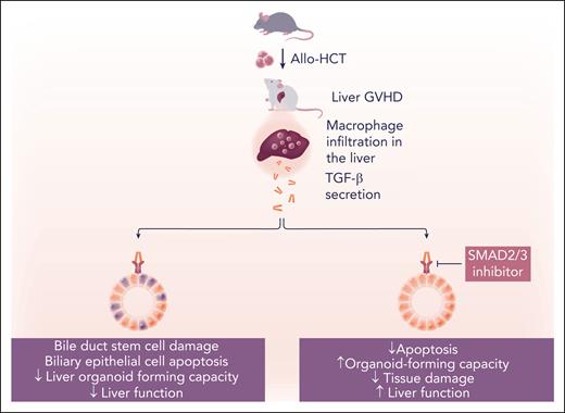
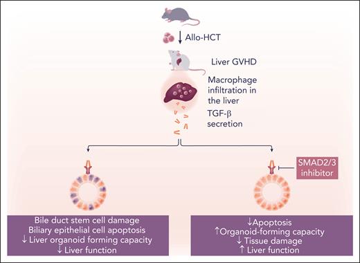
Finally, Hasegawa et al evaluated the efficacy of SMAD2/3 inhibition in 2 in vivo mouse GVHD models. Treatment with SB-431542 starting on day 14 after allo-HCT significantly restored the organoid-forming capacity of BDSCs and reduced the percentage of apoptotic biliary epithelial cells (see figure). Additionally, pathological liver GVHD scores and plasma bilirubin levels in allo-HCT recipient mice were reduced. These observations are in line with earlier reports that delayed anti-TGF-β treatment (commenced on day 14) reduced chronic GVHD in mouse models.9 Interestingly, the same study showed that early anti-TGF-β antibody administration aggravated acute GVHD, suggesting that TGF-β signaling plays diverse roles in regulating the alloimmune response depending on the posttransplant stage.
Further studies are needed to understand the fate of BDSCs and the potential applicability of TGF-β signaling inhibitors in human liver GVHD. Due to the lack of reliable specific markers for BDSCs, their abundance could only be estimated by the organoid-forming capacity of bile ducts. Although current evidence suggests that BDSC numbers are likely reduced, it is also possible that their numbers are maintained, but their proliferative function is transiently impaired. The absence of specific markers for BDSCs also hinders further studies, such as those examining their transcriptomic profile. The liver epithelium produces bile acids, and several studies have implicated bile acids as one of the drivers of intestinal GVHD.8,10 In this study, protecting the bile duct epithelium with SB-431542 did not improve intestinal GVHD histopathological scores. However, one potential explanation is the timing of SB-431542 administration. The authors treated GVHD-developing mice starting on day 14 after allo-HCT, by which time intestinal tissue damage had peaked in this specific GVHD model. Further studies might investigate whether prophylactic protection of BDSCs could support bile acid production and mitigate intestinal GVHD. Another open question is whether allogeneic T-cell infiltration, which preceded macrophage infiltration of the liver, was the initial cause of tissue damage leading to macrophage recruitment. How HLA matching and T-cell depletion strategies such as antithymocyte globulin and posttransplant cyclophosphamide affect BDSC damage in patients remains to be clarified. The study by Hasegawa et al reveals molecular insights into the pathogenesis of liver GVHD on mice and paves the way for further examinations of the potential clinical utility of TGF-β signaling inhibitors to reduce hepatic dysfunction.
BDSCs are direct targets of GVHD in a TGF-β-dependent manner. Allo-HCT in mice induces macrophage infiltration into the liver as GVHD develops. Macrophages secrete TGF-β, which induces apoptosis of biliary epithelial cells and reduces the organoid-forming and regenerative capacity of bile duct stem cells. Bile duct stem cell loss results in histological liver damage and reduced liver function. Inhibition of canonical TGF-β signaling with a SMAD2/3 inhibitor preserves bile duct stem cells, restores their organoid-forming capacity, and reduces liver dysfunction. Professional illustration by Somersault18:24.
BDSCs are direct targets of GVHD in a TGF-β-dependent manner. Allo-HCT in mice induces macrophage infiltration into the liver as GVHD develops. Macrophages secrete TGF-β, which induces apoptosis of biliary epithelial cells and reduces the organoid-forming and regenerative capacity of bile duct stem cells. Bile duct stem cell loss results in histological liver damage and reduced liver function. Inhibition of canonical TGF-β signaling with a SMAD2/3 inhibitor preserves bile duct stem cells, restores their organoid-forming capacity, and reduces liver dysfunction. Professional illustration by Somersault18:24.
Conflict-of-interest disclosure: P.A. declares no competing financial interests.


