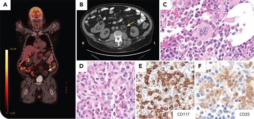
A 51-year-old man with a history of anaphylactic reaction to hornet venom presented with abdominal pain, melena, and bowel habit changes. No skin lesions or hepatosplenomegaly were identified. Positron emission and computed tomography revealed a retroperitoneal fat-containing mass in the left lower abdominal quadrant, measuring 5.7 cm in its greatest dimension (panel A-B, arrow). A needle-core biopsy of the mass showed hematopoietic elements (panel C: hematoxylin and eosin stain, 50× objective) with a small aggregate of elongated cells associated with eosinophils (panel D, 50× objective). The cells were positive for mast cell tryptase, CD117, and CD25 (panels E-F, 20× and 50× objectives, respectively). The findings were compatible with myelolipoma and an abnormal mast cell aggregate suspicious for mastocytosis. Complete blood cell count was normal. Subsequent bone marrow biopsy showed abnormal CD25+ mast cell aggregates, comprising 5% of the cellularity. There was no evidence of dysplasia or increased blasts. Cytogenetics showed a normal male karyotype 46,XY[19]. Myeloid next-generation sequencing was negative for pathogenic variants in myelodysplastic syndrome–associated genes. The tryptase level was 23.6 ng/mL (reference value, <11.5). Peripheral blood qualitative polymerase chain reaction KIT (D816V) mutation was positive. A diagnosis of indolent systemic mastocytosis was established.
Myelolipomas are rare tumorlike masses composed of mature adipose tissue and hematopoietic elements that predominantly occur in the adrenal glands. To our knowledge, this is the first reported case of systemic mastocytosis associated with myelolipoma.
For additional images, visit the ASH Image Bank, a reference and teaching tool that is continually updated with new atlas and case study images. For more information, visit https://imagebank.hematology.org.


