![A 59-year-old man presented with upper eyelid lesions and bilateral proptosis. Magnetic resonance imaging of the head (panel A) revealed bilateral enhancing intraconal masses encasing the optic nerves. A right orbital biopsy showed a diffuse, histiocytic infiltrate involving fibroconnective tissue with scant inflammatory cells, including few eosinophils (panel B; hematoxylin-eosin [H&E]; original magnification ×4 objective; panel C; H&E, original magnification ×20 objective; inset; original magnification ×40 objective). The foamy, multivacuolated histiocytes were admixed with scattered Touton-type giant cells. By immunostaining, the histiocytes were positive for CD68, CD163 (panel D; H&E, original magnification ×40 objective), factor XIIIA (panel E; H&E, original magnification ×40 objective), BRAF V600E (panel F; H&E, original magnification ×40 objective), and negative for S100 and CD1a, supporting the diagnosis of Erdheim-Chester disease (ECD).](https://ash.silverchair-cdn.com/ash/content_public/journal/blood/139/19/10.1182_blood.2022015606/3/m_bloodbld2022015606r1f1.png?Expires=1764883216&Signature=TbPvoLr5ClL5rF9or2Ctx~CNj~tsJ3Y8ekBD~vGgXOyQXoj7kKyiz7sF6lu7Ix528Bko3Vm0eXNBZJeI7Ip9hMqT75T4WqR7P5lmc6cKPBrCzOSHztyP3eL4HUnk3yrMjXiFaecPZ1GXUuNFeGJFl5~IWtkrq8Rp8A7QOZ1azwA3QpKpVjnTBGAqlXRC~xVtNpnyc2Zv~iGTOWgjc1-IBaS~ETV4hPNofeS6uHkye7zzb28KGah5FmOkbANfroy-spuUyd0gJ7-jWAA5KdDKYFm9-SKAN7jqJ3lsUXFRbT8HBX8zXPQP2J8M3Z3mkWW258oQX~rJZDPbpq5zEPLYkg__&Key-Pair-Id=APKAIE5G5CRDK6RD3PGA)
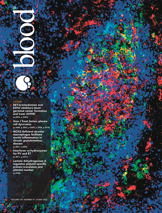
A 59-year-old man presented with upper eyelid lesions and bilateral proptosis. Magnetic resonance imaging of the head (panel A) revealed bilateral enhancing intraconal masses encasing the optic nerves. A right orbital biopsy showed a diffuse, histiocytic infiltrate involving fibroconnective tissue with scant inflammatory cells, including few eosinophils (panel B; hematoxylin-eosin [H&E]; original magnification ×4 objective; panel C; H&E, original magnification ×20 objective; inset; original magnification ×40 objective). The foamy, multivacuolated histiocytes were admixed with scattered Touton-type giant cells. By immunostaining, the histiocytes were positive for CD68, CD163 (panel D; H&E, original magnification ×40 objective), factor XIIIA (panel E; H&E, original magnification ×40 objective), BRAF V600E (panel F; H&E, original magnification ×40 objective), and negative for S100 and CD1a, supporting the diagnosis of Erdheim-Chester disease (ECD).
ECD is a multisystem, clonal non-Langerhans cell histiocytic neoplasm. ECD has myriad presentations, including bone pain, endocrinopathy, and neurological symptoms. Ocular involvement, seen in 25% of patients, is classically characterized by bilateral proptosis. A high degree of suspicion is required in assessing morphologically bland histiocytic lesions, where positivity for BRAF V600E and factor XIIIa coupled with negativity for CD1a and S100 supports a designation of ECD. Treatment options include interferon-α and BRAF inhibitors, like vemurafenib.
For additional images, visit the ASH Image Bank, a reference and teaching tool that is continually updated with new atlas and case study images. For more information, visit http://imagebank.hematology.org.