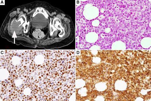
An 83-year-old woman was admitted for diplopia. She was initially diagnosed with cyclin D3–positive mantle cell lymphoma (MCL; which tested positive for CD5, CD20, and SOX11, but was negative for cyclin D1, cyclin D2, and 37% Ki-67 index) in the pterygopalatine fossa with a good response to radiation. She had a relapse in the pterygopalatine fossa and experienced bone lesions, including in the right femur (panel A, computed tomography scan), with drowsiness caused by hypercalcemia, 1 year later. Laboratory tests showed the following: albumin, 2.1 g/dL; calcium, 11.6 mg/dL; parathyroid hormone, 5 pg/mL; and parathyroid hormone–related protein (PTHrP), 3.9 pmol/L (normal, <1.1 pmol/L). Histological findings were consistent with transformation to the cyclin D3–positive blastoid variant of MCL (MCL-BV), retaining the same immunophenotype as initial diagnosis but with an increase in Ki-67 index to 93%. PTHrP expression was observed in lymphoma cells (panel B, hematoxylin and eosin, original magnification ×40; panel C, cyclin D3 immunostain, original magnification ×40; panel D, PTHrP immunostain, original magnification ×40).
A rare subtype of cyclin D1–negative MCL (positive for cyclin D2 or cyclin D3) was reported to be a part of the spectrum of MCL. MCL rarely transforms histologically into MCL-BV, which is more aggressive than typical MCL. Hypercalcemia may have been attributable to both PTHrP production by MCL-BV cells and local osteolytic metastases.
For additional images, visit the ASH IMAGE BANK, a reference and teaching tool that is continually updated with new atlas and case study images. For more information visit http://imagebank.hematology.org.


