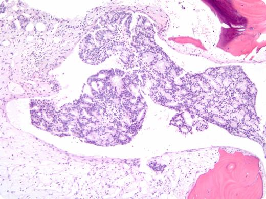
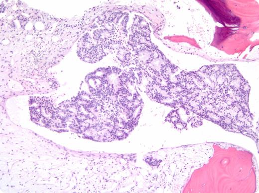
A 47-year-old African American man presented with lightheadedness and exertional dyspnea. Laboratory work revealed: hemoglobin, 2.9 g/dL; platelets, 17 × 109/L; and lactate dehydrogenase, 1492 IU/L. Folate, B12, and coagulation studies were normal. His peripheral smear showed increased schistocytes, polychromasia, and nucleated red blood cells. The patient was started on plasma exchange for presumed thrombotic thrombocytopenic purpura (TTP). However, his hematologic parameters failed to improve, and his pretreatment ADAMTS13 activity of 54% prompted further evaluation. A bone marrow biopsy demonstrated diffuse infiltration by adenocarcinoma, positive for prostate-specific antigen (PSA) and CK19 by immunohistochemistry, consistent with a diagnosis of metastatic prostate cancer. His serum PSA was elevated at 1162 ng/mL, and imaging revealed retroperitoneal lymphadenopathy with scattered bone lesions. The patient was started on combined androgen blockade with bicalutamide and leuprolide. After 8 weeks of treatment, his platelet count normalized and his hemoglobin improved to 10 g/dL.
Bone marrow involvement by metastatic carcinoma is uncommon and typically represents a late manifestation of disease. Marrow infiltration can lead to intramedullary hemolysis with significant cytopenias and schistocytes on peripheral smear, mimicking TTP. In this case, the patient’s hematologic manifestations were the presenting features of his malignancy, creating a diagnostic dilemma.
For additional images, visit the ASH IMAGE BANK, a reference and teaching tool that is continually updated with new atlas and case study images. For more information visit http://imagebank.hematology.org.


