To the editor:
Biallelic TTC7A mutations have recently been shown to cause early-onset inflammatory bowel disease or multiple intestinal atresias accompanied by severe combined immunodeficiency (MIA-SCID), a disease usually fatal in infancy without curative treatment.1-4
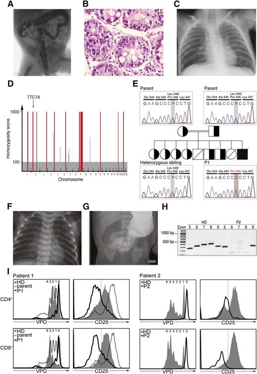
We studied a patient (P1) born in the thirty-third gestational week (to healthy Turkish consanguineous parents) who suffered from combined immunodeficiency and chronic diarrhea. Two older siblings had previously died of severe diarrhea. Within the first 2 months, P1 suffered from constipation followed by watery diarrhea without identifiable cause. Radiographic colonography revealed normal rectum diameter but hypoplastic descending and sigmoid colon and dilated small intestine and ascending and transverse colon (Figure 1A). Duodenal (Figure 1B) and colonic biopsies (supplemental Figure 1A, available online on the Blood Web site) showed graft-versus-host disease (GVHD)–like lesions. P1 experienced several pneumonia episodes (including Klebsiella pneumoniae septicemia) and pyelonephritis. Thymic tissue was hypoplastic (Figure 1C). At 3.5 months, P1 exhibited B lymphopenia and hypogammaglobulinemia (supplemental Table 1) requiring immunoglobulin substitution and trimethoprim/sulfamethoxazole prophylaxis. P1 died at age 15 months of sepsis without an identified causative agent.
Identification and intestinal histopathology of TTC7A-mutated patients. (A) Radiograph colonography with barium enema in P1 showing decreased diameter of the descending and sigmoid colon, dilated small intestine segments, and dilation of ascending and transverse colon. Rectum diameter was normal. (B) Histologic analysis of duodenal biopsy from P1 suggesting GVHD, including erosion and regeneration of the epithelial surface, focal villous atrophy, degeneration of glands, apoptotic content in crypts, and crypt loss accompanied by increase in lamina propria capillaries. (C) Chest radiograph of hypoplastic thymic tissue in P1. (D) Single nucleotide polymorphism–based mapping of the homozygous chromosomal intervals in P1 (red bars). Arrow indicates the chromosomal position of TTC7A locus within the homozygous interval on chromosome 2. (E) Identification and segregation analysis of the point mutation NM_020458:c.T1037C;p.L346P in the TTC7A gene of P1. Chromatograms display the mutation-containing TTC7A sequence in healthy heterozygous parents, 1 representative heterozygous sibling, and P1 who is homozygous for the mutation. All living siblings of P1 were heterozygous for the identified mutation. (F) Chest radiograph of P2 without visible thymic tissue. (G) Intestinal atresia included distal jejunum and the ligament of Treitz and spanned the entire distal colon. Radiographic colonography of P2 showing a small diameter of the pylorus with low passage of contrast agent. Distal intestine ends in a stoma after surgical excision of atretic tissue with no visible lumen in small bowel and colon. (H) Polymerase chain reaction amplification of indicated TTC7A exons from genomic DNA. (I) In vitro proliferation analysis of CD4+ and CD8+ T cells from patients after TCR stimulation. Proliferation history was monitored by flow cytometry–based detection of intracellular violet proliferation dye (VPD) dilution on day 3 (P1; left) and on day 4 (P2; right) after stimulation. Numbers above histograms indicate cell divisions. T-cell activation status was assessed by detection of CD25 on cell surface. HD, healthy donor.
Identification and intestinal histopathology of TTC7A-mutated patients. (A) Radiograph colonography with barium enema in P1 showing decreased diameter of the descending and sigmoid colon, dilated small intestine segments, and dilation of ascending and transverse colon. Rectum diameter was normal. (B) Histologic analysis of duodenal biopsy from P1 suggesting GVHD, including erosion and regeneration of the epithelial surface, focal villous atrophy, degeneration of glands, apoptotic content in crypts, and crypt loss accompanied by increase in lamina propria capillaries. (C) Chest radiograph of hypoplastic thymic tissue in P1. (D) Single nucleotide polymorphism–based mapping of the homozygous chromosomal intervals in P1 (red bars). Arrow indicates the chromosomal position of TTC7A locus within the homozygous interval on chromosome 2. (E) Identification and segregation analysis of the point mutation NM_020458:c.T1037C;p.L346P in the TTC7A gene of P1. Chromatograms display the mutation-containing TTC7A sequence in healthy heterozygous parents, 1 representative heterozygous sibling, and P1 who is homozygous for the mutation. All living siblings of P1 were heterozygous for the identified mutation. (F) Chest radiograph of P2 without visible thymic tissue. (G) Intestinal atresia included distal jejunum and the ligament of Treitz and spanned the entire distal colon. Radiographic colonography of P2 showing a small diameter of the pylorus with low passage of contrast agent. Distal intestine ends in a stoma after surgical excision of atretic tissue with no visible lumen in small bowel and colon. (H) Polymerase chain reaction amplification of indicated TTC7A exons from genomic DNA. (I) In vitro proliferation analysis of CD4+ and CD8+ T cells from patients after TCR stimulation. Proliferation history was monitored by flow cytometry–based detection of intracellular violet proliferation dye (VPD) dilution on day 3 (P1; left) and on day 4 (P2; right) after stimulation. Numbers above histograms indicate cell divisions. T-cell activation status was assessed by detection of CD25 on cell surface. HD, healthy donor.
Given the consanguinity, we assumed an autosomal-recessive inheritance and performed homozygosity mapping (Figure 1D) and exome sequencing. Unexpectedly, we identified a perfectly segregating homozygous missense mutation in TTC7A (NM_020458:c.T1037C;p.L346P) (Figure 1E and supplemental Figure 1B). The substitution of the highly conserved TTC7ALeu346 residue (supplemental Figure 1D) to TTC7APro346 was predicted damaging to TTC7A function by SIFT and PolyPhen-2 prediction algorithms.
Concomitantly, we investigated another patient (P2) with classical MIA-SCID manifesting in lymphopenia with near-absent CD8+ T cells, hypogammaglobulinemia, undetectable thymic tissue (supplemental Figures 1F, 2, and 3B), and extended intestinal atresia (Figure 1G). Molecular analysis revealed a large genomic deletion comprising TTC7A exons 6 through 8, causing frameshift and premature transcriptional stop codon (NM_020458:c765_1065del;p.N256Qfs*7) (Figure 1H and supplemental Figure 2B-C).
The pronounced immunodeficiency despite mild structural intestinal defects in P1 prompted us to compare the lymphocyte compartments of P1 and P2. Both showed B-cell lymphopenia and hypogammaglobulinemia (supplemental Table 1) despite the presence of naïve, marginal zone–like and class-switched memory B cells (supplemental Figure 3A). Increased proportion of transitional B cells in P1 indicated a partial developmental block (supplemental Figure 3A) reminiscent of the tonic BCR- or BAFF-signaling defects resulting in decreased B-cell survival and hypogammaglobulinemia.5,6 TTC7A is required for tethering of phosphatidylinositol 4-kinase, catalytic, alpha (PI4KCA) to the plasma membrane,2 which in turn phosphorylates phosphatidylinositol into phosphatidylinositol(4,5)P2, a second messenger in the phospholipase C gamma-2 (PLCγ2) and phosphatidylinositol 3′-kinase (PI3K) signaling cascades downstream of lymphocyte antigen receptors.7
P1 had unremarkable T-cell counts (supplemental Table 1), CD4+ and/or CD8+ T-cell distribution (supplemental Figure 3B), and T-cell repertoire (supplemental Figure 1E). Maternal cells were absent. A significant proportion of CD4+ T cells in P2 but not in P1 expressed CD25 activation marker, possibly as a compensatory mechanism that would otherwise lead to proliferative expansion8 (supplemental Figure 3B). Cell-surface expression of CD44, CD62L, and CD69 and neutrophil counts and oxidative burst were normal (not shown).
Higher TTC7A expression in thymic stroma compared with thymocytes had suggested aberrant thymic (micro)environment as a T-cell extrinsic cause for T-lymphopenia in TTC7A deficiency.1 We challenged this finding by monitoring T-cell proliferation of pulse-labeled patient peripheral blood mononuclear cells after anti-CD3 stimulation. Whereas P2’s T cells showed no proliferative response, proliferation of T cells from P1 was only partially impaired (Figure 1I). TTC7APro346 protein in HEK293 cells was stable and detectable at levels similar to those for TTC7Awildtype (supplemental Figure 3D), implying that TTC7ALeu346Pro represents a hypomorphic variant allowing for residual protein function. Furthermore, T cells from both patients were unable to upregulate CD25 expression after anti-CD3 stimulation, suggesting T-cell receptor (TCR)–signaling defects (Figure 1I). Accordingly, the majority of P1’s T cells had naïve CD45RA+ phenotype (supplemental Figure 3C). One possible explanation might be the inability of mutant TTC7A to recruit PI4KCA to the plasma membrane, required for its interaction with CD4-p56lck and downstream signal transduction.9 Because lymphocyte-specific protein tyrosine kinase (LCK) interaction with CD4 and/or CD8 is essential for T-cell development and activation,10 TTC7A might be an integral component of the TCR signalosome. Notably, in contrast to previous observations of defective T-cell proliferation1,4 and our findings, a recent study identified patients with hyperproliferative TTC7A-mutant T cells,11 underlining the phenotypic variability of TTC7A-mutant immunodeficiency.
TTC7A deficiency was recently identified as the molecular cause for MIA with or without accompanying SCID.1,3 The hallmark feature of TTC7A deficiency was varying degree of intestinal aberrations. The mildest case presented with intestinal aberrations consisting of bloody diarrhea, apoptotic enterocolitis, and acute GVHD-like symptoms, but no atresias, and the extent of the observed lymphopenia remained unclear.2 P1 had B-lymphopenia, hypogammaglobulinemia, and functional T-cell defects, but intestinal structural aberrations were mild and GVHD-like signs were discrete. Collectively, the clinical spectrum of TTC7A deficiency is considerably more variable than previously appreciated, which should alert physicians to consider TTC7A mutational analysis in (S)CID patients.
The online version of this article contains a data supplement.
Authorship
Acknowledgments: This work is supported by funding from the Austrian Science Fund (FWF) grant number P24999 (K.B.).
Contribution: S.W., E.S., C.D.C., and I.B. performed experiments; C.A., S.A., and F.O.H. provided clinical data and clinical care for patient P1; H.P., N.M.-D., E.F.-W., S.M., W.-D.H., and W.H. provided clinical data and clinical care for P2; T.L. performed chimerism analysis on blood samples from P1; K.B. conceived the study and provided laboratory resources; K.B., S.W., E.S., and I.B. planned, designed and interpreted experiments; S.W., I.B., and K.B. wrote the manuscript; and all authors critically reviewed the manuscript and agreed to its publication.
Conflict-of-interest disclosure: The authors declare no competing financial interests.
Correspondence: Kaan Boztug, CeMM Research Center for Molecular Medicine of the Austrian Academy of Sciences and Department of Pediatrics and Adolescent Medicine, Medical University of Vienna, Lazarettgasse 14, AKH BT 25.3, A-1090 Vienna, Austria; e-mail: kboztug@cemm.oeaw.ac.at.


