A 13-year-old girl with juvenile rheumatoid arthritis presented for a routine examination. Except for a short episode of a cough 1 week prior, she had no complaints. The chest examination was normal.
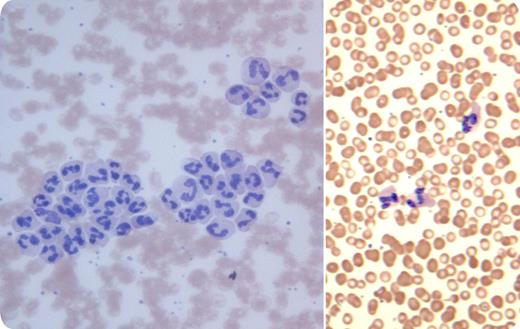
Blood cell count was performed on an automated analyzer using an ethylenediaminetetraacetic acid (EDTA) blood specimen and showed normal hemoglobin and leukocytes (7.2 × 10⁹/L), but a mild thrombocytosis (467 × 109/L). Platelet clumps were flagged by the cell counter. However, when the peripheral smear was reviewed, there was leukocyte agglutination (shown, left) rather than clumps of platelets. The cold agglutinin test was positive with relative anti-I specificity. Serology for Mycoplasma pneumoniae was positive (complement fixation test 1:32, reference value ≤ 1:16; microparticles agglutination 1:1280, reference value ≤ 1:40). When the blood was warmed at 37°C, the smear no longer showed leukoagglutination (shown, right).
The cause of leukoagglutination in this case was uncertain, but connective tissue disease and Mycoplasma infection were possibilities. Leukoagglutination has also been reported in other types of infections, inflammation, malignancies, alcoholism, and liver disease, and with blood storage. At times, an EDTA-dependent leukoagglutination can occur in healthy subjects; this possibility was not tested in this case.
Many Blood Work images are provided by the ASH IMAGE BANK, a reference and teaching tool that is continually updated with new atlas images and images of case studies. For more information or to contribute to the Image Bank, visit www.imagebank.hematology.org.


