Additional analysis of our data1 (see Table 1) on transfused patients with sickle cell disease (SCD) shows similar correlation between total lifetime transfusion (TLT) and serum ferritin (SF). This correlation improved only marginally when TLT was adjusted by age or anthropometric measurements, perhaps because SF is variable as a measure, or because the iron pool volume only partially follows changes of height or weight during growth. Inati and colleagues reported a surprisingly strong correlation between SF and liver iron concentration by R2 magnetic resonance imaging, which supports our observations that SF may be a reliable indicator of iron load, especially at lower levels, since their patients had, in general, modest iron overload, with SF levels mostly less than 1500 ng/mL and liver iron concentration less than 10 mg/g dry liver weight. We hope to see published a more detailed analysis of this data to allow readers to, for example, determine the presence of influential outliers.
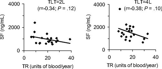
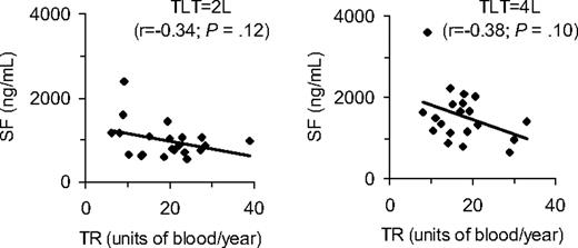
The principal observation reported by Inati et al is that SF strongly correlates with the transfusion rate (TR; defined as lifetime number of units transfused divided by the number of years on transfusion), and their graphs comparing TR with SF show a nearly linear curve. When we analyzed our patient data, we found no such correlation (Table 1). We then compared SF and TR at various fixed volumes of transfused blood, and again saw no significant trends (Figure 1). Several reasons may explain this discrepancy. In addition to having a higher iron burden, our patients received regular transfusions under close supervision, and data were collected prospectively. TR remained relatively constant throughout the duration of the transfusion regimen (mostly every 3 to 4 weeks, overall TR = 17 units per year; SD ± 7 in the data presented). It is not clear whether patients followed by Inati et al were placed on such a transfusion protocol, and the data were presumably collected retrospectively. It is also not clear whether serum alanine aminotransferase measures were obtained systematically. In addition, our patients were generally younger, and of a relatively homogenous age group, nearly all affected with SCD, hemoglobin SS phenotype. Those of Inati et al included some young children but mostly adults. Phenotype was not reported and transfusion histories of patients with fewer than 10 units/year were not described. Did this group include 4-year-old children with a couple of transfusions as well as adults with 8 to 9 transfused units per year for more than 10 years? It would be surprising if such older patients did not have measurable increases in iron load without concomitant iron losses. In our analysis, TR did not explain SF variability in regularly transfused iron overloaded children with SCD. As such, TR, which is a composite measure of iron load and transfusion history, does not appear to be a useful clinical measure of iron overload in this setting. Further carefully designed prospective studies that examine iron loading from onset of transfusion are clearly needed.
Correlation between serum ferritin and transfusion rate at given total lifetime transfusions. SF indicates serum ferritin; TR, transfusion rate; and TLT, total lifetime transfusion units (± 1 L). A unit is assumed to be 300-cc volumes. Each data point represents a different patient.
Correlation between serum ferritin and transfusion rate at given total lifetime transfusions. SF indicates serum ferritin; TR, transfusion rate; and TLT, total lifetime transfusion units (± 1 L). A unit is assumed to be 300-cc volumes. Each data point represents a different patient.
Authorship
Acknowledgments: T.V.A. was supported in part by the National Institutes of Health, National Heart, Lung, and Blood Institute (1K23HL0425101A1); National Center on Minority Health and Health Disparities (1P20MD003383-01); and Novartis Corporation (CICL670AUS06). Data were retrieved from STOP and STOP2 trials; these are registered at www.clinicaltrials.gov as #NCT00000592 and #NCT00006182.
Contribution: T.V.A. analyzed data and prepared the manuscript; and M.R.A., O.A.A., and N.O. analyzed data and reviewed the manuscript.
Conflict-of-interest disclosure: The authors declare no competing financial interests.
Correspondence: Thomas V. Adamkiewicz, MD, Morehouse School of Medicine, 1513 E. Cleveland #100, 3rd Fl, Suite 300-A, East Point, GA 30344; e-mail: tadamkiewicz@msm.edu.
References
National Institutes of Health


