Abstract
Killer immunoglobulin-like receptors (KIRs) play an essential role in the regulation of natural killer cell functions. KIR genes are highly polymorphic in nature, showing both haplotypic and allelic variations among people. We demonstrated in both in vitro and in vivo models a significant heterogeneity in function among different KIR2DL1 alleles, including their ability to inhibit YT-Indy cells from degranulation, interferon γ production, and cytotoxicity against target cells expressing the HLA-Cw6 ligand. Subsequent experiments showed that the molecular determinant was an arginine residue at position 245 (R245) in its transmembrane domain that mechanistically affects both the efficiency of inhibitory signaling and durability of surface expression. Specifically, in comparison with R245-negative alleles, KIR2DL1 that included R245 recruited more Src-homology-2 domain-containing protein tyrosine phosphatase 2 and β-arrestin 2, showed higher inhibition of lipid raft polarization at immune synapse, and had less down-regulation of cell-surface expression upon interaction with its ligand. Thus, our findings provide novel insights into the molecular determinant of KIR2DL1 and conceivably a fundamental understanding of KIR2DL1 allelic polymorphism in human disease susceptibility, transplant outcome, and donor selection.
Introduction
Natural killer (NK) cells are a part of our immune system that eliminates virally infected cells and tumor cells through cytolytic killing and cytokine secretion.1 An NK cell's responses to its targets are regulated by the balance of signals generated through various activating and inhibiting receptors.2,3 NK-cell receptors may be categorized on the basis of their ligand specificity for major histocompatibility complex class I and related molecules. Expression of various combinations of receptors on the surface of NK cells creates a diverse repertoire.4,5
In humans, one of the most important groups of receptors that regulate NK-cell function is killer immunoglobulin-like receptors (KIRs).6,7 The KIRs make up a family of diverse glycoproteins encoded by a compact cluster of genes located on human chromosome 19q13.4.8,9 KIRs expressed at the surface of NK cells regulate their response by interacting with human leukocyte antigen (HLA) class I molecules. The KIR family has both activating and inhibitory receptors. KIR inhibitory receptors suppress NK cells' function using the immunoreceptor tyrosine-based inhibitory motif in their long cytoplasmic tails. Activating KIR receptors have short cytoplasmic tails and do not contain the inhibitory motif.
Each of the KIR genes exhibits allelic variation as well as haplotypic variability in terms of the number and types of genes on the haplotypes.9,10 The allelic variations of KIR genes range from 2 to more than 30.10 The polymorphisms between the alleles of a given KIR gene can occur in its extracellular, transmembrane, or cytoplasmic domains. Polymorphism at each of these 3 domains has been associated with significant biologic consequences. For example, KIR2DS5*001 that contains proline (P) at position 111 and serine (S) at position 164 in its extracellular domain does not express on the cell surface.11 In contrast, KIR2DS5*002, which has serine and phenylalanine (F) at those positions, does express on the cell surface. Similarly, a subgroup of the 10A allele of KIR2DL4 has a transcript that lacks the first immunoglobulin-like domain and cannot express on the cell surface.12 KIR2DL4 allele 9A, which lacks a transmembrane domain, encodes a secreted receptor.
Besides KIR2DL4 and KIR2DS5, the only other KIR gene that has been studied extensively for its allelic functional polymorphism is KIR3DL1. Yawata et al13 showed that in the Japanese population, 5 common KIR3DL1 allotypes, *001, *01502, *002, *005, and *007, have distinguishable inhibitory capacity, frequency of cellular expression, and level of cell-surface expression. Studies by Carr et al14 showed that the presence of arginine (R) at position 238 in the D2 domain and isoleucine at position 320 at the transmembrane domain of the KIR3DL1*002 allele makes it a stronger receptor than the *007 allele that has glycine (G) and valine (V) at those positions, suggesting the functional consequences of polymorphism in both the extracellular and transmembrane domains. A study by Foley et al further demonstrated that the strength of signal delivered through KIR3DL1 was dependent not only on KIR polymorphism, but also on the effectiveness of different Bw4+ ligands. The Bw4+ HLA-A*2301, for instance, can uniquely inhibit NK cells expressing KIR3DL1*005 allele.15 Other studies have also indicated that KIR3DL1 polymorphism affects the function of KIR3DL1.16
In this study, we investigated for the first time the functional differences among KIR2DL1 alleles. We selected KIR2DL1 because of its importance in a wide variety of human disease and in transplantation.17-19 KIR2DL1 has 14 alleles that contain polymorphisms in their extracellular, transmembrane, and cytoplasmic domains. Data obtained from the crystal structures of KIR2DL1 and its ligand interaction suggest no functional importance of the allelic diversity of KIR2DL1 receptors in extracellular domain and ligand recognition,20,21 because all the amino acids that are important for receptor ligand interaction are conserved among the 14 alleles. Therefore, we focused our investigation on the functional consequences of polymorphism in the stem and transmembrane domains. Our findings indicate a significant functional heterogeneity among KIR2DL1 alleles and a pivotal role of arginine245 in the transmembrane domain.
Methods
DNA constructs
Peripheral blood mononuclear cells were obtained from healthy human donors with informed consent under a protocol approved by our institutional review board at St Jude Children's Research Hospital, in accordance with the Declaration of Helsinki. KIR2DL1-positive samples were screened by flow cytometry using anti-KIR2DL1 (EB6.B; BD Biosciences) monoclonal antibody,22 and total RNA was extracted from cells expressing KIR2DL1 using RNA extraction kits (QIAGEN). cDNA of various alleles of KIR2DL1 was generated from total RNA and cloned into mammalian expression vector pcDNA3 (Invitrogen). The identities of the KIR2DL1 alleles were confirmed by sequencing. Specific amino acids were substituted into KIR2DL1 using recombinant polymerase chain reaction. To create a C-terminally FLAG-tagged KIR2DL1 allele, the appropriate cDNA was amplified with FLAG tag fused in reverse primers. The FLAG-tagged KIR2DL1 alleles were cloned into retroviral vector MSCV-IRES-GFP (received from vector laboratory, St Jude Children's Research Hospital). HLA-Cw6 and HLA-Cw7 cDNA were amplified from a normal human peripheral blood cDNA pool as described for KIR2DL1 alleles, confirmed by sequencing, and cloned into retroviral vector MMP-IC-GFP-W (received from vector laboratory, St Jude Children's Research Hospital) by replacing green fluorescent protein (GFP) with HLA-Cw6.
Cell lines, culture, and transfection
YT-Indy cells (a generous gift from Dr Zacharie Brahmi, Indiana University) were cultured in RPMI 1640 supplemented with 10% fetal bovine serum (FBS), 1mM penicillin/streptomycin, 2mM l-glutamine, 1mM sodium pyruvate, and 1% minimum essential medium nonessential amino acids (Invitrogen). B-lymphoblastic cell line 721.221 was purchased from the International Histocompatibility Working Group and cultured in RPMI 1640 supplemented with 20% FBS and 1mM penicillin/streptomycin. YT-Indy cells were transfected with pcDNA3 vector containing various KIR2DL1 alleles by electroporation (Gene Pulser II; Bio-Rad). Stable cell lines were generated by selection in Geneticin (Invitrogen). The 721.221 cells were transduced with retroviral vector MMP-IC-GFP-W containing HLA-Cw6. High-expressing cells were sorted by flow cytometric cell sorting using the monoclonal antibody (mAb) against HLA-Cw6 (One Lambda). YT-Indy cells expressing FLAG-tagged KIR2DL1 alleles were generated by transduction with retroviral vector MSCV-IRES-GFP containing FLAG-tagged KIR2DL1 alleles.
Cytolytic assays
A cell-killing assay was performed using DELFIA BATDA reagent (PerkinElmer Life and Analytical Sciences) according to the manufacturer's instructions. In brief, target cells, 721.221 (untransduced) and 721.221 transduced with HLA-Cw6 (721.221-Cw6), were labeled with DELFIA BATDA reagent. YT-Indy transfectants were used as effector cells and mixed with labeled target cells, followed by incubation in a cell culture incubator for 2 hours. Cells were spun down, supernatant was added to DELFIA Europium solution, and the fluorescence was measured using a Wallac Victor 2 Counter Plate Reader (both from PerkinElmer Life and Analytical Sciences).
CD107 mobilization assay
YT-Indy cells expressing a KIR2DL1 allele as well as control cells (YT-Indy transfected with vector pcDNA3) were mixed with target 721.221 untransduced and transduced cells at a 1:2 (E/T) ratio with phycoerythrin-conjugated CD107 mAb (lysome-associated membrane protein-1), clone H4A3 (BD Pharmingen). After 1 hour of incubation at 37°C in 5% CO2, 5mM Golgi stop (BD Biosciences) was added, followed by another 3 hours of incubation. YT-Indy transfectants without target cells were used as background. YT-Indy KIR2DL1 was gated based on the expression of KIR2DL1 using mAb EB6.B (Beckman Coulter). For control, YT-Indy cells transfected with vector were strained with CD20 mAb (clone B9E9; Beckman Coulter) and gated on their CD20− cell population because YT-Indy lacks expression of CD20. Cell-surface expression of CD107 was detected by flow cytometry (LSR II cytometer; BD Biosciences). Relative CD107 cell-surface expression was calculated as follows: [(YT-Indy transfectants + 721.221-Cw6) − (YT-Indy transfectants without target cells)/(YT-Indy transfectants + 721.221) − (YT-Indy transfectants without target cells)] × 100
Relative cell-surface expression of CD107 was further normalized with vector control.
Detection of interferon-γ production
Interferon γ (IFN-γ) production in YT-Indy transfectants was measured after stimulation with target cells 721.221-Cw6. To separate the cells in the coculture, YT-Indy transfectants were first labeled with common pan-leukocyte marker CD45. Labeled YT-Indy cells were cocultured with 721.221-transduced cells at a 1:2 ratio. After 1 hour of coculturing, Golgi stop (BD Biosciences) was added to a final concentration 5 mM, followed by 2 hours of incubation. Cells were washed, fixed, and permeabilized in Cytofix Cytoperm buffer (BD Biosciences) following the manufacturer's instructions. The permeabilized cells were labeled with anti–IFN-γ antibody (BD Biosciences). The CD45+ cell population was gated, and IFN-γ production was measured by flow cytometry (LSR II cytometer).
In vivo experiment
YT-Indy cells expressing various alleles of KIR2DL1 were mixed with target 721.221-Cw6 at a 10:1 (E/T) ratio and injected subcutaneously into 6- to 8-week-old nonobese diabetic/severe combined immunodeficient (NOD-scid) interleukin-2 receptor γnull (IL2Rγnull) mice (The Jackson Laboratory). The mice were handled by animal care facilities of St Jude Children's Research Hospital according to institutional animal care and use committee regulations. Thirty mice (in 6 groups of 5) were given the injections. When tumors in positive control mice (given only 721.221-Cw6 cells) reached 20% of the body weight of the mice, the mice were humanely killed by CO2 and cervical dislocation, dissected, and photographed.
Immunoprecipitation and Western blotting
YT-Indy cells expressing FLAG-tagged KIR2DL1*00401 and KIR2DL1*010 alleles were stimulated with target 721.221-Cw6 cells at a 1:1.5 ratio for 0, 1, 2, and 5 minutes. The stimulation was ended by adding lysis buffer (1% Triton X-100, 150mM NaCl, and 50mM tris(hydroxymethyl)aminomethane-HCl, pH 7.4). FLAG-tagged KIR2DL1 and associated proteins were immunoprecipitated using a FLAG Tagged Protein Immunoprecipitation Kit following the manufacturer's instructions (Sigma-Aldrich). Proteins were eluted by 3× FLAG Peptide (Sigma-Aldrich). The eluted proteins were electrophoresed on 4% to 12% NuPAGE Bis-Tris gel (Invitrogen). Separated proteins were blotted with goat anti–β-arrestin 2 (Abcam) and rabbit anti–Src-homology-2 domain-containing protein tyrosine phosphatase 2 (SHP-2; Cell Signaling Technologies) antibodies using a Western blotting protocol as described previously.23 The membrane was stripped with Restore Plus Western Blot Stripping Buffer (Thermo Scientific) and reblotted with anti-FLAG antibody (Sigma-Aldrich) to use as a loading control.
Slide preparation and microscopy
Slides were prepared following a protocol adapted from Fassett et al.24 Effector (YT-Indy/2DL1*00401 or YT-Indy/2DL1*010) and target (721.221 or 721.221-Cw6) cells were suspended in 1 mL of RPMI supplemented with 10% FBS and 1% penicillin/streptomycin to a final concentration of 5 × 10⁶ and 2.5 × 10⁶ cells/mL, respectively. Effector cells were stained for 1 hour in ice with Alexa 488–conjugated cholera toxin β subunit diluted to 40 μg/mL in phosphate-buffered saline (PBS) according to the manufacturer's instructions (Molecular Probes). Then, 25 μL of labeled effector cells (5 × 10⁶ cells/mL) was mixed with 25 μL of target cells (2.5 × 10⁶ cells/mL) to achieve a 2:1 cell ratio and coincubated at 37°C for 15 minutes. Cells were fixed by adding 8% paraformaldehyde (final concentration 4%) directly into the cell mixture. Cells were put in slide by cytospin at 50g for 3 minutes, followed by blocking with 4% bovine serum albumin. Cells were washed with PBS and stained with anti–human KIR2DL1 mAb (clone 143211; R&D Systems) for 1 hour at room temperature. Cells were again washed with PBS and stained with Alexa 594 anti–mouse secondary antibody (Molecular Probes). Finally, slides were sealed with coverslips. Image analysis was performed on a Nikon Eclipse E800 microscope equipped with a Nikon DXM 1200 camera using a 40×/1.3 numeric aperature oil objective. Image collection was performed with Nikon NIS-Elements software (Version AR3.0).
KIR down-regulation experiment
Down-regulation of the expression of KIR2DL1 alleles on the surface of YT-Indy cells was assessed by flow cytometry. YT-Indy transfectants were cocultured with 721.221, 721.221-Cw6, and 721.221-Cw7 in the presence of 10 μg/mL of cycloheximide (Sigma-Aldrich) for various durations. After coculture, cells were harvested, and cell-surface expression of KIR2DL1 alleles was detected using mAb EB6.B (Beckman Coulter).
Results
KIR2DL1 allelic polymorphism
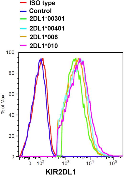
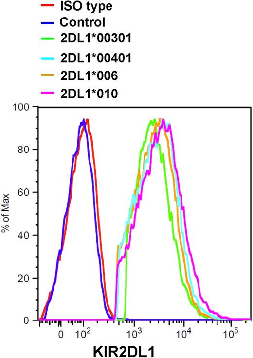
To study the differences among various KIR2DL1 alleles and the functional significance of allelic polymorphism, we aligned them using the Immuno Polymorphism Disease (IPD) KIR database (http://www.ebi.ac.uk/ipd/kir).25 In the 14 KIR2DL1 alleles, polymorphisms were found in the extracellular domain as well as in the transmembrane, stem, and cytoplasmic domains (Table 1). The specificity between KIR2DL1 and its ligand is conferred through a specificity pocket in KIR2DL1 for Lys80 of HLA-C2.21 This specificity pocket is formed by Met44, Ser184, Glu187, and several other amino acids in the extracellular domain of KIR2DL1. None of the polymorphisms in the extracellular domain of KIR2DL1 occurs in amino acids that are involved in forming the specificity pocket and are conserved among all KIR2DL1 alleles. KIR2DL1*005 and *009 alleles are polymorphic at positions 312 and 282 in their cytoplasmic domain (Table 1), but the full sequence of both alleles is unknown. We then decided to focus on the functional consequences of allelic polymorphism in the stem and transmembrane domains of KIR2DL1. Among the 14 alleles of KIR2DL1, 9 (KIR2DL1*001, *002, *00301, *00302, *00303, *005, *006 *008, and *009) have lysine (K) at position 216 in their stem domain, and 5 (KIR2DL1*0040101, *0040102, *00402, *007, and *010) contain glutamate (E) at that position (Table 1). On the other hand, 9 alleles (KIR2DL1*001, *002, *00301, *00302, *00303, *005, *008, *009, and *010) contain arginine (R) at position 245 in the transmembrane domain, whereas 5 (KIR2DL1*0040101, *0040102, *00402, *006, and *007) contain cysteine (C) at that position. We grouped KIR2DL1 alleles on the basis of differences and similarities in their stem and transmembrane domains in numeric order. Members of group 1 had lysine and arginine at position 216 and 245 in their stem and transmembrane domains, respectively (Table 1). Group 2 had glutamate and cysteine, group 3 had lysine and cysteine, and group 4 had glutamate and arginine in those respective positions. cDNA libraries were created from RNA extracted from donor blood. One member of each group of KIR2DL1 alleles was selected for further investigation (KIR2DL1*00301 from group 1, KIR2DL1*00401 from group 2, KIR2DL1*006 from group 3, and KIR2DL1*010 from group 4). The NK-like YT-Indy cell line, which lacks the expression of KIRs, was stably transfected with the 4 selected KIR2DL1 alleles, and similarly expressing cells were sorted using flow cytometric cell sorting (Figure 1). YT-Indy cells transfected with the vector were used as a control.
Generation of stable YT-Indy cell line expressing KIR2DL1 alleles. YT-Indy cells were stably transfected with pcDNA3 vector containing various alleles of KIR2DL1 (*00301, *00401, *006, and *010) or with pcDNA3 vector alone as control. Expression of alleles of KIR2DL1 in sorted and stably transfected YT-Indy cells was confirmed to be similar as assessed by flow cytometry.
Generation of stable YT-Indy cell line expressing KIR2DL1 alleles. YT-Indy cells were stably transfected with pcDNA3 vector containing various alleles of KIR2DL1 (*00301, *00401, *006, and *010) or with pcDNA3 vector alone as control. Expression of alleles of KIR2DL1 in sorted and stably transfected YT-Indy cells was confirmed to be similar as assessed by flow cytometry.
Functional diversity among KIR2DL1 alleles in inhibition of YT-Indy cells
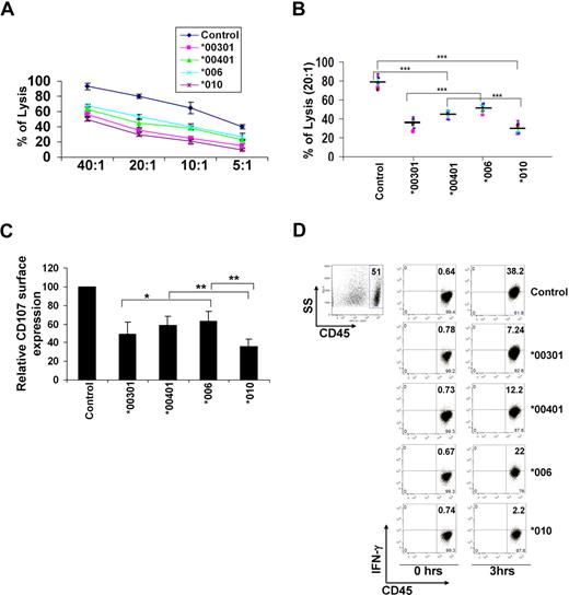
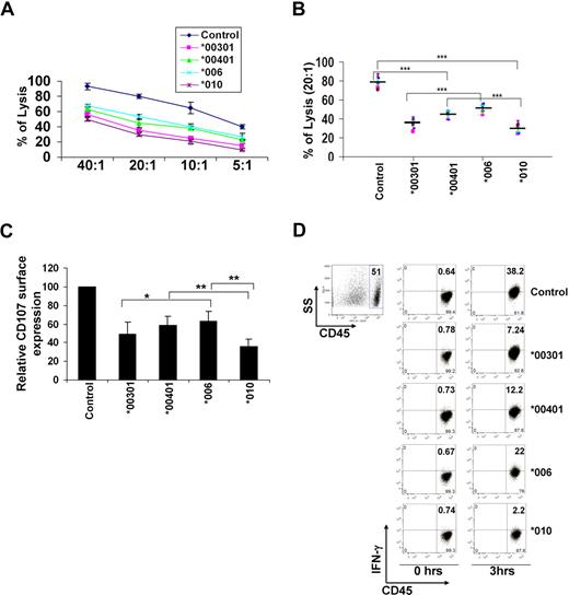
The cytotoxicity of YT-Indy cells expressing various KIR2DL1 alleles was assessed by a BATDA release assay using 721.221 (untransduced) or 721.221-Cw6 (transduced) as target cells. As expected, YT-Indy cells transfected with different KIR2DL1 alleles or with the control vector showed similar killing against 721.221 untransduced cells with no inhibitory ligand (supplemental Figure 1, available on the Blood website; see the Supplemental Materials link at the top of the online article). However, YT-Indy cells expressing various alleles of KIR2DL1 showed differential killing against 721.221-Cw6 (Figure 2A) at various effector-target (E/T) ratios. The order of inhibition of killing is KIR2DL1*010 > KIR2DL1*00301 > KIR2DL1*00401 > KIR2DL1*006 (Figure 2A). The cytotoxicity experiments were repeated 9 times at a 20:1 E/T ratio, and the results were similar (Figure 2B).
KIR2DL1 alleles differentially inhibit the cytotoxicity, degranulation, and cytokine production of YT-Indy cells. YT-Indy cells transfected with empty vector were used as control. (A) Specific lysis by YT-Indy cells expressing various alleles of KIR2DL1 (*00301, *00401, *006, and *010) were assessed against 721.221-Cw6 at various ratios of effector to target cells using a BADTA release assay. The E/T ratios were 40:1, 20:1, 10:1, and 5:1. Data shown are average of 3 independent experiments. (B) Specific lysis of target 721.221-Cw6 cells by YT-Indy transfectants was assessed at E/T = 20:1. Data shown are average of 9 independent experiments. ***P < .01. (C) Relative expression of CD107 at the surface of YT-Indy cells expressing various alleles of KIR2DL1 was detected by flow cytometry after challenge with target 721.221-Cw6 cells. The results represent the mean of 3 independent experiments; *P = .22, **P < .05. Error bars represent SD. (D) Production of IFN-γ in YT-Indy transfectants was assessed after stimulation with target 721.221-Cw6 cells. To separate effector from target cells, YT-Indy transfectants were first stained with CD45 antibody, followed by incubation with target cells. Cell mixtures were fixed, permeabilized, and stained with IFN-γ antibody. YT-Indy transfectants were gated based on CD45 (left panel), and IFN-γ production was assessed (right panels). Data shown are representative of 3 independent experiments.
KIR2DL1 alleles differentially inhibit the cytotoxicity, degranulation, and cytokine production of YT-Indy cells. YT-Indy cells transfected with empty vector were used as control. (A) Specific lysis by YT-Indy cells expressing various alleles of KIR2DL1 (*00301, *00401, *006, and *010) were assessed against 721.221-Cw6 at various ratios of effector to target cells using a BADTA release assay. The E/T ratios were 40:1, 20:1, 10:1, and 5:1. Data shown are average of 3 independent experiments. (B) Specific lysis of target 721.221-Cw6 cells by YT-Indy transfectants was assessed at E/T = 20:1. Data shown are average of 9 independent experiments. ***P < .01. (C) Relative expression of CD107 at the surface of YT-Indy cells expressing various alleles of KIR2DL1 was detected by flow cytometry after challenge with target 721.221-Cw6 cells. The results represent the mean of 3 independent experiments; *P = .22, **P < .05. Error bars represent SD. (D) Production of IFN-γ in YT-Indy transfectants was assessed after stimulation with target 721.221-Cw6 cells. To separate effector from target cells, YT-Indy transfectants were first stained with CD45 antibody, followed by incubation with target cells. Cell mixtures were fixed, permeabilized, and stained with IFN-γ antibody. YT-Indy transfectants were gated based on CD45 (left panel), and IFN-γ production was assessed (right panels). Data shown are representative of 3 independent experiments.
Another way to assess the function of NK cells is to measure the mobilization of CD107 (lysome-associated membrane protein) at the cell surface by flow cytometry. CD107 is a marker for intracytoplasmic cytolytic granule, and cell-surface expression of CD107 can be used as an indicator of effector cell degranulation.26 In the CD107 mobilization assay, YT-Indy cells with KIR2DL1*006 and KIR2DL1*00401 alleles showed higher expression of CD107, and those with KIR2DL1*010 and KIR2DL1*00301 alleles showed lower cell-surface expression (Figure 2C), indicating that members of group 1 and group 4 had stronger inhibitory effects on YT-Indy cytotoxicity than did members of group 3 and group 4. These findings corroborate our observations in the cytotoxicity assay.
In our next experiments, we measured the production of IFN-γ by YT-Indy cells expressing various alleles of KIR2DL1 stimulated with target 721.221-Cw6 cells. At 0 hours, YT-Indy transfected with vectors and the 4 KIR2DL1 alleles showed very little intracellular IFN-γ (Figure 2D left panel). After 3 hours of coculture with target 721.221-Cw6 cells, control (YT-Indy transfected with vector) cells showed the highest production of IFN-γ (Figure 2D right panel). Among the 4 KIR2DL1 alleles, IFN-γ production is KIR2DL1*006 > KIR2DL1*00401 > KIR2DL1*00301 > KIR2DL1*010 (Figure 2D).
In vivo data confirm the in vitro functional differences among KIR2DL1 alleles
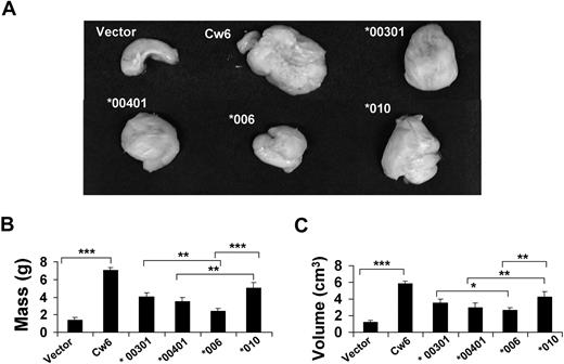
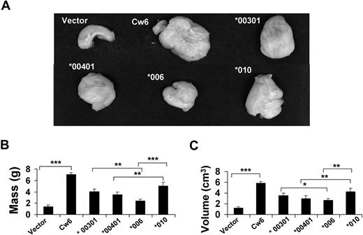
To investigate the functional differences among KIR2DL1 alleles in vivo, we subcutaneously injected the YT-Indy cells expressing either KIR2DL1 *00301, *00401, *006, or *010, together with 721.221-Cw6 cells into 6- to 8-week-old NOD-scid IL2Rγnull mice at a 10:1 E/T ratio (100 000 effectors and 10 000 target cells). The control mice were subcutaneously given injections of the same number of YT-Indy cells stably transfected with vector together with 721.221-Cw6 cells. Mice given only 721.221-Cw6 cells were used as positive controls. After the tumors reached 20% of the body weight of the positive control mice (considered terminal, according to St Jude guidelines), the mice were humanely killed, and the tumors were excised. In mice that were given YT-Indy cells transfected with vector lacking the inhibitory receptor, very small tumors were produced (Figure 3A vector). In contrast, positive control mice had the biggest tumors (Figure 3A Cw6). When we measured the mass and volume of the tumors, those in mice that were given only target cells (721.221-Cw6) were approximately 6 times bigger than those in the mice that were given YT-Indy cells containing control vector together with 721.221-Cw6 cells (Figure 3B and C, vector and Cw6, respectively). The mass and volume of the tumors in mice that were given YT-Indy cells with KIR2DL1*010 were approximately 2 times higher than in mice given those with KIR2DL1*006 (Figure 3B and C, tumor *010 and *006, respectively). YT-Indy cells expressing KIR2DL1*00401 killed more 721.221-Cw6 cells than did the YT-Indy cells expressing KIR2DL1*00301 and KIR2DL1*010 but fewer than did the YT-Indy cells expressing KIR2DL1*006 in vivo (Figure 3A, B, and C, tumor *00401, *00301, *010, and *006).
Functional differences among KIR2DL1 alleles observed in vitro are confirmed in vivo. YT-Indy cells expressing different alleles of KIR2DL1 were mixed with 721.221-Cw6 at a 10:1 (E/T) ratio and injected subcutaneously into NOD-scid IL2Rγnull mice. A total of 30 mice, 5 mice for each group, were given injections. After the tumors reached 20% of body mass in the positive control group (mice injected with only target cells), the mice were humanely killed, dissected, and photographed. The image of a representative tumor from each group is shown. (A) Vector, *00301, *00401, *006, and *010 are tumors from mice given injections of YT-Indy transfected with empty vector, KIR2DL1*00301, KIR2DL1*00401, KIR2DL1*006, or KIR2DL1*010, together with 721.221 expressing HLA-Cw6. Cw6 was a tumor from a mouse given only 721.221 expressing HLA-Cw6. Tumor mass (B) and tumor volume (C) were also measured. The experiment was repeated 3 times. *P = .08, **P < .05, ***P < .01. Error bars represent SD.
Functional differences among KIR2DL1 alleles observed in vitro are confirmed in vivo. YT-Indy cells expressing different alleles of KIR2DL1 were mixed with 721.221-Cw6 at a 10:1 (E/T) ratio and injected subcutaneously into NOD-scid IL2Rγnull mice. A total of 30 mice, 5 mice for each group, were given injections. After the tumors reached 20% of body mass in the positive control group (mice injected with only target cells), the mice were humanely killed, dissected, and photographed. The image of a representative tumor from each group is shown. (A) Vector, *00301, *00401, *006, and *010 are tumors from mice given injections of YT-Indy transfected with empty vector, KIR2DL1*00301, KIR2DL1*00401, KIR2DL1*006, or KIR2DL1*010, together with 721.221 expressing HLA-Cw6. Cw6 was a tumor from a mouse given only 721.221 expressing HLA-Cw6. Tumor mass (B) and tumor volume (C) were also measured. The experiment was repeated 3 times. *P = .08, **P < .05, ***P < .01. Error bars represent SD.
Arginine, not cysteine, in the transmembrane domain of KIR2DL1 plays an important role in its inhibitory function
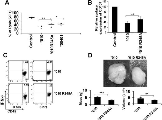
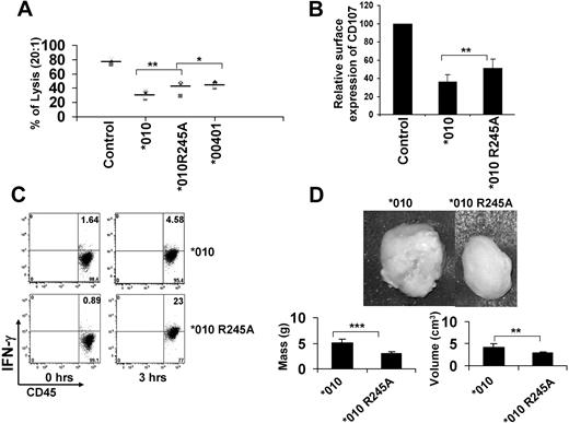
In our next experiment, we investigated whether the stem domain, the transmembrane domain, or both are important for the functional differences observed among KIR2DL1 alleles. First, we checked the role of the stem domain by replacing glutamate in the stem domain of KIR2DL1*010 alleles with alanine. We chose the KIR2DL1*010 allele because it was the strongest inhibitor among the KIR2DL1 alleles investigated. We found no significant functional differences between wild-type KIR2DL1*010 and the mutant (KIR2DL1*010 E216A; supplemental Figure 2A-B). This suggests that polymorphism in the stem domain does not play an important role in observed functional discrepancies among KIR2DL1 alleles. We then investigated whether the transmembrane domain of KIR2DL1 played any role in the functional discrepancies among its alleles. An arginine residue (R245) is present in the transmembrane domain of KIR2DL1*00301 and KIR2DL1*010, and a cysteine residue is present at those respective positions on KIR2DL1*00401 and KIR2DL1*006 (Table 1). Moreover, the only difference among the 5 domains of KIR2DL1*00401 and KIR2DL1*010 is the cysteine instead of arginine in the transmembrane domain (Table 1). If the transmembrane domain is important for the observed functional differences, then we speculate either arginine or cysteine should be responsible for it, because that is the only difference between the 2 alleles. If our speculation is true, then there are 2 possibilities for this functional discrepancy. In one scenario, the presence of a basic charged amino acid, arginine, makes KIR2DL1*010 a stronger inhibitor than KIR2DL1*00401, which does not have arginine at the same position. On the other hand, the presence of cysteine may make KIR2DL1*00401 a less inhibitory allele. To address this question, we replaced the arginine residue with the small and neutral amino acid alanine. Cytotoxic function assessed by a BADTA release assay showed that the YT-Indy cell expressing mutated KIR2DL1*010 reversed the inhibition of target 721.221-Cw6 cell killing to a level comparable with that of KIR2DL1*00401-transfected YT-Indy cells (Figure 4A). Because the mutation of arginine in *010 brought its inhibitory ability to a level comparable with that of *00401, which has cysteine at the same position, it indicates that the presence of arginine, not the absence of cysteine, made *010 more inhibitory. Both the surface expression of CD107 (Figure 4B) and the production of IFN-γ (Figure 4C) were higher in YT-Indy expressing mutated KIR2DL1*010 than in wild-type KIR2DL1*010. These results were verified in vivo (Figure 4D).
Arginine residue in the transmembrane domain of KIR2DL1 plays an important role in its inhibitory function. Arginine in the transmembrane domain of KIR2DL1*010 was replaced with alanine. A stable YT-Indy cell line expressing the KIR2DL1*010 mutant was generated. YT-Indy transfected with vector was used as control. (A) Specific killing of YT-Indy expressing wild-type KIR2DL1*010 (*010), KIR2DL1*010 mutants (*010 R245A), and KIR2DL1*00401 (*00401) was assessed against 721.221-Cw6 by BADTA release assay. *P = .62, **P < .05. (B) Relative expression of CD107 at the surface of YT-Indy cells expressing wild-type (*010) and mutated (*010 R245A) KIR2DL1*010 was assessed by CD107 mobilization assay. **P < .05. (C) Production of IFN-γ was assessed after YT-Indy cells expressing wild-type (top) and mutated (bottom) KIR2DL1*010 were stimulated with target 721.221-Cw6 cells. Results are representative of 3 independent experiments. (D) YT-Indy cells expressing wild-type and mutated KIR2DL1*010 alleles were mixed with target 721.221-Cw6 cells at a 10:1 (E/T) ratio and injected into mice subcutaneously. A total of 10 mice, 5 for each group, were given injections. After tumors reached 20% of the body mass in some mice, the mice were humanely killed, dissected, and photographed. The image of a representative tumor from each group is shown. The tumor shown at left is from a mouse that was given YT-Indy expressing wild-type KIR2DL1*010 (*010), and the tumor shown at right is from a mouse that was given YT-Indy cells expressing the mutated KIR2DL1*010 (*010 R245A) allele. Mass and volume of these tumors are shown at bottom. **P < .05, ***P < .01. The experiments were repeated 3 times. Error bars represent SD.
Arginine residue in the transmembrane domain of KIR2DL1 plays an important role in its inhibitory function. Arginine in the transmembrane domain of KIR2DL1*010 was replaced with alanine. A stable YT-Indy cell line expressing the KIR2DL1*010 mutant was generated. YT-Indy transfected with vector was used as control. (A) Specific killing of YT-Indy expressing wild-type KIR2DL1*010 (*010), KIR2DL1*010 mutants (*010 R245A), and KIR2DL1*00401 (*00401) was assessed against 721.221-Cw6 by BADTA release assay. *P = .62, **P < .05. (B) Relative expression of CD107 at the surface of YT-Indy cells expressing wild-type (*010) and mutated (*010 R245A) KIR2DL1*010 was assessed by CD107 mobilization assay. **P < .05. (C) Production of IFN-γ was assessed after YT-Indy cells expressing wild-type (top) and mutated (bottom) KIR2DL1*010 were stimulated with target 721.221-Cw6 cells. Results are representative of 3 independent experiments. (D) YT-Indy cells expressing wild-type and mutated KIR2DL1*010 alleles were mixed with target 721.221-Cw6 cells at a 10:1 (E/T) ratio and injected into mice subcutaneously. A total of 10 mice, 5 for each group, were given injections. After tumors reached 20% of the body mass in some mice, the mice were humanely killed, dissected, and photographed. The image of a representative tumor from each group is shown. The tumor shown at left is from a mouse that was given YT-Indy expressing wild-type KIR2DL1*010 (*010), and the tumor shown at right is from a mouse that was given YT-Indy cells expressing the mutated KIR2DL1*010 (*010 R245A) allele. Mass and volume of these tumors are shown at bottom. **P < .05, ***P < .01. The experiments were repeated 3 times. Error bars represent SD.
Transmembrane domain arginine-positive KIR2DL1*010 recruits more SHP-2 and β-arrestin 2
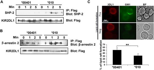
Once we identified the molecular determinant for functional heterogeneity was R245, we sought to determine the mechanisms by examining its effects on inhibitory signaling and kinetics of surface expression. β-Arrestin 2 is known to associate with KIR2DL1 and mediate recruitment of tyrosine phosphatase SHP-2, which leads to downstream signaling.27 In our next experiments, we investigated the association of KIR2DL1*00401 and KIR2DL1*010 alleles with SHP-2 and β-arrestin 2. If the R245 is important for KIR2DL1 inhibitory signaling, we would expect to see less recruitment of β-arrestin 2 and SHP-2 by the KIR2DL1*00401 allele. YT-Indy cells were transduced with FLAG-tagged KIR2DL1*00401 and KIR2DL1*010 alleles. Transduced YT-Indy cells expressing similar levels of KIR2DL1*00401 and KIR2DL1*010 were collected by flow cytometric cell sorting. YT-Indy cells expressing both KIR2DL1 alleles were stimulated with target 721.221-Cw6 cells for 0, 1, 2, and 5 minutes. The greatest amount of SHP-2 immunoprecipitated with KIR2DL1*010 allele was detected after 2 minutes of stimulation (Figure 5A lane 7). The association lessened after 5 minutes of stimulation (Figure 5A lane 8). In contrast, we found barely any association between KIR2DL1*00401 and SHP-2 (Figure 5A left 4 lanes). We observed similar results with β-arrestin 2 and alleles KIR2DL1*00401 and KIR2DL1*010 (Figure 5B). These findings clearly suggest that the arginine residue in the transmembrane domain of KIR2DL1 significantly affects the downstream inhibitory signaling pathway.
KIR2DL1 alleles *00401 and *010 showed different signaling intensity and inhibition of lipid raft polarization at immune synapse upon stimulation with 721.221-Cw6 cells. YT-Indy cells were retrovirally transduced with KIR2DL1 alleles *00401 and *010 fused with FLAG tag. Similarly expressing cells were collected by flow cytometry cell sorting. YT-Indy cells expressing FLAG-tagged KIR2DL1 alleles *00401 and *010 were stimulated with target 721.221-Cw6 cells at a 1:2 (E/T) ratio for the indicated times. The stimulated cells were lysed and immunoprecipitated (IP) with anti-FLAG antibody, followed by immunoblotting (IB) with antibodies to (A) SHP-2 and (B) β-arrestin 2. Membranes were stripped and reprobed with FLAG antibody for loading control. (C) Positive and negative are representatives of lipid raft polarization and inhibition of lipid raft polarization, respectively. 2DL1 indicates cells stained with KIR2DL1 antibody, GM1 for lipid raft, and BF for conjugate formation. Calculated percentages of lipid raft polarization in YT-Indy/KIR2DL1*00401401 and YT-Indy/KIR2DL1*010010 in the presence of 721.221-Cw6 are shown in the lower panel. Images were obtained by a Nikon Eclipse E800 fluorescence microscope. The experiment was repeated 3 times. **P < .05. Error bars represent SD.
KIR2DL1 alleles *00401 and *010 showed different signaling intensity and inhibition of lipid raft polarization at immune synapse upon stimulation with 721.221-Cw6 cells. YT-Indy cells were retrovirally transduced with KIR2DL1 alleles *00401 and *010 fused with FLAG tag. Similarly expressing cells were collected by flow cytometry cell sorting. YT-Indy cells expressing FLAG-tagged KIR2DL1 alleles *00401 and *010 were stimulated with target 721.221-Cw6 cells at a 1:2 (E/T) ratio for the indicated times. The stimulated cells were lysed and immunoprecipitated (IP) with anti-FLAG antibody, followed by immunoblotting (IB) with antibodies to (A) SHP-2 and (B) β-arrestin 2. Membranes were stripped and reprobed with FLAG antibody for loading control. (C) Positive and negative are representatives of lipid raft polarization and inhibition of lipid raft polarization, respectively. 2DL1 indicates cells stained with KIR2DL1 antibody, GM1 for lipid raft, and BF for conjugate formation. Calculated percentages of lipid raft polarization in YT-Indy/KIR2DL1*00401401 and YT-Indy/KIR2DL1*010010 in the presence of 721.221-Cw6 are shown in the lower panel. Images were obtained by a Nikon Eclipse E800 fluorescence microscope. The experiment was repeated 3 times. **P < .05. Error bars represent SD.
KIR2DL1*010 allele shows higher inhibition of lipid raft formation at the interface of YT-Indy cells and target cells expressing its ligand compared with KIR2DL1*00401 allele
Lipid raft polarized at the immune synapse region of NK cells with sensitive target cells but not with resistant target cells.28 KIR2DL1 signaling at the inhibitory NK immune synapse inhibits the migration of lipid raft to the interface of NK cells and target cells.24,28 Moreover, overexpression of dominant-negative SHP-1 reverses KIR-mediated inhibition of raft polarization.28 Because the KIR2DL1*00401 allele recruited less SHP-2 and β-arrestin 2, we hypothesized that it also had less inhibition of lipid raft polarization. In our next experiments, we investigated the inhibition of lipid raft polarization in YT-Indy cells expressing KIR2DL1 alleles *00401 and *010 in the presence of 721.221 and 721.221-Cw6. Representative pictures of lipid raft polarization and nonpolarization are shown (Figure 5C). Approximately 65% of YT-Indy/KIR2DL1*00401 and YT-Indy/KIR2DL1*010 showed lipid raft polarization at their immune synapse with untransduced 721.221. However, similar raft polarization percentages at the contact of 721.221-Cw6 and YT-Indy/KIR2DL1*00401 were 31%, and of 721.221-Cw6 and YT-Indy/KIR2DL1*010 were 24%. We normalized the percentage of lipid raft polarization at the immune synapse of YT-Indy transfectants and 721.221-Cw6 by considering raft polarization at the contact site of untransduced 721.221 and YT-Indy transfectants to be 100%. After normalization, raft polarization percentages at the contact sites of YT-Indy/KIR2DL1*00401 and 721.221-Cw6 were approximately 50% and YT-Indy/KIR2DL1*010 and 721.221-Cw6 were 34% (Figure 5C). This result further proved that the arginine in the transmembrane domain of KIR2DL1 is important for its inhibitory signal.
Arginine-positive transmembrane domain prevents down-regulation of KIR expression upon interaction with its ligand
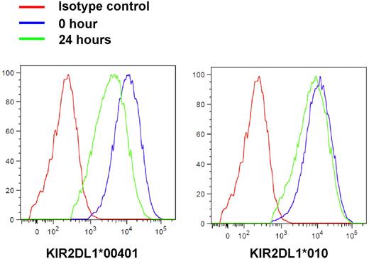
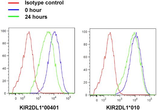
In addition to its effect in short-term signaling, we investigated whether R245 may have another mechanism that affects KIR2DL1 function by altering its durability of cell-surface expression. In CD8+ T cells, surface expression of KIRs is regulated by the engagement of T-cell receptor (TCR) in the presence of its ligand. In the absence of TCR engagement, KIRs expressed on CD8+ T cells are slowly internalized after interaction with its ligands on antigen-presenting cells.29 In our next experiments, we investigated whether this also happened to YT-Indy cells expressing KIR2DL1 ligands and the effect of transmembrane domain arginine. YT-Indy cells expressing KIR2DL1 allele *00401 and *010 were cocultured with 721.221-Cw6 cells for various durations. The expression of KIR2DL1*00401 was down-regulated much more than the expression of KIR2DL1*010 when YT-Indy cells expressing them were cocultured with 721.221-Cw6 for 24 hours (Figure 6), indicating that the presence of R245 helps KIR2DL1 to sustain its cell-surface expression, which may lead to its durable inhibitory effect. The down-regulation of KIR2DL1 was ligand-specific, as KIR2DL1*00401 and *010 did not significantly down-regulate after 24 hours of coculturing with 721.221 or 721.221 carrying the unrecognized ligand HLA-Cw7 (721.221-Cw7; supplemental Figure 3). To understand the functional significance of down-regulation of KIR2DL1 surface expression, we analyzed the ability to degranulate by high versus low KIR2DL1*00401-expressing YT-Indy cells. The low KIR2DL1*00401-expressing cells showed greater CD107 mobilization compared with high expressing cells when challenged with 721.221-Cw6, suggesting that KIR surface density modulation may fine-tune its inhibitory effect (supplemental Figure 4).
Transmembrane arginine prevents down-regulation of cell-surface expression of KIR2DL1 after interaction with its ligand. YT-Indy cells expressing KIR2DL1 alleles *00401 and *010 were coincubated with target 721.221-Cw6 cells for the indicated times. Cell-surface expression of KIR2DL1 was detected using KIR2DL1 mAb. Left panel shows cell-surface expression of KIR2DL1*00401, and right panel shows KIR2DL1*010. Data shown are representative of 3 independent experiments.
Transmembrane arginine prevents down-regulation of cell-surface expression of KIR2DL1 after interaction with its ligand. YT-Indy cells expressing KIR2DL1 alleles *00401 and *010 were coincubated with target 721.221-Cw6 cells for the indicated times. Cell-surface expression of KIR2DL1 was detected using KIR2DL1 mAb. Left panel shows cell-surface expression of KIR2DL1*00401, and right panel shows KIR2DL1*010. Data shown are representative of 3 independent experiments.
Discussion
Our functional studies of the 4 groups of KIR2DL1 alleles characterized by polymorphism at positions 216 and 245 in the stem and transmembrane domains clearly showed that various KIR2DL1 alleles inhibit YT-Indy cells differently. In position 216 of the stem domain, some alleles have lysine where others have glutamate. The mutation that replaces the glutamate of KIR2DL1*010 has shown no significant functional effect, suggesting that lysine or glutamate in the stem domain of KIR2DL1 may not be important for its function. However, we cannot rule out the possibility of a small synergistic effect of the glutamate together with the arginine in the transmembrane domain, because both *010 and *00301 have arginine in the same position and *010 shows a small but significantly higher inhibition than *00301, indicating that the combination of glutamate and arginine is more inhibitory than the combination of lysine and arginine at positions 216 and 245, respectively. The synergistic effect of 2 amino acids also has precedence in KIR. The presence of an arginine and a cysteine at positions 16 and 148 of KIR2DL2*001, respectively, makes it a stronger receptor for HLA-C than KIR2DL3*001, which has proline and arginine at those positions.30
In position 245 of the transmembrane domain of KIR2DL1, some alleles contain arginine, whereas others have cysteine. The presence of polar residues (ie, arginine) at the transmembrane domain is interesting because previous studies showed that transmembrane polar residues mediate peptide-peptide interactions within the lipid bilayer.31 Moreover, polar residues in the transmembrane domain of the TCR play an important role in its assembly, cell-surface expression, and signaling,32,33 underscoring the biologic significance of these residues. Alignment of KIR2DL1 alleles *00401 and *010 showed only a single mismatch at position 245 in the transmembrane domain (cysteine and arginine, respectively). However, the *00401 allele was much less inhibitory than *010, strongly suggesting either arginine or cysteine plays a critical role in inhibition by KIR2DL1. A similar difference has been observed between KIR2DL1 alleles *00301 and *006. The presence of basic amino acid lysine at the transmembrane domain of noninhibitory KIR is critical for interaction with DNAX-activating protein of 12 kDa.34,35 In addition, the idea of polar residues in the transmembrane domain forming a multisubunit complex and interacting with oppositely charged amino acids has precedence in other biologic systems.23,36 Therefore, it is reasonable to predict that arginine at position 245 in the transmembrane domain of KIR2DL1 may form multisubunit complexes by interacting with other proteins that are important for its function. On the other hand, cysteine has also been shown to play an important role in the function of many other proteins by forming a disulfide bond. Nevertheless, replacing the transmembrane arginine residue of the *010 allele with alanine reverses its inhibitory function to a level similar to that of the *00401 allele, indicating that in KIR2DL1 arginine but not cysteine plays a critical role in inhibition.
Once we established R245 as the molecular determinant, we sought to identify its mechanisms by examining the effects on inhibitory signaling and surface expression. SHP-2 and β-arrestin 2 are 2 important signaling molecules in inhibition by KIR2DL1, and we found that KIR2DL1 allele *010 with R245 recruits more SHP-2 and β-arrestin 2 than allele *00401, which lacks R245. Moreover, the *010 allele is a stronger inhibitor of lipid raft polarization. Because inhibitory signaling regulates the lipid raft polarization at the immune synapse of NK cells,24,28 these results indicate the importance of transmembrane arginine in the KIR2DL1 signaling pathway. This also explains why KIR2DL1 alleles with transmembrane arginine residues are stronger inhibitors. Another possible mechanism for the greater inhibition by KIR2DL1*010 is that it sustains longer at the surface of YT-Indy than the *00401 allele upon interacting with its ligand. It is possible that the presence of a charged amino acid at the transmembrane domain of some KIR2DL1 alleles leads to interaction with other proteins that help it to stay at the cell surface, which in turn makes the inhibitory effect more durable than that of those without it.
In addition to biologic significance, our findings may also have clinical implications. Previous studies have shown that the risk of many human diseases was associated with differences in KIR gene content, including autoimmune diseases, inflammatory disorders, infectious diseases, immunodeficiency, cancer, and reproductive disorders.17 KIR2DL1, for instance, was specifically associated with tuberculoid leprosy,37 microscopic polyangiitis,38 endometriosis,39 immunodeficiency,40 birdshot chorioretinopathy,41 solid tumors,42,43 leukemia,44 and graft-versus-host disease.45 Mechanistically, the risk may be related to the strength of interaction between the KIR gene and its HLA ligand.17 However, no studies have been performed to examine disease risk and transplant outcome in relation to functional allelic polymorphism. Both C245 and R245 KIR2DL1 allelic groups as well as their ligand (HLA-C group 2) are well represented in the general population with variable frequency among different ethnic groups (supplemental Table 1).46,47 Overall, the gene frequency of C245 and R245 KIR2DL1 is approximately 0.15 and 0.75, respectively, and the HLA-Cw*0602 ligand used in our study is the fourth most frequent HLA-C allele.47 HLA-C group 2 (Lys80) allotypes are found in approximately 50% of the people. Thus, our finding of a significant difference in inhibition by alleles with polymorphisms in the transmembrane and stem domains should encourage future research to test the hypothesis that KIR2DL1 allelic polymorphism is important in disease susceptibility, transplant outcome, and donor selection.
The online version of this article contains a data supplement.
The publication costs of this article were defrayed in part by page charge payment. Therefore, and solely to indicate this fact, this article is hereby marked “advertisement” in accordance with 18 USC section 1734.
Acknowledgments
We thank David Galloway for scientific editing.
This work was supported in part by research grants from the National Institutes of Health (P30 CA-21765-24, CA-21765), the Assisi Foundation of Memphis, and the American Lebanese Syrian Associated Charities (ALSAC).
National Institutes of Health
Authorship
Contribution: R.B. designed and performed research, analyzed data, and wrote the paper; W.-H.L., Q.P.V., and W.K.C. designed and performed research; T.B., N.D.G., M.H., and B.R. performed research; and W.L. designed research, analyzed data, and wrote the paper.
Conflict-of-interest disclosure: The authors declare no competing financial interests.
Correspondence: Wing Leung, 262 Danny Thomas Pl, Mail Stop 260, Memphis, TN 38105; e-mail: wing.leung@stjude.org.

