Abstract
Previous studies have shown that during the lifespan of red blood cells (RBCs) 20% of hemoglobin is lost by shedding of hemoglobin-containing vesicles. However, the fate of these vesicles is unknown. To study this fate we used a rat model, after having established that rat RBCs lose hemoglobin in the same way as human RBCs, and that RBC-derived vesicles are preferentially labeled by
Introduction
In previous studies, we have shown that a fractionation method combining counterflow centrifugation and subsequent Percoll gradient centrifugation is superior to any of these methods alone with respect to analyzing differences in cell age-related parameters of the red blood cell (RBC) fractions.1 Using this fractionation method, we obtained data that strongly suggest that during the RBC lifespan the mean corpuscular volume (MCV), mean corpuscular hemoglobin (MCH) content, and mean surface area decrease by 30%, 20%, and 20% respectively, whereas the mean corpuscular hemoglobin concentration (MCHC) increases by 14%.1-3 This means that loss of hemoglobin and of membrane parts occurs from intact RBCs in vivo. In a later study it could be shown that these losses are caused by shedding of hemoglobin-containing vesicles.4 Moreover, the results of these studies indicated that vesicles are produced by RBCs of all ages with vesicles derived from older RBCs containing more hemoglobin. However, the fate of these vesicles is unknown. Due to practical reasons this issue is virtually impossible to study in humans. We, therefore, studied the kinetics of vesicle elimination in a rat model that is analogous to the murine model used by one of us (T.J.C.v.B.) in a study, in which the fate of damaged RBCs was investigated.5 In addition this rat model enabled us to study the putative involvement of the mononuclear phagocyte system and its receptors. First, however, we had to establish that circulating rat RBCs lose hemoglobin by vesiculation in a similar manner to human RBCs.
Materials and methods
Materials
Sodium 51Cr-chromate was obtained from Amersham (Buckinghamshire, United Kingdom). Collagenase (type IV), bovine serum albumin (BSA, fraction V), 3,3′-diaminobenzidine, and polyinosinic acid (poly-I) were obtained from Sigma (St Louis, MO). Bovine brain phosphatidylserine (PS) and egg yolk phosphatidylcholine (PC) were purchased from Fluka (Buchs, Switzerland).
Rats
Ten- to 12-week-old male Wistar rats weighing between 250 and 300 g were used. Rats were kept in our local facility under low pathogen conditions with free access to food and water.
Preparation of vesicles
Approximately 9 mL blood was drawn from a male Wistar rat in a tube containing 18 mg K-EDTA (potassium-ethylenediaminetetraacetic acid). Plasma was obtained by centrifugation for 10 minutes at 1900g. Blood cell vesicles were obtained from plasma by 30 minutes of centrifugation at 16 000g and vesicles were washed 3 times with phosphate-buffered saline (PBS) containing 10% sodium citrate (10 minutes at 11 200g). Labeling of the isolated vesicles was carried out with 50 μCi (1.85 MBq) sodium 51Cr-chromate in 500 μL vesicle suspension for 60 minutes at 37°C. Vesicles were washed 3 times for 10 minutes at 11 200g to remove free chromate.
Flow cytometry
The presence of vesicles was determined by side-scatter–forward-scatter (SSC/FSC) dot plots. The expression of HIS49 and exposure of PS on, and the presence of hemoglobin in vesicles, were examined by flow cytometry (Becton Dickinson, FACScan Research Software version 2.1, San José, CA and Beckman Coulter, Fullerton, CA, respectively). The following reagents were used: anti-HIS496 mouse monoclonal antibodies specific for rat RBCs and their vesicles (IQProducts, Groningen, The Netherlands) in combination with goat polyclonal F(ab′)2 anti–murine immunoglobins labeled with fluorescein isothiocyanate (FITC; Dako Cytomation, Glostrup, Denmark), annexin V labeled with FITC or R-phycoerythrin (PE; Southern Biotechnology Associates, Birmingham, AL), and anti-Hb antibody labeled with FITC (clone 4B8, IgG2b; Wallac, Akron, OH). For each analysis 1 × 10⁴ events were collected using Beckman Coulter software EXPO32 on the flow cytometer. In the SSC/FSC dot plot a gate was set around the area with small particles. To identify the presence of hemoglobin, vesicles were blocked, fixed, permeabilized, and washed as described before.4 The sorting of human vesicles was performed in the presence of antibodies specific for RBC-derived and platelet-derived vesicles by a Coulter Epics Elite (Coulter Beckman, Hialeah, FL). The following reagents were used: anti–glycophorin A (GPA) labeled with R-PE (clone JC159, IgG1; Dako Cytomation, Glostrup, Denmark) and an antibody against platelet protein IIIa (CD-61) labeled with FITC (clone Y2/561, IgG1, Dako). After sorting, the isolated RBC-derived vesicles and platelet-derived vesicles were separately labeled with sodium 51Cr-chromate. After washing the pellet per 105 sorted events the label was preferentially bound to the RBC-derived vesicles in comparison to the platelet-derived vesicles (ratio 16.7: 1).
Hemoglobin fractionation
Hemoglobin fractionation was performed by means of ion exchange chromatography (Protein-Pak, SP8HR, Millipore, Billerica, MA). The elution buffers used were based on the buffer system described by Jeppson et al.7 The composition of buffer A was 20 mM malonic acid and 1 mM NaCN, pH 5.9, in high-performance liquid chromatography (HPLC)–grade water. Buffer B had the same composition as buffer A with the addition of 380 mM NaNO3. Sodium cyanide was used as a stabilizer of hemoglobin. Before use, the buffers were passed through a 0.22-μm filter and degassed. The column was kept at 30°C. The hemoglobin components were separated with the use of a buffer gradient and monitored at 417 nm with a photodiode array detector. The relative amount of each component was calculated from the peak area of that component at 417 nm divided by the total area of all components. With the use of this technique, 11 fractions were separated. After incubation of rat RBCs in a glucose medium (overnight incubation of 1 mL packed cells in 3 mL 5% glucose at 37°C) peaks 5 and 7 proved to be the most pronounced glycated hemoglobin components (mean increase 6.2% and 2.1% of total hemoglobin, respectively). Therefore, these peaks were used as markers of RBC age.
RBC fractionation according to cell volume
RBC fractionation according to cell volume was performed using counterflow centrifugation in a Dijkstra Curame 3000 elutriation system (Dijkstra Vereenigde, Lelystad, The Netherlands) as described before for human RBCs.4
The separation was performed at a flow rate of 10 mL/min at 20°C, using an isotonic buffer containing albumin and glucose (GASP): 9 mM Na2HPO4, 1.3 mM NaH2PO4, 140 mM NaCl, 5.5 mM glucose, and 0.8 g/L BSA (fraction V, pH 7.4; Sigma). Blood was diluted 1: 3 with GASP buffer, and 2 mL of this suspension was introduced into the buffer flow to the elutriation chamber. RBCs with increasing volume were eluted from the elutriation chamber at 8 different speeds of rotation from 2200 to 1500 rpm, yielding 8 fractions.
RBC parameters
MCH content, MCV, and RBC count were determined on an ADVIA (Bayer-Technicon, Tarrytown, NY).
Liver uptake, blood decay, and tissue distribution of 51Cr-labeled vesicles in rats
Rats were anesthetized with a subcutaneous injection of a mixture of Hypnorm (6.75 mg fluanisone/kg and 0.21 mg fentanyl citrate/kg) and midazolam (3.38 mg/kg). The abdomens were opened, and 480 μL 51Cr-labeled vesicles were injected via the inferior vena cava with or without preinjection of competitors. At the indicated times, liver lobules were tied off, excised, and weighed. The total amount of liver tissue tied off during the experiment did not exceed 10% of the total liver weight. Blood samples (300 μL) were taken, and radioactivity was measured. At 30 minutes, the rats were killed and tissues were excised, weighed, and the radioactivity was determined. The radioactivity in liver and other tissue samples was corrected for blood present at the time of sampling, according to earlier determinations with 125I-BSA.
Isolation of liver cells and light microscopy
At 30 minutes after injection of 51Cr-labeled vesicles, the liver was preperfused at 8°C for 10 minutes with Hanks buffer, pH 7.4, with HEPES (N-2-hydroxyethylpiperazine-N′-2-ethanesulfonic acid; 6.7 mM), at a flow rate of 14 mL/min and subsequently with 50 mL Hanks buffer with HEPES (6.7 mM) containing 20 mg collagenase type VI. Parenchymal, endothelial, and Kupffer cells were isolated by differential centrifugation and counterflow elutriation, as described earlier.5,8 The endothelial fraction contained more than 95% of endothelial cells, the percentage of Kupffer cells in the Kupffer cell fraction being 85% ± 5% (n = 3). The cell fractions were stained with 3,3′-diaminobenzidine to determine the peroxidase activity as an indication for the presence of endogenous peroxidase and hemoglobin by light microscopy. Microscope information is as follows: make, Leica; model, DMRE; lenses, Leica PL Fluotar 100 × 1.30; camera, Nikon FX 90; image processing was not performed.
Liposomes
Unilamellar liposomes were prepared with egg yolk PC, bovine brain PS, and cholesterol in a phospholipid-to-cholesterol molar ratio of 2: 1.5 Lipids in chloroform were mixed and evaporated under nitrogen. The lipid film was resuspended in sterile PBS at a concentration of 3 mM total lipid and sonicated for 30 minutes with an MSE soniprep 150 (amplitude 16 μm) at 52°C under a constant stream of argon.
Statistical analysis
Data are shown as mean ± SEM. Statistical significance was calculated using the student t test (2-tailed).
Results
Vesicles in rats and humans
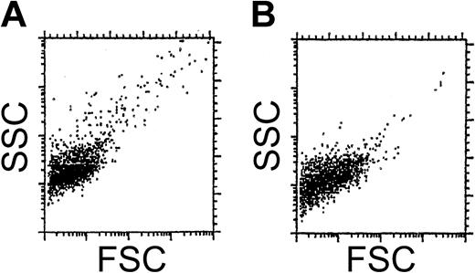
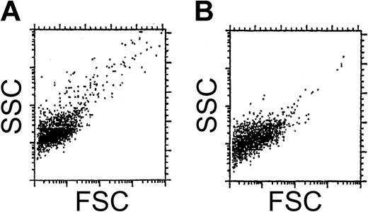
Visual inspection of the pellets obtained after centrifugation indicated that vesicle-like material can be obtained from rat plasma. Flow cytometry studies confirmed this (Figure 1).
Flow cytometry of rat vesicles. SSC/FSC dot plots of vesicles of rats (A) and humans (B). Both determinations were performed in the presence of annexin V-PE.
Flow cytometry of rat vesicles. SSC/FSC dot plots of vesicles of rats (A) and humans (B). Both determinations were performed in the presence of annexin V-PE.
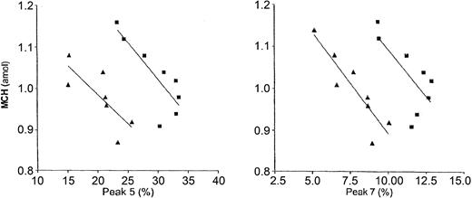
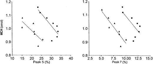
Studies with antibodies against RBC-specific antigen HIS49,6 antihemoglobin antibodies, and annexin V demonstrated that at least 21% of all vesicles were derived from RBCs, of which 66% contained hemoglobin and 67% exposed PS. In RBC fractions obtained by counterflow centrifugation, there was an inverse relationship between MCH content and the glycated hemoglobins peak 5 and peak 7 that were used as cell age markers (Figure 2). This means that the hemoglobin content decreases as the RBCs become older. The decrease in the percentage of peak 5 and peak 7 in the fractions with the lowest MCH content, that is, in the fractions containing the oldest cells, is explained by presuming that the decrease of these glycated hemoglobin components from these cells cannot be compensated for by their generation within these cells. This decrease of glycated hemoglobins in RBC fractions with the lowest MCH content has been described by us in humans.4 Likewise, it has been described that in the first 80 days after infusion of 59Fe-labeled transferrin a linear rise of the hemoglobin A1c percentage and thereafter leveling off and even a decrease occur.9
The relationship between MCH content and the glycated hemoglobins 5 and 7. Determinations in rat 1 (▴) and rat 2 (▪) show an inverse relationship between MCH and peak 5 (left) and peak 7 (right). The lines are the regression lines.
The relationship between MCH content and the glycated hemoglobins 5 and 7. Determinations in rat 1 (▴) and rat 2 (▪) show an inverse relationship between MCH and peak 5 (left) and peak 7 (right). The lines are the regression lines.
When the relative decrease of the MCH content and the relative increase of the most pronounced glycated hemoglobin components were compared between rats and humans, a striking similarity was found despite the obvious differences between rat and human RBC parameters (Table 1). These differences are remarkable because rat MCH content and MCV are approximately half of those in humans, and the RBC count in rats is twice as high.
Vesicle clearance and liver uptake
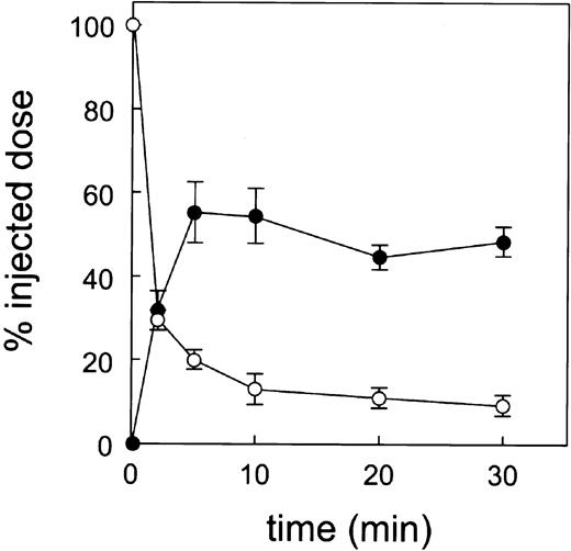
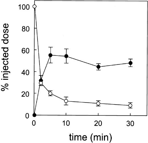
Vesicles were isolated, labeled with sodium 51Cr-chromate, and injected into recipient rats. Within 5 minutes, 80% of the labeled vesicles were removed from the circulation with a concomitant uptake by the liver of 55% of the injected radioactive dose. At 30 minutes, 91% of the vesicles were cleared, the liver uptake being unchanged (Figure 3).
Blood decay and liver uptake of 51Cr vesicles (6 experiments). At time 0 a single bolus of donor blood cell-derived vesicles was injected into recipient rats and at various times after injection a liver lobule and a blood sample were taken. Blood decay is indicated by ○ and liver uptake by ⬡.
Blood decay and liver uptake of 51Cr vesicles (6 experiments). At time 0 a single bolus of donor blood cell-derived vesicles was injected into recipient rats and at various times after injection a liver lobule and a blood sample were taken. Blood decay is indicated by ○ and liver uptake by ⬡.
To analyze which liver cell type is responsible for the avid liver uptake, we isolated parenchymal, endothelial, and Kupffer cells at 30 minutes after injection of the sodium 51Cr-chromate–labeled vesicles. Nearly all liver activity was found in the Kupffer cells (Table 2).
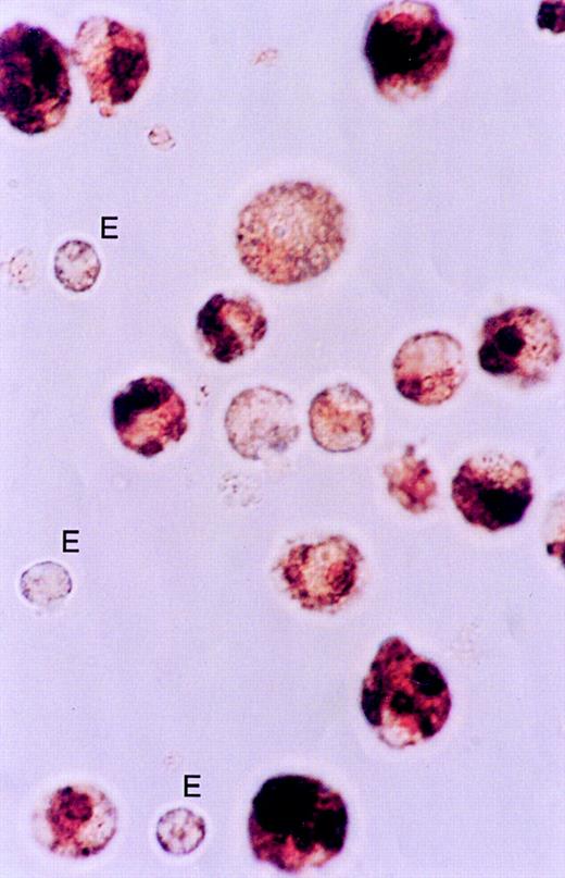
Light microscopy showed that most of the Kupffer cells contained a considerable amount of hemoglobin (dark brown staining in Figure 4). Kupffer cells of uninjected animals only contained very slight staining, as compared to the vesicle-injected animals. Liposome uptake by Kupffer cells did not influence the slight staining of the cells,10 indicating that the dark brown color within the Kupffer cells is indeed the consequence of hemoglobin uptake.
Histochemistry of Kupffer cells for the presence of peroxidase activity at 30 minutes after injection of vesicles. Cells were isolated as described in “Materials and methods.” Cytospins were fixed in acetone and stained with 3,3′-diaminobenzidine. The large quantities of stainable material in the Kupffer cells indicate the presence of hemoglobin. The endothelial cells (E) are slightly stained indicating the presence of endogenous peroxidase. Original objective magnification × 100. The procedure was repeated 2 times with very similar results.
Histochemistry of Kupffer cells for the presence of peroxidase activity at 30 minutes after injection of vesicles. Cells were isolated as described in “Materials and methods.” Cytospins were fixed in acetone and stained with 3,3′-diaminobenzidine. The large quantities of stainable material in the Kupffer cells indicate the presence of hemoglobin. The endothelial cells (E) are slightly stained indicating the presence of endogenous peroxidase. Original objective magnification × 100. The procedure was repeated 2 times with very similar results.
Effect of preinjection of PC and PS liposomes and poly-I
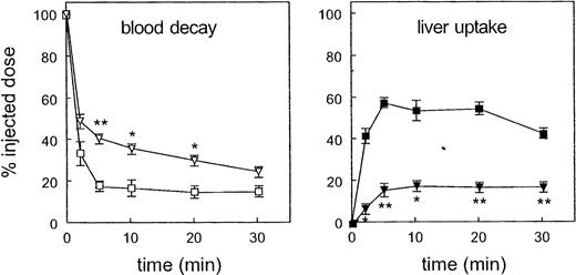
Preinjection of PC liposomes did not alter the blood decay and liver uptake of 51Cr vesicles. In contrast, PS liposomes inhibited the liver uptake of vesicles by 40% of the total radioactive dose with a concomitant slowing down of the blood decay (Figure 5). Also, preinjection of poly-I inhibited the liver uptake by 40% of the total radioactive dose and was accompanied by a slower removal of the vesicles from the blood circulation (Figure 6).
Blood decay and liver uptake of 51Cr vesicles after preinjection of PC and PS liposomes (2 experiments). Left, blood decay after preinjection of PC liposomes (□) and PS liposomes (▿). Right, liver uptake after preinjection of PC liposomes (▪) and PS liposomes (▾). *Significantly different (.01 < P < .05) from controls. **Significantly different (P < .01) from controls.
Blood decay and liver uptake of 51Cr vesicles after preinjection of PC and PS liposomes (2 experiments). Left, blood decay after preinjection of PC liposomes (□) and PS liposomes (▿). Right, liver uptake after preinjection of PC liposomes (▪) and PS liposomes (▾). *Significantly different (.01 < P < .05) from controls. **Significantly different (P < .01) from controls.
Blood decay and liver uptake of 51Cr vesicles after preinjection of 2 mg poly-I in comparison to controls (2 experiments). Left, blood decay after preinjection of poly-I (▵) in comparison with controls (○). Right, liver uptake after preinjection of poly-I (▴) compared to controls (⬡). *Significantly different (0.01 < P < .05) from controls. **Significantly different (P < .01) from controls.
Blood decay and liver uptake of 51Cr vesicles after preinjection of 2 mg poly-I in comparison to controls (2 experiments). Left, blood decay after preinjection of poly-I (▵) in comparison with controls (○). Right, liver uptake after preinjection of poly-I (▴) compared to controls (⬡). *Significantly different (0.01 < P < .05) from controls. **Significantly different (P < .01) from controls.
Tissue distribution of vesicles
At 30 minutes after injection, tissue radioactivity was found in declining order in liver (44.9%), bone (22.5%), skin (9.7%), muscle (5.8%), spleen (3.4%), kidney (2.7%), and lung (1.8%). The blood still contained 9.1% of the radioactivity. The same pattern was seen after preinjection of PC liposomes. In contrast, preinjection of poly-I and PS liposomes affected mainly the uptake of radioactivity in the liver, whereas after preinjection of poly-I an increase in bone and lung activity was seen. After preinjection of both poly-I and PS liposomes more labeled vesicles were still present in the blood as compared to the control situation (Figure 7).
Tissue distribution of 51Cr vesicles at 30 minutes after injection. The bars represent control rats (▪), rats with preinjection of PC liposomes (□), rats with preinjection of poly-I (▨), and rats with preinjection of PS liposomes (▥). The recovery of the injected dose in the various tissues and blood was for the various groups: controls, 100.0% ± 10.5%; PC liposomes, 107.9% ± 0.2%; poly-I, 98.8% ± 6.3%; and PS liposomes, 90.8% ±0.8%. *Significantly different (P = .038) from controls. **Significantly different (P < .01) from controls.
Tissue distribution of 51Cr vesicles at 30 minutes after injection. The bars represent control rats (▪), rats with preinjection of PC liposomes (□), rats with preinjection of poly-I (▨), and rats with preinjection of PS liposomes (▥). The recovery of the injected dose in the various tissues and blood was for the various groups: controls, 100.0% ± 10.5%; PC liposomes, 107.9% ± 0.2%; poly-I, 98.8% ± 6.3%; and PS liposomes, 90.8% ±0.8%. *Significantly different (P = .038) from controls. **Significantly different (P < .01) from controls.
Discussion
Using counterflow centrifugation as fractionation procedure of rat RBC populations, and 2 glycated hemoglobin components as cell age markers, we showed that circulating RBCs in the rat lose relatively as much hemoglobin as human RBCs. As in human plasma, vesicles were also present in rat plasma. It was demonstrated that at least 21% of vesicles were derived from RBCs; 66% of these vesicles contained hemoglobin. Taking these data together we conclude that not only in humans but also in rats hemoglobin loss from circulating RBCs results from vesiculation.
To study the fate of the blood cell-derived vesicles in the rat we chose sodium 51Cr-chromate because in RBCs this label is bound to hemoglobin. Possibly this label also binds to platelet-derived vesicles because 51Cr binds to cytoplasmic constituents such as mitochondria in intact platelets.11 However, this label binds preferentially to human RBC-derived vesicles in comparison to platelet-derived vesicles, as revealed by sorting experiments with anti-GPA antibodies and anti-CD61 antibodies. The difference in binding of the 51Cr label to RBC-derived vesicles and platelet-derived vesicle is, despite the difference in numbers, so large (ratio, 16.7: 1) that more than 80% of the radioactive label is bound to RBC-derived vesicles, and as a consequence the hemoglobin-containing RBC-derived vesicles contribute to more than 50% of total radioactivity (Table 3).
Therefore, it is not surprising that not only approximately 50% of the injected radioactive dose but also a considerable amount of hemoglobin is present in the Kupffer cells at 30 minutes after injection. Both phenomena are a strong indication for a large turnover of this RBC constituent. When 51Cr-labeled donor rat vesicles were injected into recipient rats, we found a biphasic blood decay. Most vesicles (approximately 85%) were removed from the plasma within minutes, the remaining much more slowly. The reason for this dichotomy might be explained by the ratio of labeled RBC-derived versus platelet-derived vesicles or PS exposure or both. Also, differences in release from tissues, in which vesicles are trapped, could play a role. The liver uptake showed the inverse pattern with a steep rise within minutes followed by a plateau phase. After fractionation of liver tissue more than 90% of liver radioactivity was found in the Kupffer cells. The uptake of vesicles by Kupffer cells was inhibited by preinjection of PS liposomes and poly-I, equivalent to 40% of the injected dose. In accordance, 67% of RBC-derived vesicles exposed PS. When these data were compared to those of the study in which the kinetics of oxidized murine RBCs were investigated,5 a marked similarity was noted. This suggests that the same mechanism is responsible for the elimination from the circulation of both RBC-derived vesicles and oxidatively damaged RBCs.
Tissue distribution of radioactivity after preinjection with poly-I showed not only slower removal of circulating vesicles from the plasma, but also an increased uptake in bone and lung, possibly due to a compensatory activity of bone marrow and lung macrophages. No such increased uptake was seen after preinjection with PS liposomes, suggesting inhibition of PS-binding macrophages in bone marrow and lungs besides the inhibition of macrophages in the liver (Kupffer cells). Putative receptors that are operative in these processes consist of scavenger receptor A, CD68, CD 36, or LOX-1.12 Possibly the PS-specific receptor, which plays such a crucial role in the uptake of apoptotic cells, is also of importance in the uptake of PS-exposing vesicles by Kupffer cells.13
We conclude that in rats RBC-derived vesicles are rapidly removed from the circulation mainly by liver Kupffer cells and to a lesser extent by other macrophages of the mononuclear phagocyte system. In this process receptors that can be blocked by PS liposomes and poly-I, but not by PC-liposomes, play an important role. Because hemoglobin loss from RBCs in humans is similar to that in rats, the same mechanism is probably operative for human RBC-derived vesicles.
Approximately 20% of hemoglobin and 20% of surface area are lost from circulating human RBCs during their lifespan. Therefore, vesiculation can be conceived as the first step of an important pathway of RBC breakdown. It also means that by using flow cytometric sorting of RBC-derived vesicles the premortal substrate of 20% of any RBC is at our disposal. The results presented here, together with preliminary data indicating that human RBC-derived and platelet-derived vesicles expose PS in vivo,14 support the hypothesis that exposure of PS on both circulating cells and vesicles leads to their removal by Kupffer cells and other macrophages of the mononuclear-phagocyte system.
Prepublished online as Blood First Edition Paper, November 18, 2004; DOI 10.1182/blood-2004-04-1578.
The publication costs of this article were defrayed in part by page charge payment. Therefore, and solely to indicate this fact, this article is hereby marked “advertisement” in accordance with 18 U.S.C. section 1734.
We thank Mr Arie Pennings and Dr Wim van den Broek of the University Medical Center Nijmegen, The Netherlands for assistance with the combined sorting and labeling experiments.

