Abstract
AML1/Runx1 is a frequent target of human leukemia–associated gene aberration and encodes a transcription factor with nonredundant biologic functions in initial development of definitive hematopoiesis, T-cell development, and steady-state platelet production. AML1/Runx1 and 2 closely related family genes, AML2/Runx3 and AML3/Runx2/Cbfa1, present in mammals, comprise the Runt-domain transcription factor family. Although they have similar structural and biochemical properties, gene-targeting experiments have identified distinct biologic roles. To directly determine the presence of functional overlap among runt-related transcription factor (Runx) family molecules, we replaced the C-terminal portion of acute myeloid leukemia 1 (AML1) with that derived from its family members, which are variable in contrast to conserved Runt domain, using the gene knock-in method. We found that C-terminal portions of either AML2 or AML3 could functionally replace that of AML1 for myeloid development in culture and within the entire mouse. However, while AML2 substituted for AML1 could effectively rescue lymphoid lineages, AML3 could not, resulting in a smaller thymus and lymphoid deficiency in peripheral blood. Substitution by the C-terminal portion of AML3 also led to high infantile mortality and growth retardation, suggesting that AML1 has as yet unidentified effects on these phenotypes. Thus, the C-terminal portions of Runx family members have both similar and distinct biologic functions.
Introduction
The runt-related transcription factor (Runx) family is a newly established gene family consisting of 3 molecules so far identified in mammalian cells: acute myeloid leukemia 1 (AML1)/Runx1, AML2/Runx3, and AML3/Runx2/core-binding factor α 1 (Cbfα1).1-5 Runx gene is known to encode a DNA-binding subunit of the CBF transcription factor complex, which is a heterodimer complex consisting of one Runx molecule and one molecule of the common non–DNA-binding subunit CBFβ.6,7 The association between these subunits is mediated through the signature domain of 128 amino acid residues near the N-terminus of Runx molecules, known as the Runt domain.8,9 This domain is tightly conserved through evolution with homology to the Drosophila orthologue Runt and also functions in the sequence-specific DNA binding to the core DNA sequence, TGT/cGGT.8-10 This sequence is present in a number of candidate target genes although critical target(s) responsible for their biologic functions are not yet thoroughly identified.
In contrast to the Runt domains, whose DNA and amino acid sequences are highly homologous among all Runx family molecules with over 90% identity, their C-termini show substantial differences, with only about 60% of the sequences identical.1-5 Nevertheless, these C-terminal portions share biochemically equivalent components, each of which is responsible for their respective properties as transcriptional factors, such as nuclear translocation, nuclear matrix targeting, trans-activation, auto-inhibition, and trans-repressive activities.11-13 These subdomains serve as binding sites to recruit functional cofactors. Thus, all Runx molecules have modular configurations that are biochemically equivalent, and it has been experimentally demonstrated that they function as transcriptional activators and trans-repressors in a similar context-dependent fashion.13,14
AML1 is the originally isolated member of the Runx family molecules1 and is known as a frequent target of human leukemia–related chromosomal translocations.15,16 Most of these genomic rearrangements result in the production of corresponding abnormal fusion proteins with strong dominant-negative activity against wild-type AML1/CBF functioning.17-23 In addition, it has recently been reported that point, deletion, and insertion mutations of the AML1 gene locus are associated with occasional leukemia cases and with the congenital preleukemic status of familial platelet disorder with predisposition to acute myeloblastic leukemia.24-28 The structural alteration of this molecule is thus profoundly associated with leukemic transformation of hematopoietic progenitor cells.
Consistent with the observation that this gene is frequently involved in hematopoietic malignancies, AML1's crucial function in hematopoietic regulation has been established in that knockout mice of this gene completely lacked definitive hematopoiesis, leading to midgestational death.29,30 The site where AML1 functions has been narrowed down to the hemogenic endothelium of great vessels in early gestation from which hematopoietic stem cells initially emerge.31,32 In addition, AML1 is involved in adult thrombopoiesis and thymocyte development.33-35
In contrast to AML1's involvement in hematopoietic regulation, its related molecules, AML2 and AML3, appear to have distinct nonredundant biologic roles although their structural and biochemical properties are closely related. Targeted inactivation of AML2 results in abnormal proliferation of gastric mucosa36 and severe limb ataxia due to abnormal spinal motor neurons,37,38 indicating in which cell lineages AML2 is essential. Disruption of AML3 on the other hand leads to the complete loss of bone formation,39,40 signifying that AML3 is necessary for ossification. Although it is widely thought that the biologic differences stem from their distinct expression patterns, no detailed investigation has been conducted as to whether the subtle structural differences among these molecules influences their biologic effects.
We have shown that loss of definitive hematopoiesis manifested in mouse embryonic stem (ES) cells or entire mice as the result of AML1 deficiency could be undone by re-expression of its cDNA through a gene knock-in procedure.34,41 We previously used this experimental system to characterize the C-terminal domains of AML1 by demonstrating that the trans-activation domain of AML1 was necessary for its biologic activity, whereas the C-terminal trans-repressive subdomain was dispensable for this activity but appeared to have a function in thymocyte development.34,41 To directly assess functional overlap among Runx family molecules and to further characterize biologic activities of the C-terminal subdomains of AML1, we generated ES cells and germ line mice that contain knock-in alleles that produce chimeric AML1 cDNA whose C-terminal portion was replaced with that of one of its family members through the knock-in procedure. Biologic consequences of these substitutions revealed shared and distinct roles of the Runx family molecules.
Materials and methods
Construction of the vectors
To construct replacement-type vectors for generating knock-in alleles, we first cloned full-length cDNAs for murine AML2/Runx3 and AML3/Runx2 by recombinant reverse-transcription polymerase chain reaction (RT-PCR) using messenger RNAs isolated from in vitro–differentiated ES cells as the template. Both strands of the cDNAs thus obtained were sequenced to confirm their integrity.
Replacement-type vectors to introduce targeted mutations were generated by using a 12-kb genomic DNA fragment containing exon 4 of AML1 gene29 as the backbone of the construct. First, silent mutations at nucleotides 381 and 495 (amino acid residues 127 and 165) were introduced via site-directed mutagenesis into AML2 and AML3 cDNAs, respectively, to create artificial SacII cleavage sites that correspond to the SacII site of exon 4 of the murine AML1 gene. Downstream sequences from the artificial SacII site of either cDNA (AML2 or AML3) were then inserted at the SacII site of the genomic DNA fragment of AML1 so that the reading frame was open. The poly-adenylation signal sequences from rabbit globin gene (pA) and a puromycin resistance cassette (puro) were then inserted at 3′ to this artificial exon, and a diphtheria toxin–A suicide cassette was put at the 3′ end of the DNA fragment in the sense orientation.
Eukaryotic expression vectors transiently expressing the engineered AML1 molecules were prepared by inserting the cDNAs into the pRc/CMV vector (Invitrogen, Carlsbad, CA).
Western blot analysis for the transiently expressed AML1 proteins in COS-7 cells
COS-7 cells were transfected by the diethylaminoethyl (DAEA)–dextran method41 with the plasmid DNA of each of the pRc constructs. After a 72-hour culture, cells were lysed, electrophoretically separated, and then transferred to a nitrocellulose membrane. Proteins were detected by using rabbit antisera against a synthesized peptide corresponding to the N-terminus of the AML1 (RIPVDASTSRRFTPPSC) and secondary donkey antisera against rabbit immunoglobulin G (IgG) conjugated with horseradish peroxidase (Amersham Biosciences, Piscataway, NJ), resulting in visualization with enhanced chemiluminescence (ECL) substrate (Amersham Biosciences) according to the manufacturer's instruction.
Luciferase reporter assay
The pRc vector construct(s) of the AML1-related molecule(s) to be studied and either of the firefly luciferase reporter constructs, pM-CSF-R-luc42 (a gift from Dr Dong-Er Zhang) or pGL3-basic plasmid (Promega, Madison, WI) containing -147/+13 promoter DNA of mouse osteocalcin 2 gene (pGL-mOG2), were transfected into HeLa cells (Riken Cell Bank, Tsukuba, Japan) with the calcium precipitation method. A control plasmid, ph RL-TK (Promega), was cotransfected at a fixed ratio to provide internal transfection efficiency. After a 48-hour culture, the cells were lysed and subjected to the double-luciferase reaction with specific substrates (Promega), according to the manufacturer's instructions. Firefly luciferase activity relative to Renila luciferase activity was measured with a luminometer (Berthorld Detection Systems, Portzheim, Germany).
Introducing knock-in alleles into murine ES cells
Each of the targeting vectors was linearized and transfected by electroporation into embryonic day-14 (E14)–derived ES cells. Puromycin-resistant clones were screened for homologous recombination by serial Southern blot analysis with 5′ and 3′ outside probes. Knock-in clones into ES cells of the AML1-deficient genotype were used for in vitro experiments on hematopoietic differentiation, whereas those introduced into wild-type ES cells were used for the generation of germ line mice.
In vitro hematopoietic differentiation of ES cells clones
In vitro hematopoietic differentiation was performed with the methods originally described by Keller et al43 with some modifications.41,44 This procedure allows plated ES cells to form embryoid bodies (EBs) that consist of tight cell aggregates containing all 3 germ cell populations. All experiments were performed in triplicate. EBs were scored for hematopoietic differentiation in the presence of a combination of colony-stimulating factors41,44 on day 14 with an inverted microscope (CK40, Olympus, Tokyo, Japan) equipped with a digital camera (C-5050 Zoom, Olympus).
Generation of mutant mice
Manipulated ES cells were used to generate chimera mice with the conventional blastocyst injection method.29,34 Germ line transmission of the mutated alleles into F1 agouti mice and their progeny was monitored by means of Southern blot analysis as described in “Introducing knock-in alleles into ES cells.” All procedures of the animal experiments performed in this study had been approved by the Committee for Animal Research, Kyoto Prefectural University of Medicine.
Semiquantitative RT-PCR
Total RNA was used as the template for the random hexamer–primed RT reaction with Moloney murine leukemia virus (MMLV) reverse transcriptase (Invitrogen). Serially diluted synthesized cDNA samples were examined by PCR for the presence of messages from the gene of interest. In parallel, the expression of the housekeeping gene hypoxantin-guanine phosphoribosyl transferase (HPRT) within the specimens was also evaluated with a specific primer pair, as described elsewhere,34 to set a standard for the quantity and integrity of the RNA samples. Primer sequences are available upon request.
FACS analysis
Hematopoietic progenitor assay for fetal liver cells
Fetal livers of E12.5 embryos were dissected and mechanically disaggregated into single-cell suspensions. Cells were cultivated in an in vitro culture as previously described.29,34 Colonies thus grown were counted after 12 days of incubation,29,34 and the total number of the progenitors was estimated in proportion to the total number of cells obtained from each liver.
T-cell proliferation assay
Splenic T cells were stimulated with 0.1 μg/mL plate-bound anti-CD3 antibody (BD Biosciences, San Jose, CA) in the presence or absence of 2 μg/mL anti-CD28 (BD Biosciences) and 20 μg/mL murine interleukin-2 (m–IL-2; Roche Diagnostics, Mannheim, Germany). Cells were cultured in triplicate for 48 hours and were pulsed with [3H]-thymidine (0.5 μCi/well [0.0185 MBq/well]) for the last 14 hours. The incorporation of [3H]-thymidine was measured with a β-counter.
Skeletal preparation
Skeletal bone structures were examined with Alcian blue/alizarin red double-staining according to the methods described by Hogan et al.45 In brief, newborn mice were skinned, eviscerated, and fixed in 100% ethanol for 24 hours. The carcasses were first stained in 0.015% of Alcian blue (Sigma Chemical Company, St Louis, MO) solution for 24 hours. After rinsing with 95% ethanol, they were immersed in 2% KOH overnight. The specimens were then restained with 0.005% alizarin red (Sigma Chemical Company) solution in 1% KOH for 24 hours and kept in 20% glycerol/1% KOH until the skeletons became clearly visible. They were stored in 50% and finally 100% glycerol.
Results
Generation of knock-in alleles expressing chimeric AML1 genes with heterologous C-terminal portion
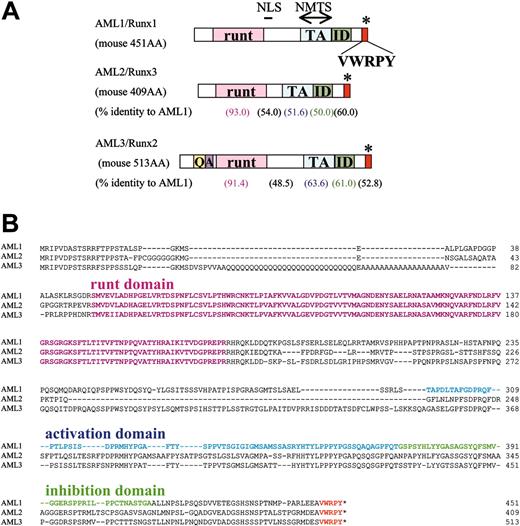
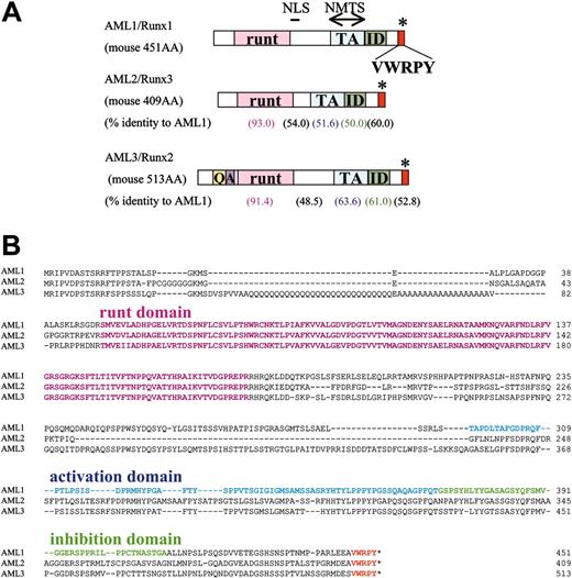
To investigate functional overlap among Runx transcription factor family members and to further analyze biologic functions performed by AML1 mediated through its C-terminal subdomains, we directly examined whether C-terminal portions from AML2 or AML3 could functionally replace that of AML1 in hematopoietic regulation in vitro and in vivo. In contrast to the tightly conserved Runt domain, the C-terminal portions of Runx family member molecules vary (Figure 1A-B). We first cloned murine cDNAs for AML2 and AML3 and then engineered chimeric murine AML1b cDNAs whose C-terminal portion was replaced by one from either AML2 or AML3 by taking advantage of the artificially introduced SacII cleavage site within the Runt domain of AML2 or AML3 to keep the amino acid sequences of the domain unchanged (Figure 2A). The chimeric AML1 gene with the AML2-derived C-terminus (and the generated knock-in allele with this cDNA) is hereafter referred to as “AML1-2” and that with the AML3-derived one as “AML1-3.” When these molecules were exogenously expressed in COS-7 cells, protein bands of the expected sizes were recognized by antisera against N-terminal AML1 (Figure 2B), thus indicating the integrity of the cDNAs. In addition, both AML1-2 and AML1-3 could activate the M-CSF receptor gene reporter construct, pM-CSF-R-P-luc, though the activity of AML1-3 was somewhat weaker than that of AML1b or AML1-2 for this promoter (Figure 2C). This observation indicates that these chimeric proteins retain their biochemical properties.
Structure and amino acid sequences of Runx family molecules. (A) Schematic representation of the structure of Runx molecules. runt indicates the Runt domain; NLS, nuclear localizing signal; NMTS, nuclear matrix targeting signal; TA, trans-activation domain; ID, auto-inhibitory domain; VWRPY, VWRPY-motif; Q, glutamine (Q)-residue stretch; A, alanine (A)-residue stretch. Numerals within parentheses below the AML2 and AML3 schemes indicate the percentages of amino acid (AA) identity of the subdomains in comparison with those of AML1. (B) Comparison of amino acid sequences of the Runx molecules represented by the 1-letter code. Manual alignment was performed visually. Runt domain sequences are highlighted in pink. Dashes (–) indicate gaps. Numerals to the right of the columns represent the positions of the amino acid residues.
Structure and amino acid sequences of Runx family molecules. (A) Schematic representation of the structure of Runx molecules. runt indicates the Runt domain; NLS, nuclear localizing signal; NMTS, nuclear matrix targeting signal; TA, trans-activation domain; ID, auto-inhibitory domain; VWRPY, VWRPY-motif; Q, glutamine (Q)-residue stretch; A, alanine (A)-residue stretch. Numerals within parentheses below the AML2 and AML3 schemes indicate the percentages of amino acid (AA) identity of the subdomains in comparison with those of AML1. (B) Comparison of amino acid sequences of the Runx molecules represented by the 1-letter code. Manual alignment was performed visually. Runt domain sequences are highlighted in pink. Dashes (–) indicate gaps. Numerals to the right of the columns represent the positions of the amino acid residues.
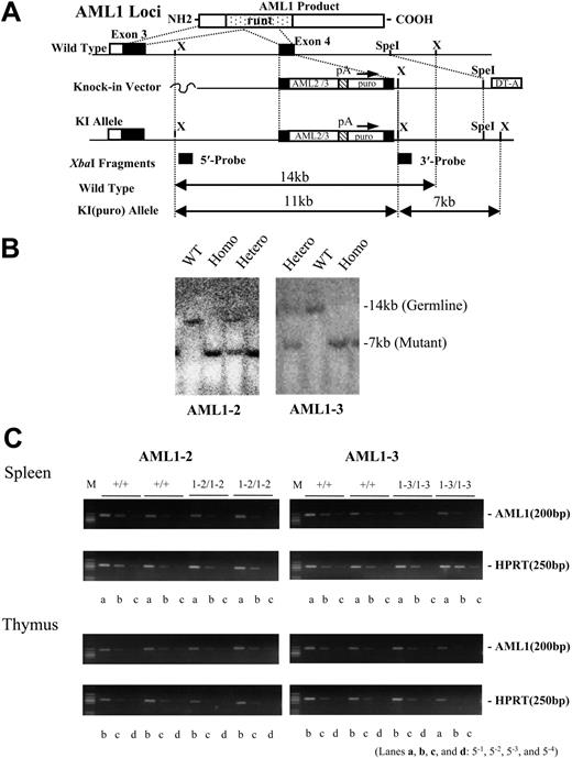
With these cDNAs we constructed replacement-type vectors for generating knock-in alleles in order to introduce targeted insertion at the murine AML1 gene locus so that chimeric protein(s), either AML1-2 or AML1-3, would be expressed under the control of endogenous promoter activities of this gene locus (Figure 2D-E). Our AML1-deficient ES cell clones had been generated by serial targeting of exon 4: one allele was replaced with a hygromycin-resistance cassette (hygr) and the other with a neomycin-resistance cassette (neo)29 (Figure 2D). Multiple ES clones that underwent homologous recombination for either allele were successfully isolated for both AML1-2 and AML1-3 knock-in mutations and then analyzed for in vitro differentiation.
C-terminal portion of AML2 or AML3 can functionally replace that of AML1 for hematopoietic rescue of mouse ES cell in culture
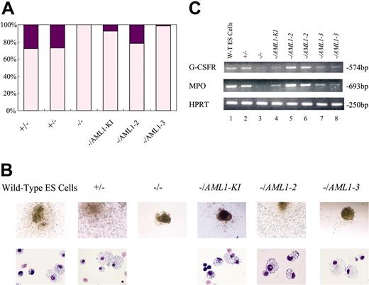
ES cells develop EBs with differentiating primitive and definitive hematopoietic cells under optimal culture conditions.42 AML1-deficient ES cells lose their ability to develop definitive hematopoietic cells, and this hematopoietic defect can be overcome if wild-type AML1 cDNA is re-expressed within these AML1-deficient cells by means of the knock-in approach employed in our study.41 As the representative results shown in Figure 3 indicate, ES cell clones with one knock-in allele, either AML1-2 or AML1-3, develop definitive hematopoietic cells as observed in control clones of heterozygously knock-out (AML1+/-), AML1 knock-in (AML1-/AML1-KI), or wild-type genotypes (Figure 3A). This hematopoietic rescue was observed reproducibly in multiple clones for either AML1-2 or AML1-3, regardless of which allele, Neo or Hygr, was targeted (data not shown). An important finding is that in May-Grünwald-Giemsa–stained cytospin preparations the morphology of cells developed in vitro from knock-in clones for AML1-2 or AML1-3, including granulocytes, macrophages, and definitive erythroid precursor cells, was indistinguishable from that of control clones (Figure 3B). In addition, semiquantitative RT-PCR analysis showed that the clones rescued by either AML1-2 or AML1-3 allele regained expression of the AML1-dependent genes, including the G-CSF receptor (G-CSFR) and myeloperoxidase (MPO) genes, in contrast to the drastic reduction in the AML1-deficient ES cells (Figure 3C). These results indicate that the C-terminal portions of the runt-related transcription factor family members have the same biologic function, at least in terms of supporting the initiation of definitive hematopoiesis in vitro.
Targeting strategy to introduce knock-in alleles expressing chimeric AML1 genes with heterologous C-terminal portion. (A) Structure of Runx family molecules and chimeric AML1 with AML2- or AML3-derived C-terminus (AML1-2 and AML1-3, respectively). Numerals indicate the positions of amino acid residues of the molecules as shown in Figure 1B. □ indicates parts derived from AML1, ▦ from AML2, and  from AML3. (B) Integrity of the mutant construct was confirmed by Western blot analysis of transiently transfected COS-7 cells. Arrows indicate protein bands detected by rabbit antisera against N-terminal peptide of AML1. (C) Biochemical activities of AML1 or the mutant molecules were examined by means of reporter assay experiments. Each of pRc effector constructs and pM-CSF-R-luc were cotransfected into HeLa cells with CBFβ and phRL-TK control vectors. The height of the columns indicates the increase in relative luciferase activity on the M-CSF receptor gene promoter (see “Materials and methods”). Bars indicate standard deviations of triplicate experiments. Both of the chimera proteins retained trans-activation activity although the activity of AML1-3 for this promoter construct was somewhat weaker than that of AML1b or AML1-2. Three independent experiments were performed with basically the same results. Representative results are shown. (D) Targeting strategy to introduce knock-in allele for AML1-2 or AML1-3 into ES cell of AML1-deficient genotype. Both alleles of exon 4, which corresponds to the middle of the Runt domain, had been disrupted by insertion of KO(hygr) (the hygromycin-resistance cassette) and KO(neo) (the neomycin-resistance cassette). Replacement-type (knock-in) vectors were designed to introduce KI(puro) (the knock-in allele). □ indicates non-coding regions of the exons; ▪, coding exons;
  , runt domain; ▧, poly-adenylation signal sequences; ▤, DNA fragments for the probes. (E) Clones subjected to homologous recombination are detectable by Southern blot analysis.
Targeting strategy to introduce knock-in alleles expressing chimeric AML1 genes with heterologous C-terminal portion. (A) Structure of Runx family molecules and chimeric AML1 with AML2- or AML3-derived C-terminus (AML1-2 and AML1-3, respectively). Numerals indicate the positions of amino acid residues of the molecules as shown in Figure 1B. □ indicates parts derived from AML1, ▦ from AML2, and  from AML3. (B) Integrity of the mutant construct was confirmed by Western blot analysis of transiently transfected COS-7 cells. Arrows indicate protein bands detected by rabbit antisera against N-terminal peptide of AML1. (C) Biochemical activities of AML1 or the mutant molecules were examined by means of reporter assay experiments. Each of pRc effector constructs and pM-CSF-R-luc were cotransfected into HeLa cells with CBFβ and phRL-TK control vectors. The height of the columns indicates the increase in relative luciferase activity on the M-CSF receptor gene promoter (see “Materials and methods”). Bars indicate standard deviations of triplicate experiments. Both of the chimera proteins retained trans-activation activity although the activity of AML1-3 for this promoter construct was somewhat weaker than that of AML1b or AML1-2. Three independent experiments were performed with basically the same results. Representative results are shown. (D) Targeting strategy to introduce knock-in allele for AML1-2 or AML1-3 into ES cell of AML1-deficient genotype. Both alleles of exon 4, which corresponds to the middle of the Runt domain, had been disrupted by insertion of KO(hygr) (the hygromycin-resistance cassette) and KO(neo) (the neomycin-resistance cassette). Replacement-type (knock-in) vectors were designed to introduce KI(puro) (the knock-in allele). □ indicates non-coding regions of the exons; ▪, coding exons;
  , runt domain; ▧, poly-adenylation signal sequences; ▤, DNA fragments for the probes. (E) Clones subjected to homologous recombination are detectable by Southern blot analysis.
Embryoid body differentiation experiments for knock-in clones. (A) Results of EB differentiation. Incidence of hematopoietic differentiation of day-14 EBs derived from ES cell clones of wild-type (+/+), heterozygous for AML1-disruption (+/-), homozygous for the disruption (-/-), one knock-in allele for wild-type AML1 cDNA (-/AML1-KI), one knock-in allele for AML1-2 (-/AML1-2), and one knock-in allele for AML1-3 (-/AML1-3) genotypes in a representative experiment. Purple parts of the columns indicate the proportion of grown EBs with hematopoietic differentiation whereas pink parts represent those without hematopoietic elements. ES clones of the AML1-2 or AML1-3 knock-in allele developed hematopoietic cells in vitro as did control clones. (B) Appearance of representative day-14 EBs derived from the ES cell clones (top row). Morphology of the hematopoietic cells developed from ES cells of AML1-2 or AML1-3 knock-in clones showed no marked abnormalities of component cells examined with May-Grünwald-Giemsa staining (bottom row). Original magnifications: top row, × 20; bottom row, × 132. (C) Semiquantitative RT-PCR analysis of the total RNA from embryoid bodies was employed to identify recovered hematopoietic gene expression in the rescued clones. Messages for the G-CSF receptor (G-CSFR) and myeloperoxidase (MPO) genes were detected in the AML1-2– and AML1-3 clone–derived ES cells (lanes 5-8; results for 2 independent clones for each mutation are shown) as was the case for positive control clones (lanes 1, 2, and 4). On the other hand, a profound decrease in the expression for these genes was observed in the AML1-deficient (-/-) clone (lane 3). Expression of a housekeeping gene, HPRT, was assessed to provide a standard in parallel for the messenger RNA within the specimen. A dilution of 5-0 of the RT specimen was used for PCR of G-CSFR and MPO and 5-3 dilution for PCR of HPRT (see “Materials and methods”). bp indicates base pair.
Embryoid body differentiation experiments for knock-in clones. (A) Results of EB differentiation. Incidence of hematopoietic differentiation of day-14 EBs derived from ES cell clones of wild-type (+/+), heterozygous for AML1-disruption (+/-), homozygous for the disruption (-/-), one knock-in allele for wild-type AML1 cDNA (-/AML1-KI), one knock-in allele for AML1-2 (-/AML1-2), and one knock-in allele for AML1-3 (-/AML1-3) genotypes in a representative experiment. Purple parts of the columns indicate the proportion of grown EBs with hematopoietic differentiation whereas pink parts represent those without hematopoietic elements. ES clones of the AML1-2 or AML1-3 knock-in allele developed hematopoietic cells in vitro as did control clones. (B) Appearance of representative day-14 EBs derived from the ES cell clones (top row). Morphology of the hematopoietic cells developed from ES cells of AML1-2 or AML1-3 knock-in clones showed no marked abnormalities of component cells examined with May-Grünwald-Giemsa staining (bottom row). Original magnifications: top row, × 20; bottom row, × 132. (C) Semiquantitative RT-PCR analysis of the total RNA from embryoid bodies was employed to identify recovered hematopoietic gene expression in the rescued clones. Messages for the G-CSF receptor (G-CSFR) and myeloperoxidase (MPO) genes were detected in the AML1-2– and AML1-3 clone–derived ES cells (lanes 5-8; results for 2 independent clones for each mutation are shown) as was the case for positive control clones (lanes 1, 2, and 4). On the other hand, a profound decrease in the expression for these genes was observed in the AML1-deficient (-/-) clone (lane 3). Expression of a housekeeping gene, HPRT, was assessed to provide a standard in parallel for the messenger RNA within the specimen. A dilution of 5-0 of the RT specimen was used for PCR of G-CSFR and MPO and 5-3 dilution for PCR of HPRT (see “Materials and methods”). bp indicates base pair.
Both AML1-2 and AML1-3 knock-in alleles rescue embryonic lethal phenotype of AML1 deficiency
In order to further examine if the hematopoietic rescue observed in the in vitro study described in “C-terminal portion of AML2 or AML3 can functionally replace that of AML1 for hematopoietic rescue of mouse ES cell in culture” can be extended to entire mice, we used the same targeting vectors to transmit these artificial knock-in alleles into mouse germ line (Figure 4A-B). With conventional blastocyst injection, 2 independent mouse lines were established for each of the mutations (Table 1). Phenotypes specific to the mutations were assessed only when they were reproducibly observed in both mouse lines for each of the introduced artificial alleles. Mice heterozygous for each of the mutated alleles were healthy and fertile, and live pups homozygous for both knock-in alleles were born from intercrossed heterozygous parents. The mutated alleles segregated according to the Mendelian ratio for both mutations (Table 1), as was also observed for the knock-in allele with wild-type AML1 cDNA (AML1-KI),34 and both males and females proved to be fertile. Semiquantitative RT-PCR analysis showed that the amounts of messenger RNA for the knock-in genes in the thymus and spleen appeared to be comparable to those for the endogenous AML1 gene (Figure 4C).
AML1-deficient mice die in utero due to complete absence of fetal liver hematopoiesis,29,30 whereas the hematopoietic progenitor cells emerge in the fetal liver of AML1-KI mice as previously reported.34 This AML1 activity during initial development of definitive hematopoiesis appears to be dose dependent: in heterozygous mice the simple disruption of this gene locus results in the reduction by about half of the number of the progenitors per liver.30 To assess embryonic hematopoiesis in AML1-2 and AML1-3 mice, we cultured fetal liver cells from the mutant embryos and their control littermates on E12.5. Under conditions optimal for hematopoietic growth of definite origin, we found that both AML1-2 and AML1-3 embryos retained hematopoietic progenitor cells in their fetal livers (Table 2) as was also observed for the animals rescued by the AML1-KI allele reported.34 Homozygous embryos of AML1-2 tended to have approximately twice the number of progenitors per liver as their wild-type littermates, whereas homozygous embryos of AML1-3 tended to have approximately 30% to 40% fewer progenitor cells than those of their wild-type littermates. The differences were, however, not statistically significant.
Transmission of knock-in alleles to mouse germ line. (A) Targeting strategy for introducing knock-in alleles, AML1-2 and AML1-3, into wild-type ES cells. Refer to the legend of Figure 2D for symbols. (B) Genotype of the germ line mice harboring the knock-in allele was determined by Southern blot analysis using XbaI-digested genomic DNA. The mutated allele was detected as a 7-kb fragment that hybridizes with the 3′ outside probe whereas the wild-type allele yields a band of 14 kb. Representative results are shown. WT indicates wild type; Homo, homozygous; and Hetero, heterozygous. (C) Semiquantitative RT-PCR analysis was used to evaluate the expression level of the knock-in genes in comparison with that of a housekeeping gene, HPRT. Results are shown for 2 wild-type and 2 homozygous mice that were littermates for each of the mutations (+/+, wild-type). Serially diluted cDNA pools were analyzed (see “Materials and methods”). Lanes a, b, c, and d indicate 5-1, 5-2, 5-3, and 5-4 dilutions, respectively. M indicates size marker. Expression levels of the exogenous genes from the knock-in alleles in the homozygous animals were almost the same as those observed for the endogenous AML1 gene in wild-type littermates.
Transmission of knock-in alleles to mouse germ line. (A) Targeting strategy for introducing knock-in alleles, AML1-2 and AML1-3, into wild-type ES cells. Refer to the legend of Figure 2D for symbols. (B) Genotype of the germ line mice harboring the knock-in allele was determined by Southern blot analysis using XbaI-digested genomic DNA. The mutated allele was detected as a 7-kb fragment that hybridizes with the 3′ outside probe whereas the wild-type allele yields a band of 14 kb. Representative results are shown. WT indicates wild type; Homo, homozygous; and Hetero, heterozygous. (C) Semiquantitative RT-PCR analysis was used to evaluate the expression level of the knock-in genes in comparison with that of a housekeeping gene, HPRT. Results are shown for 2 wild-type and 2 homozygous mice that were littermates for each of the mutations (+/+, wild-type). Serially diluted cDNA pools were analyzed (see “Materials and methods”). Lanes a, b, c, and d indicate 5-1, 5-2, 5-3, and 5-4 dilutions, respectively. M indicates size marker. Expression levels of the exogenous genes from the knock-in alleles in the homozygous animals were almost the same as those observed for the endogenous AML1 gene in wild-type littermates.
These findings demonstrate that the C-terminal portions of AML2 or AML3 can functionally replace that of AML1 in the initial development of fetal liver hematopoiesis in vivo.
Influence on myeloid hematopoiesis by AML1-2 and AML1-3 alleles
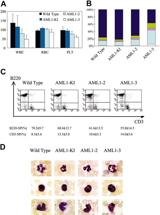
It has recently been demonstrated that AML1 performs an essential function in megakaryocyte/platelet lineage. Induced disruption of this gene locus by means of the Mx-Cre–transgenic approach with the floxed AML1 allele in adult animals resulted in the reduction of the number of platelets in peripheral blood by one third to one sixth due to impaired maturation of megakaryocytes.35 To determine whether AML1-2 or AML1-3 allele could overcome this defect, we analyzed the hematopoietic status of these mutations in adult knock-in mice. Platelet counts in AML1-2 or AML1-3 animals tended to be fewer than those in controls, but this difference was minor and not statistically significant (Figure 5A). This indicates that both AML1-2 and AML1-3 alleles can largely, if not completely, overcome the hematopoietic defect of this lineage resulting from the loss of AML1. In addition, the red blood cell counts for these animals showed no difference from those for the controls (Figure 5A).
In contrast, AML1-2 and AML1-3 mice had considerably fewer white blood cells than did their wild-type littermates or AML1-KI control animals and this difference was significant, especially for the AML1-3 mutation (Figure 5A). The leukocyte differential showed that the reduction in the white blood cells of AML1-3 was due to a decrease in the lymphoid cell population, whereas the quantity of myeloid components was maintained (Figure 5B). However, there was no change in the composition of T- and B-cell populations detectable by flow cytometric analysis in the peripheral blood lymphocytes of the AML1-3 mice (Figure 5C). In addition, the morphologic appearance of the circulating blood cells, including granulocytes, monocytes, lymphocytes, platelets, and erythrocytes, of these mutant animals was indistinguishable from that of the control animals (Figure 5D). Furthermore, microscopic examination of the hematoxylin-eosin (HE)–stained bone marrow sections from AML1-2 or AML1-3 homozygous mice showed no marked changes in the architecture or cellularity compared with those from their wild-type littermates (data not shown). Thus, myeloid hematopoiesis of these knock-in mice appeared to be minimally influenced by the knock-in mutant alleles.
Abnormal thymus size in AML1-3 mice
T lymphocytes constitute another cell lineage affected by AML1 in adult individuals. T-cell–specific inactivation of AML1 by means of the lck-Cre–transgenic approach with the floxed AML1 gene locus demonstrated that AML1 has essential roles during early thymocyte development.33,35 In the thymus of the conditionally targeted mice, the number of the thymocytes was markedly reduced to less than 10% of controls. Moreover, the CD4 gene was derepressed in the early double-negative stage and a variegated expression of CD8 gene was observed.33,35 We found that thymi obtained from AML1-3 mice were smaller than those from wild-type or heterozygous littermates. This difference was detectable at birth and persisted through the age of 5 weeks. The average number of cells in the thymus was about one quarter to half of that of the controls (Figure 6A; data not shown). Although AML1-3 homozygous mice tended to have smaller body size (see “Infant mortality and abnormal body weight in AML1-3 mice”), the number of thymocytes per body weight was still significantly smaller than in controls (Figure 6B). However, the size of most other organs, such as kidney or heart, appeared to be proportional in relation to body size (Figure 6C; not shown). Although the spleen appeared small in infancy (Figure 6C), it became proportional to the body size later in their life (not shown). Flow cytometric analysis of the thymocytes showed no obvious changes in the proportions of CD4- and/or CD8-expressing thymocytes in these mice (Figure 6D). In addition, HE-stained sections of the thymus showed no remarkable abnormalities in the architecture other than slightly less developed medulla in the AML1-3 mutant mice (Figure 6E). In contrast, the thymus of AML1-2 mice was comparable in size to that of controls from birth up to 3 weeks of age. Although we detected a slight decrease in the number of thymocytes in these animals at the age of 5 weeks to about 70% of that of controls, it was not statistically significant (data not shown). Splenic T cells of the AML1-3 animal, nevertheless, proliferated in vitro in response to T-cell receptor (TCR) stimulation with or without CD28 costimulation or exogenous IL-2 treatment. The extent of proliferation was comparable to that observed for the cells from control mice (Figure 6F). Thus, both AML1-2 and AML1-3 alleles could functionally replace AML1 for T-cell development, as was observed in the case of AML1-KI.34 However, the rescue by the AML1-3 allele did not appear to be complete, since the number of thymocytes in AML1-3 mice was still lower.
Findings for peripheral blood of knock-in mice. (A) Peripheral blood cell counts of the mutant mice are indicated by the height of the columns. Lines above the columns indicate standard deviations. WBC indicates white blood cell count; RBC, red blood cell count; and PLT, platelet count. (B) Bar graphs showing differences of peripheral leukocytes from knock-in and control mice. Twenty mice were examined for each mutation, and means of the results are shown. Lymphoid population in peripheral blood of AML1-3 mice is reduced. Dark blue indicates lymphocytes; green, monocytes; pink, eosinophils; light blue, seg (segmented neutrophils); and white, stab (stab neutrophils). (C) Flow cytometric analysis of peripheral T- and B-lymphoid populations, showing no marked differences in the composition of T- and B-cell populations for the mutant and control animals. Six mice were analyzed for each of the genotypes. Numerals below the charts show means and standard deviations for single-positive (SP) populations. (D) Microscopic appearance of representative blood cells from mutant and control animals, showing no marked abnormalities in granulocytes (top row), lymphocytes (middle row), or monocytes (bottom row). Smear samples were stained with May-Grünwald-Giemsa, and the photographs were taken by using a microscope (BH-2, Olympus) equipped with a digital camera (C-5050 Zoom, Olympus). Original magnification, × 300.
Findings for peripheral blood of knock-in mice. (A) Peripheral blood cell counts of the mutant mice are indicated by the height of the columns. Lines above the columns indicate standard deviations. WBC indicates white blood cell count; RBC, red blood cell count; and PLT, platelet count. (B) Bar graphs showing differences of peripheral leukocytes from knock-in and control mice. Twenty mice were examined for each mutation, and means of the results are shown. Lymphoid population in peripheral blood of AML1-3 mice is reduced. Dark blue indicates lymphocytes; green, monocytes; pink, eosinophils; light blue, seg (segmented neutrophils); and white, stab (stab neutrophils). (C) Flow cytometric analysis of peripheral T- and B-lymphoid populations, showing no marked differences in the composition of T- and B-cell populations for the mutant and control animals. Six mice were analyzed for each of the genotypes. Numerals below the charts show means and standard deviations for single-positive (SP) populations. (D) Microscopic appearance of representative blood cells from mutant and control animals, showing no marked abnormalities in granulocytes (top row), lymphocytes (middle row), or monocytes (bottom row). Smear samples were stained with May-Grünwald-Giemsa, and the photographs were taken by using a microscope (BH-2, Olympus) equipped with a digital camera (C-5050 Zoom, Olympus). Original magnification, × 300.
Infant mortality and abnormal body weight in AML1-3 mice
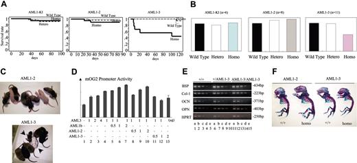
Knock-in mice generated for this study also showed some additional unexpected phenotypes. First, mice homozygous to the AML1-3 mutation showed a high infant mortality rate. The Kaplan-Meier survival curve showed that almost half of the AML1-3 mice died within the first 3 weeks after birth (Figure 7A). They died suddenly without any severe sick appearance so far observed. The survivors beyond the first 3 weeks achieved a normal lifespan. In addition, AML1-3 mice showed abnormalities in their body size and weight. In contrast to the AML1 knock-in mice or AML1-2 mutants, AML1-3 mutants were smaller than their control littermates throughout their lifetime (Figure 7B-C). The body weight of the AML1-3 mutants was about 60% of that of the wild-type or heterozygous littermates (Figure 7B). It was demonstrated that AML1-3 had no obvious dominant-negative effects on AML3's trans-activating activity, at least as far as could be detected by luciferase reporter assay experiments in a nonosseous cell line, on the promoter of an AML3's osseous target gene, mouse osteocalcin (Figure 7D). In addition, the homozygous embryo of the AML1-3 allele expressed comparable amounts of the messenger RNAs of known AML3-dependent genes, including bone sialoprotein, collagen I alpha 1, osteocalcin, and osteopontin (Figure 7E). Consistent with these results so far showing no evidence of the disturbed transcriptional regulation for osseous genes present in the mutant animal, Alcian blue and alizarin red double-staining of the skeletal structure of newborn mice with the AML1-3 genotype showed no marked abnormality in their bone ossification (Figure 7F). In addition, they displayed no obvious difficulties in suckling milk, and their blood sugar level was normal (not shown). Although most of the details remain to be clarified, AML1-3 mice showed the presence of such a few additional phenotypes that are likely to be caused by incomplete rescue of these mice from AML1 deficiency.
Smaller thymus observed in AML1-3 mice. (A) Macroscopic appearance of dissected thymi from representative 1-, 3-, and 5-week-old litters obtained from intercrossed parents heterozygous for AML1-2 or AML1-3 knock-in mutations. *Homozygous mutants. Photographs were taken with a digital camera (Coolpix 4300, Nikon, Tokyo, Japan). (B) The number of thymocytes per thymic lobe per body weight (g) is indicated by columns for 1-week-old AML1-3 homozygous mice compared with the numbers for wild-type littermates. Bars indicate standard deviations. Thymi from AML1-3 homozygous mice were smaller than those of controls even if reduced body size was taken into account (see “Abnormal thymus size in AMLI-3 mice.”). (C) Appearance of the spleens and kidneys of the 1-week-old AML1-3 litter together with their thymi. Most organs other than thymi, including kidneys and hearts (not shown), were proportional to body weight (BW). Spleens appeared small in infancy, but they became proportional to the body size later in their life (not shown). (D) Results of flow cytometric analysis of the CD4 and CD8 expression for thymocytes. Numbers in graph quadrants indicate fractions of the cells analyzed by percentile. (E) Hematoxylin-eosin–stained sections of the thymi. Note that slightly less developed medulla is observed in the thymus from AML1-3 mice. No obvious bleeding or degenerative lesions were detected in these specimens. A microscope (BH-2, Olympus) with a digital camera (C-5050 Zoom, Olympus) was used. Original magnification, × 100. (F) In vitro proliferation assay on the splenic T cells isolated from mice of each genotype. Cells (1.5 × 10⁵) were stimulated with appropriate antibodies and/or cytokines (light blue, 0.1 μg/mL CD3; medium blue, CD3 plus CD28; dark blue, CD3 plus CD28 plus IL-2) for 48 hours, and were pulsed with [3H]-thymidine for the last 14 hours. The [3H]-thymidine uptake (counts per minute) is shown with standard deviation. The T cells from AML1-3 animals proliferated equally upon stimulation.
Smaller thymus observed in AML1-3 mice. (A) Macroscopic appearance of dissected thymi from representative 1-, 3-, and 5-week-old litters obtained from intercrossed parents heterozygous for AML1-2 or AML1-3 knock-in mutations. *Homozygous mutants. Photographs were taken with a digital camera (Coolpix 4300, Nikon, Tokyo, Japan). (B) The number of thymocytes per thymic lobe per body weight (g) is indicated by columns for 1-week-old AML1-3 homozygous mice compared with the numbers for wild-type littermates. Bars indicate standard deviations. Thymi from AML1-3 homozygous mice were smaller than those of controls even if reduced body size was taken into account (see “Abnormal thymus size in AMLI-3 mice.”). (C) Appearance of the spleens and kidneys of the 1-week-old AML1-3 litter together with their thymi. Most organs other than thymi, including kidneys and hearts (not shown), were proportional to body weight (BW). Spleens appeared small in infancy, but they became proportional to the body size later in their life (not shown). (D) Results of flow cytometric analysis of the CD4 and CD8 expression for thymocytes. Numbers in graph quadrants indicate fractions of the cells analyzed by percentile. (E) Hematoxylin-eosin–stained sections of the thymi. Note that slightly less developed medulla is observed in the thymus from AML1-3 mice. No obvious bleeding or degenerative lesions were detected in these specimens. A microscope (BH-2, Olympus) with a digital camera (C-5050 Zoom, Olympus) was used. Original magnification, × 100. (F) In vitro proliferation assay on the splenic T cells isolated from mice of each genotype. Cells (1.5 × 10⁵) were stimulated with appropriate antibodies and/or cytokines (light blue, 0.1 μg/mL CD3; medium blue, CD3 plus CD28; dark blue, CD3 plus CD28 plus IL-2) for 48 hours, and were pulsed with [3H]-thymidine for the last 14 hours. The [3H]-thymidine uptake (counts per minute) is shown with standard deviation. The T cells from AML1-3 animals proliferated equally upon stimulation.
Discussion
AML1 plays an essential and nonredundant role in the initial development of definitive hematopoiesis during early gestation.29,30 In this study, we have shown that the C-terminal portion of either AML2 or AML3, which are molecules related to AML1, could functionally replace that of AML1 for this biologic activity as indicated by hematopoietic rescue of AML1 deficiency. ES cells containing a knock-in artificial allele that expresses chimeric AML1 whose C-terminal portion was replaced by corresponding sequences derived from either AML2 or AML3 (referred to as AML1-2 and AML1-3, respectively, as described in “Results”), could regain the ability to differentiate in vitro into definitive hematopoiesis via embryoid body formation. Moreover, germ line mice homozygously carrying either the AML1-2 or AML1-3 allele were born healthy, thus avoiding embryonic death caused by the loss of AML1, and the mutant alleles segregated in a manner consistent with Mendelian inheritance, as was reported for the germ line mice rescued by the knock-in expression of wild-type cDNA of AML1.34
In contrast to the tightly conserved Runt domains, the C-terminal portions of members of the Runx family vary considerably in their amino acid as well as in their genomic organization. AML3, for example, has an additional stretch of a few amino acid residues encoded by an additional exon that is localized between the Runt domain and the trans-activation subdomain.3,5 AML2, on the other hand, has fewer C-terminal exons, making it the smallest molecule of the 3 because it lacks nearly 40 amino acid residues at the region just downstream to the Runt domain.3,4 Nevertheless, our results indicate that the C-terminal portions of AML2 or AML3 are biologically equivalent to that of AML1 within the entire mouse for the initial step in hematopoietic development. These results are compatible with the fact that the C-terminal portions of the Runx molecules have similar biochemical characteristics despite the fact that variations exist.13,14
Each of the Runx molecules has N-terminal domains consisting of a few amino acid residues upstream to the Runt domain. Of note is that AML3 has unique alanine (A)–residue and glutamine (Q)–residue stretches inserted between the N-terminal and Runt domains, which does not occur in AML1 or AML2.1-5 The precise biologic significance of these N-terminal portions is not yet clear. One hint comes from Goyama et al,50 whose study was reported while this manuscript was being prepared, who transduced whole cDNA of AML1 (human AML1b) into cultured tissues of the P-Sp (para-aortic splanchnopleure) region of AML1-deficient murine embryos and found that emergence of the round cells, which are hematopoietic precursors, was recovered. They then showed that both AML2 and AML3 are capable of this in vitro hematopoietic rescue for whole molecules, including their N-terminal domains. Together with our results, these findings strongly support the notion that AML/Runx family members are functionally interchangeable, at least in terms of AML1's biologic activity in the initial step of the emergence of hematopoietic stem cells. In other words, AML1's noninterchangeable role appears to be based on its unique expression pattern, which is distinct from other family members, as was suggested by a series of reports on their expressing profiles.3,31,51 In addition, our findings are also noteworthy in that interplay among Runx molecules can occur where more than one Runx molecule is coexpressed. In this connection, it was recently demonstrated that AML1 and AML2 had combinatorial effects on thymocyte development52 and that loss of AML2 aggravated abnormal bone formation resulting from inactivation of AML3.53
High infant mortality and growth retardation in AML1-3 mice. (A) Kaplan-Meier survival curves for the wild-type–AML1–knock-in (AML1-KI), AML1-2, and AML1-3 mutants. The solid line indicates the survival curve for homozygous mutants; the broken line, heterozygous; and the dotted line, wild-type littermates. Homo indicates homozygous; and hetero, heterozygous. (B) Comparative body weight for littermates according to genotype is indicated by columns. For randomly extracted litters, mean body weight of heterozygous or homozygous mice relative to that of wild-type littermates is shown. Six AML1-KI, 8 AML1-2, and 11 AML1-3 litters were analyzed. (C) Gross appearance of representative 1-week-old AML1-2 and AML1-3 litters. Arrows indicate pups homozygous for the AML1-3 mutant. (D) Effects of the AML1-3 molecule on the AML3's trans-activation activity were examined by reporter promoter assay experiments using an AML3-dependent gene, the mouse osteocalcin gene 2 (mOG2) promoter reporter construct. A DNA fragment (-147 to +13) of the mOG2 promoter was isolated by means of PCR as described in the literature46 and then subcloned into a firefly luciferase construct, pGL3-basic (pGL3-mOG2). Relative activities of the various combinations of AML3-related effector plasmids on the pGL3-mOG2 in the presence of exogenous CBFβ in HeLa cells are indicated by the columns (see “Materials and methods”). Bars signify standard deviation. This mOG2 promoter DNA fragment contained one binding site for AML3,46,47 and AML3 molecule exerted a somewhat weak but reproducibly consistent activity on this promoter construct (lanes 2-4), as previously reported for nonosseous cells.46,47 Addition of AML1-3 had no obvious effects on this AML3's trans-activation activity (lanes 11-13), whereas addition of AML1b or AML1-2 to AML3 appeared to augment the promoter activity (lanes 5-10). Three independent experiments were performed and similar results were obtained. (E) Semiquantitative RT-PCR for AML3-dependent genes39,40,46,48,49 was performed for AML1-3 embryos. Total RNA samples were carefully isolated from maxillae, mandibles, and calvariae of E18 embryos from intercrossed AML1-3 heterozygotes as described.48 Expression levels comparable to those of the controls were observed in the RNA samples from AML1-3 homozygous animals (lanes 11-15) for the genes examined, including bone sialoprotein (BSP), collagen I alpha 1 (Col-1), osteocalcin (OCN), and osteopontin (OPN), in accordance with the standard levels for HPRT. (F) Skeleton of newborn mice homozygous for AML1-2 (left) and AML1-3 (right) mutants with their respective wild-type littermates. Skeletons were stained with Alcian blue and alizarin red. Photographs for (C) and (F) were taken with a digital camera (Coolpix 4300, Nikon) equipped with (C) or without (F) a macro-light (SL-1, Nikon).
High infant mortality and growth retardation in AML1-3 mice. (A) Kaplan-Meier survival curves for the wild-type–AML1–knock-in (AML1-KI), AML1-2, and AML1-3 mutants. The solid line indicates the survival curve for homozygous mutants; the broken line, heterozygous; and the dotted line, wild-type littermates. Homo indicates homozygous; and hetero, heterozygous. (B) Comparative body weight for littermates according to genotype is indicated by columns. For randomly extracted litters, mean body weight of heterozygous or homozygous mice relative to that of wild-type littermates is shown. Six AML1-KI, 8 AML1-2, and 11 AML1-3 litters were analyzed. (C) Gross appearance of representative 1-week-old AML1-2 and AML1-3 litters. Arrows indicate pups homozygous for the AML1-3 mutant. (D) Effects of the AML1-3 molecule on the AML3's trans-activation activity were examined by reporter promoter assay experiments using an AML3-dependent gene, the mouse osteocalcin gene 2 (mOG2) promoter reporter construct. A DNA fragment (-147 to +13) of the mOG2 promoter was isolated by means of PCR as described in the literature46 and then subcloned into a firefly luciferase construct, pGL3-basic (pGL3-mOG2). Relative activities of the various combinations of AML3-related effector plasmids on the pGL3-mOG2 in the presence of exogenous CBFβ in HeLa cells are indicated by the columns (see “Materials and methods”). Bars signify standard deviation. This mOG2 promoter DNA fragment contained one binding site for AML3,46,47 and AML3 molecule exerted a somewhat weak but reproducibly consistent activity on this promoter construct (lanes 2-4), as previously reported for nonosseous cells.46,47 Addition of AML1-3 had no obvious effects on this AML3's trans-activation activity (lanes 11-13), whereas addition of AML1b or AML1-2 to AML3 appeared to augment the promoter activity (lanes 5-10). Three independent experiments were performed and similar results were obtained. (E) Semiquantitative RT-PCR for AML3-dependent genes39,40,46,48,49 was performed for AML1-3 embryos. Total RNA samples were carefully isolated from maxillae, mandibles, and calvariae of E18 embryos from intercrossed AML1-3 heterozygotes as described.48 Expression levels comparable to those of the controls were observed in the RNA samples from AML1-3 homozygous animals (lanes 11-15) for the genes examined, including bone sialoprotein (BSP), collagen I alpha 1 (Col-1), osteocalcin (OCN), and osteopontin (OPN), in accordance with the standard levels for HPRT. (F) Skeleton of newborn mice homozygous for AML1-2 (left) and AML1-3 (right) mutants with their respective wild-type littermates. Skeletons were stained with Alcian blue and alizarin red. Photographs for (C) and (F) were taken with a digital camera (Coolpix 4300, Nikon) equipped with (C) or without (F) a macro-light (SL-1, Nikon).
Recent studies of AML1's functions using conditional knockout techniques have demonstrated that AML1 performs nonredundant biologic roles not only in the early development of definitive hematopoiesis but also in thymocyte development and steady-state thrombopoiesis in adult individuals.33-35 Our observations indicate that the C-terminal portions from AML2 and AML3 appear to be able to mostly, if not completely, compensate for the loss of the effect of AML1 on thrombopoiesis. Although the animals homozygous for either AML1-2 or AML1-3 allele tended to have slightly fewer platelets in their peripheral blood, the difference was not statistically significant. This is in sharp contrast to the marked reduction to 20% to 30% in circulating platelets observed in mice with induced disruption of AML1.35
As for T-cell lineage, we found that homozygous mice for the AML1-3 allele had a smaller thymus than did controls, and the average number of thymocytes per body weight was less than half that of the control littermates. While mouse lines with T-cell–specific disruption of AML1 were described to have abnormalities in CD4 and CD8 expression in addition to thymocyte reduction to less than 10% of the controls,33,35 AML1-3 mice did not show any abnormalities in the proportion of the thymocyte subpopulation as determined by the CD4/CD8-expressing profile in the newborn or adult mice. Since knock-in mice with C-terminally deleted AML1, known as Δ446, which lost its C-terminal trans-repression subdomain but retained all other subdomains of AML1, showed similar thymus abnormalities to those reported for AML1-3 mice,34 and since the AML1-3 mice manifest this phenotype only when they homozygously carry the allele, this phenotype is likely to be the result of incomplete recovery of the AML1 function. In addition, however, AML1-3 mice showed a reduction in circulating lymphocytes not observed in the Δ446 mutant mice,34 which implies that these mutations compromise AML1's action in a different way. Although the cellular or molecular basis of this phenotype remains to be further elucidated, it is possible that AML1-to-AML3, not AML1-to-AML2, substitution at the C-terminal portion resulted in the loss of some regulatory elements important for T-cell proliferation from the molecule. These findings may help to further characterize the novel functional subdomain localized on the C-terminal Runx molecules.
In addition to thymocyte abnormality, AML1-3 mice showed high mortality rates during infancy and lower body weight than their wild-type or heterozygous littermates. AML3 disruption results in the incomplete bone formation and neonatal lethality as previously established.39,40 However, the possibility that AML3 function was dominantly repressed by the AML1-3 molecule seems to be unlikely, since AML1-3 did not interfere with AML3's trans-activation in the luciferase promoter assay experiments and the target genes we examined were expressed in the AML1-3 mutant animal to a degree comparable to that seen in controls. In addition, heterozygous mice did not develop this phenotype. Thus, this phenomenon is likely to be caused by incomplete compensation for AML1 action. Although the cause of the death is not clear and the molecular mechanism for these phenotypes remains to be defined in detail, this phenotype seems to point to the sites where AML1 performs its nonredundant biologic function(s). Consistently, genome-modified mice having disrupted AML2 and haplo-insufficient AML1 also showed similar phenotype,52 suggesting that this phenotype relates to impaired Runx functioning and that interplay of Runx molecules exists on these manifestations.
In summary, we have shown that the C-terminal portions of Runx family members have common and different roles in terms of hematopoietic regulation by AML1. Our findings suggest that there can be functional interplay in cell lineages where more than one Runx molecule coexists, whereas the nature of distinct functional subdomains remains to be characterized in more detail. These novel findings can be expected to contribute to a more thorough understanding of the molecular basis of regulatory mechanisms and leukemic transformation of hematopoietic stem cells.
Prepublished online as Blood First Edition Paper, February 15, 2005; DOI 10.1182/blood-2004-08-3372.
Supported by grants from the Ministry of Education, Sports, Culture, Science, and Technology, Japan, as well as by Grants-in-Aid for Cancer Research from the Ministry of Health, Welfare, and Labor, Japan. T.O. received research grants from The Shimizu Foundation for the Promotion of Immunology Research, The Yamanouchi Foundation for Research on Metabolic Disorders, and Japan Leukemia Research Fund.
The publication costs of this article were defrayed in part by page charge payment. Therefore, and solely to indicate this fact, this article is hereby marked “advertisement” in accordance with 18 U.S.C. section 1734.
We wish to thank Drs Motohiro Nishimura, Mitsushige Nakao, and Yasuko Fujita of the laboratory, for, respectively, instructing us in the procedures for manipulating mouse embryos, commenting on plasmid construction, and discussing RT-PCR conditions for cloning the Runx genes. We also thank Dr Dong-Er Zhang for providing us with the pM-CSF-R-luc construct. Finally, we wish to express our appreciation to the Core Animal Resource Center of Kyoto Prefectural University of Medicine, supervised by Dr Masakazu Kita, for providing us with an outstanding environment for mouse husbandry.




![Figure 6. Smaller thymus observed in AML1-3 mice. (A) Macroscopic appearance of dissected thymi from representative 1-, 3-, and 5-week-old litters obtained from intercrossed parents heterozygous for AML1-2 or AML1-3 knock-in mutations. *Homozygous mutants. Photographs were taken with a digital camera (Coolpix 4300, Nikon, Tokyo, Japan). (B) The number of thymocytes per thymic lobe per body weight (g) is indicated by columns for 1-week-old AML1-3 homozygous mice compared with the numbers for wild-type littermates. Bars indicate standard deviations. Thymi from AML1-3 homozygous mice were smaller than those of controls even if reduced body size was taken into account (see “Abnormal thymus size in AMLI-3 mice.”). (C) Appearance of the spleens and kidneys of the 1-week-old AML1-3 litter together with their thymi. Most organs other than thymi, including kidneys and hearts (not shown), were proportional to body weight (BW). Spleens appeared small in infancy, but they became proportional to the body size later in their life (not shown). (D) Results of flow cytometric analysis of the CD4 and CD8 expression for thymocytes. Numbers in graph quadrants indicate fractions of the cells analyzed by percentile. (E) Hematoxylin-eosin–stained sections of the thymi. Note that slightly less developed medulla is observed in the thymus from AML1-3 mice. No obvious bleeding or degenerative lesions were detected in these specimens. A microscope (BH-2, Olympus) with a digital camera (C-5050 Zoom, Olympus) was used. Original magnification, × 100. (F) In vitro proliferation assay on the splenic T cells isolated from mice of each genotype. Cells (1.5 × 10⁵) were stimulated with appropriate antibodies and/or cytokines (light blue, 0.1 μg/mL CD3; medium blue, CD3 plus CD28; dark blue, CD3 plus CD28 plus IL-2) for 48 hours, and were pulsed with [3H]-thymidine for the last 14 hours. The [3H]-thymidine uptake (counts per minute) is shown with standard deviation. The T cells from AML1-3 animals proliferated equally upon stimulation.](https://ash.silverchair-cdn.com/ash/content_public/journal/blood/105/11/10.1182_blood-2004-08-3372/6/m_zh80110579090006.jpeg?Expires=1766026514&Signature=rvcjZpbkzz~YylZRKwsz8gKsM-GPKKfBeBGyy7nBFnKT5lB0nTcMfueylc651epePIvBJbkMxd9-ldug5B7jPNNMDx8HXtvrIrKhrK~Yc6hLKNzL-6hjGMWEgwfE2pRfCQVaWHFdSHqZNTXTCyAjYFXKKx8Y0y8AKGDSX3HKcvi5atWlR9bxwm~w5x9quoyPVvDuNsbr4L4wfLfn1J3LQKsvAWYRcNNYWZuPSs9aiv-mHaulivyL2EIO2UHGJB69IEqQzHs7TWpxofaD5g4s5V-Q7b-Tgcdlmxv8GzlzZNBVL~61wX9dpt6GtE90~MpOPTzQZLIek48YIUexMyBEgg__&Key-Pair-Id=APKAIE5G5CRDK6RD3PGA)



 from AML3. (B) Integrity of the mutant construct was confirmed by Western blot analysis of transiently transfected COS-7 cells. Arrows indicate protein bands detected by rabbit antisera against N-terminal peptide of AML1. (C) Biochemical activities of AML1 or the mutant molecules were examined by means of reporter assay experiments. Each of pRc effector constructs and pM-CSF-R-luc were cotransfected into HeLa cells with CBFβ and phRL-TK control vectors. The height of the columns indicates the increase in relative luciferase activity on the M-CSF receptor gene promoter (see “Materials and methods”). Bars indicate standard deviations of triplicate experiments. Both of the chimera proteins retained trans-activation activity although the activity of AML1-3 for this promoter construct was somewhat weaker than that of AML1b or AML1-2. Three independent experiments were performed with basically the same results. Representative results are shown. (D) Targeting strategy to introduce knock-in allele for AML1-2 or AML1-3 into ES cell of AML1-deficient genotype. Both alleles of exon 4, which corresponds to the middle of the Runt domain, had been disrupted by insertion of KO(hygr) (the hygromycin-resistance cassette) and KO(neo) (the neomycin-resistance cassette). Replacement-type (knock-in) vectors were designed to introduce KI(puro) (the knock-in allele). □ indicates non-coding regions of the exons; ▪, coding exons;

![Figure 6. Smaller thymus observed in AML1-3 mice. (A) Macroscopic appearance of dissected thymi from representative 1-, 3-, and 5-week-old litters obtained from intercrossed parents heterozygous for AML1-2 or AML1-3 knock-in mutations. *Homozygous mutants. Photographs were taken with a digital camera (Coolpix 4300, Nikon, Tokyo, Japan). (B) The number of thymocytes per thymic lobe per body weight (g) is indicated by columns for 1-week-old AML1-3 homozygous mice compared with the numbers for wild-type littermates. Bars indicate standard deviations. Thymi from AML1-3 homozygous mice were smaller than those of controls even if reduced body size was taken into account (see “Abnormal thymus size in AMLI-3 mice.”). (C) Appearance of the spleens and kidneys of the 1-week-old AML1-3 litter together with their thymi. Most organs other than thymi, including kidneys and hearts (not shown), were proportional to body weight (BW). Spleens appeared small in infancy, but they became proportional to the body size later in their life (not shown). (D) Results of flow cytometric analysis of the CD4 and CD8 expression for thymocytes. Numbers in graph quadrants indicate fractions of the cells analyzed by percentile. (E) Hematoxylin-eosin–stained sections of the thymi. Note that slightly less developed medulla is observed in the thymus from AML1-3 mice. No obvious bleeding or degenerative lesions were detected in these specimens. A microscope (BH-2, Olympus) with a digital camera (C-5050 Zoom, Olympus) was used. Original magnification, × 100. (F) In vitro proliferation assay on the splenic T cells isolated from mice of each genotype. Cells (1.5 × 10⁵) were stimulated with appropriate antibodies and/or cytokines (light blue, 0.1 μg/mL CD3; medium blue, CD3 plus CD28; dark blue, CD3 plus CD28 plus IL-2) for 48 hours, and were pulsed with [3H]-thymidine for the last 14 hours. The [3H]-thymidine uptake (counts per minute) is shown with standard deviation. The T cells from AML1-3 animals proliferated equally upon stimulation.](https://ash.silverchair-cdn.com/ash/content_public/journal/blood/105/11/10.1182_blood-2004-08-3372/6/m_zh80110579090006.jpeg?Expires=1766028426&Signature=CoOY4umxiKgSJwILkywsxcGSNXzv7SpkB9EC9Da00dRbsN8fxtwpjnnGyaugp6yhn3ura-xYRcBkGCvMiBZL-k8hvOr~8uX3EBmk9ZdZU5ZR5PFAsLrQ0zNNF9b6KRMchqMNceqmbVf97K5NW1-iiKb4cRyXzFbqCpZOsbDIJXNuuqnAueJogtwOMuteBQEqutk7wegeQi7YvGr5HU4mJTI7QP1GDBCsQPfucXrtxw8rVBw8A7YzM4-cpllfYegMDd-A3ix54wectoYHeOqpGFon9Y0XMJUDBzrQ~ti1Jqw-XblKSL8EGgKlPKmJqg1SQqnRsXmDgH-SZZBDaCGY3A__&Key-Pair-Id=APKAIE5G5CRDK6RD3PGA)
