Key Points
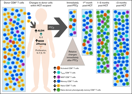
CD8+ T cells increase drug effluxing and aldehyde dehydrogenase expression in allogeneic reactions, enhancing resistance to cyclophosphamide.
Common γ-chain cytokines and the proliferative state of the cell modulate these resistance pathways.
Abstract
Mechanisms of T-cell survival after cytotoxic chemotherapy, including posttransplantation cyclophosphamide (PTCy), are not well understood. Here, we explored the impact of PTCy on human CD8+ T-cell survival and reconstitution, including what cellular pathways drive PTCy resistance. In major histocompatibility complex (MHC)-mismatched mixed lymphocyte culture (MLC), treatment with mafosfamide, an in vitro active cyclophosphamide analog, preserved a relatively normal distribution of naïve and memory CD8+ T cells, whereas the percentages of mucosal-associated invariant T (MAIT) cells and phenotypically stem cell memory (Tscm) T-cell subsets were increased. Activated (CD25+) and proliferating CD8+ T cells were derived from both naïve and memory subsets and were reduced but still present after mafosfamide. By contrast, cyclosporine-A (CsA) or rapamycin treatment preferentially maintained nonproliferating CD25− naïve cells. Drug efflux capacity and aldehyde dehydrogenase-1A1 expression were increased in CD8+ T cells in allogeneic reactions in vitro and in patients, were modulated by common γ-chain cytokines and the proliferative state of the cell, and contributed to CD8+ T-cell survival after mafosfamide. The CD8+ T-cell composition early after hematopoietic cell transplantation (HCT) in PTCy-treated patients was dominated by CD25+ and phenotypically memory, including Tscm and MAIT, cells, consistent with MLC. Yet, MHC-mismatched murine HCT studies revealed that peripherally expanded, phenotypically memory T cells 1 to 3 months after transplant originated largely from naïve-derived rather than memory-derived T cells surviving PTCy, suggesting that initial resistance and subsequent immune reconstitution are distinct. These studies provide insight into the complex immune mechanisms active in CD8+ T-cell survival, differentiation, and reconstitution after cyclophosphamide, with relevance for post-HCT immune recovery, chemotherapy use in autologous settings, and adoptive cellular therapies.
Introduction
The therapeutic efficacy of allogeneic hematopoietic cell transplantation (HCT) is limited by graft-versus-host disease (GVHD). Posttransplantation cyclophosphamide (PTCy) reduces the risk of severe GVHD1 and the associated need for post-HCT immunosuppression2 without compromising relapse or survival outcomes.3-5 Despite the recent burgeoning use of PTCy worldwide, PTCy’s immunologic impact and the immune landscape after PTCy are only beginning to be understood.6-17
CD8+ T cells recover much more rapidly after PTCy than do CD4+ T cells, with the exception of CD4+Foxp3+ regulatory T cells (Tregs) that are highly enriched after PTCy.7,8,12,15 Early after HCT in patients treated with PTCy, naïve CD8+ T cells rapidly acquire CD95-positivity, conferring a T stem cell memory (Tscm)-like phenotype, and this population contributes substantially to T-cell reconstitution after PTCy.9,10 Yet, T-cell receptor (TCR) sequencing studies suggested that the early posttransplant T-cell repertoire appears derived from more rare clones, but after 2 to 3 months, the T-cell repertoire becomes dominated by a resurgence of CD8+, often cytomegalovirus (CMV)-specific, T cells derived from the memory compartment of the donor.12
How CD8+ T cells survive PTCy, why their reconstitution is more robust than conventional CD4+ T cells, and how this may differ from standard GVHD prophylaxis are not understood. Our work in CD4+ T cells suggested increased expression of aldehyde dehydrogenase (ALDH), the major in vivo detoxifying enzyme for cyclophosphamide,18 by Tregs in allogeneic reactions contributes to their resistance to PTCy.7 Yet, what cellular pathways drive CD8+ T-cell resistance to PTCy have not been explored.
Methods
Patients
Peripheral blood mononuclear cells (PBMCs) from patients treated in a prospective study of allogeneic HCT using PTCy (NCT02579967)19 were isolated by density-gradient centrifugation and used fresh for flow-cytometric or functional studies. Patient samples were obtained after informed consent on a protocol approved by the institutional review board of the National Institutes of Health.
Mice
C57BL/6J, B6.SJL-PtprcaPepcb/BoyJ, B6.PL-Thy1a/CyJ, and BALB/cJ mice were obtained from The Jackson Laboratory and housed in specific pathogen-free conditions at the NCI (National Cancer Institute) or Johns Hopkins University and treated under protocols approved by Institutional Animal Care and Use Committees. Mice were given food and water ad libitum.
Mixed lymphocyte cultures
T cells were isolated from the peripheral blood (PB) of healthy donors using density-gradient centrifugation followed by immunomagnetic negative selection (Pan T-Cell Isolation Kit, Miltenyi). CD3− PBMCs were irradiated (30 Gy) and cocultured (mixed lymphocyte culture [MLC]) with T cells from a human leukocyte antigen (HLA)-mismatched donor from days 0 to 7 as previously described.7 For some experiments, T cells were labeled with carboxyfluorescein succinimidyl ester or CellTrace Violet (ThermoFisher) before coculture. Mafosfamide (Baxter Oncology) was reconstituted in phosphate-buffered saline and administered at 7.5 µg/mL for a 1-hour incubation on MLC day 3, followed by 2 washings to remove the residual drug.7 Rapamycin (15 ng/mL; Sigma) or cyclosporine-A ([CsA] 600 ng/mL; Sigma) treatment was from MLC days 0 to 7 unless otherwise noted.7 Unless otherwise noted, culture media consisted of RPMI with l-glutamine, 10% fetal bovine serum (FBS; Gibco), and 1% penicillin-streptomycin (Gibco). DEAB (diethylaminobenzaldehyde) and PK11195 were obtained from Sigma. Cytokines were obtained from the NCI Preclinical Repository Biological Resources Branch Developmental Therapeutics Program (interleukin-2 [IL-2], IL-7, and IL-15) or R&D Systems (IL-4, IL-6, IL-9, IL-17, IL-21, and IL-23). For some experiments, Dynabeads Human T-Activator CD3/CD28 beads (ThermoFisher) were used at a concentration of 1 bead per cell.
Flow cytometry and quantitative polymerase chain reaction
Details are in the supplemental Methods.
Rhodamine-123 (Rh-123) effluxing
Cells were incubated in Rh-123 loading buffer (RPMI with l-glutamine, 1% bovine serum albumin [Sigma], and 10 μg/mL Rh-123 [Sigma]) for 30 minutes on ice. After washing, samples were split in half and incubated with or without CsA 600 ng/mL for 30 minutes at 37°C. Effluxing was quenched with cold buffer, and samples were kept on ice for subsequent flow-cytometric staining and acquisition.
Murine HCT
Eight- to 12-week-old female recipient BALB/cJ mice were irradiated (7.75 Gy split into 2 fractions 8 hours apart) on the day of HCT. Bone marrow (BM) from 8- to 12-week-old female B6.SJL-PtprcaPepcb/BoyJ (CD45.1+) donor mice was T-cell depleted, as previously described.14 From C57BL/6J and B6.PL-Thy1a/CyJ 8- to 12-week-old female mice, T cells were isolated from spleens and lymph nodes by mechanical disruption and underwent CD4+ and CD8+ T-cell positive selection using Dynabeads FlowComp Mouse CD4 and Mouse CD8 kits (ThermoFisher) before flow-cytometric sorting. Recipient mice received sulfamethoxazole/trimethoprim- or levofloxacin-treated water from days 0 to 14. Cyclophosphamide (Baxter Oncology) 33 mg/kg per day was given intraperitoneally on days +3 and +4.
Statistical analysis
Percentage and numerical data underwent arcsin and natural logarithmic transformations, respectively, before statistical analysis with a t test or one-way ANOVA as appropriate. Untransformed data are shown for ease of comprehension by the reader. GraphPad Prism version 8.4.3 was used for statistical analysis and data presentation. For all comparisons, 2-sided P values were used, and P < .05 was considered statistically significant.
Results
The composition of surviving CD8+ T cells is distinct following mafosfamide as compared with CsA or rapamycin treatment
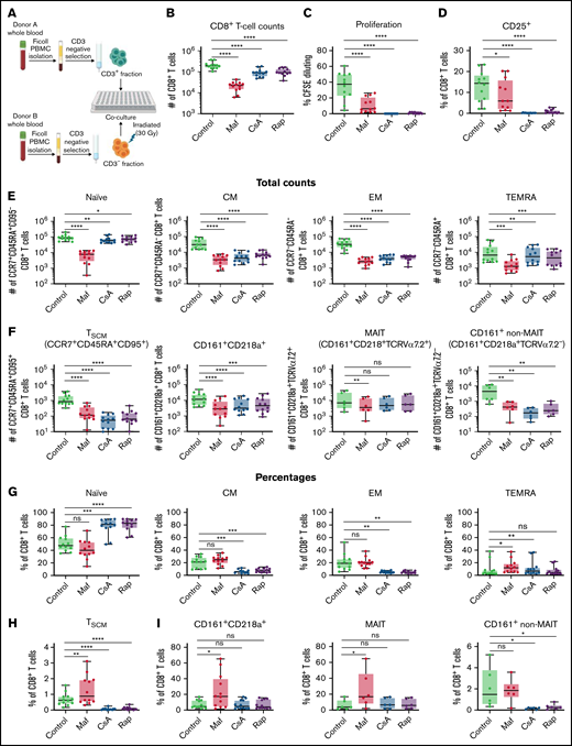
To study the impact of PTCy on CD8+ T-cell subsets, we used MLC as an in vitro model of HCT (Figure 1A). Since cyclophosphamide is a prodrug that requires hepatic activation, we used masfosfamide, a cyclophosphamide analog that becomes active upon exposure to aqueous solution. Total CD8+ T-cell counts at MLC day 7 were significantly reduced by all 3 treatments compared with the vehicle-treated control, with the most significant drop seen after mafosfamide (Figure 1B). Moreover, proliferation was significantly blunted in cultures treated with any of the 3 drugs. However, a subset of surviving mafosfamide-treated cells still had proliferated, in contrast with completely abolished proliferation after CsA or rapamycin treatment (Figure 1C). Correspondingly, CD25+ CD8+ T cells persisted after mafosfamide but not CsA or rapamycin (Figure 1D; supplemental Figure 1 in the data supplement). Activated and proliferating cells were derived both from the naïve and memory compartments in these HLA-mismatched MLCs (supplemental Figure 2).
Mafosfamide treatment on day 3 of human mixed lymphocyte culture kills many CD8+ T cells, but the surviving cellular composition on day 7 is more similar to the compositions within vehicle-treated cultures than within cultures treated with CsA or rapamycin. Human CD3+ T cells and CD3− cells were obtained from the fresh blood of healthy donors via Ficoll density–gradient separation and immunomagnetic selection. CD3+ T cells first were labeled with CFSE 2.5 µM and then placed in mixed lymphocyte cultures (MLCs) with irradiated (30 Gy) allogeneic MHC-mismatched CD3− PBMCs in a 1:1 ratio of 1 × 10⁵ of each cell type per well. Cells were either vehicle (PBS)-treated, treated with mafosfamide (Maf) 7.5 µg/ml as a 1-hour incubation on day 3, or treated with CsA (600 ng/mL) or rapamycin (Rap, 15 ng/mL) from days 0 to 7. Six wells per treatment group were pooled for analyses. Total numbers or percentages of different CD8+ T-cell subsets were determined using the gating schema shown in supplemental Figure 1. Combined results of 2 independent experiments are shown with n = 12 per group for parts (B-H) except for the MAIT and CD161+ non-MAIT total numbers and percentages in parts (F) and (I) (n = 6 per group). *P < .05, **P < .01, ***P < .001, ****P < .0001, and ns, not significantly different on repeated-measure one-way ANOVA followed by the Holm-Sidak post hoc test compared with the control group. CFSE, carboxyfluorescein succinimidyl ester; CM, central memory; EM, effector memory; MAIT, mucosal-associated-invariant-T cells; PBMC, peripheral blood mononuclear cells; TEMRA, terminally differentiated effector memory expressing CD45RA.
Mafosfamide treatment on day 3 of human mixed lymphocyte culture kills many CD8+ T cells, but the surviving cellular composition on day 7 is more similar to the compositions within vehicle-treated cultures than within cultures treated with CsA or rapamycin. Human CD3+ T cells and CD3− cells were obtained from the fresh blood of healthy donors via Ficoll density–gradient separation and immunomagnetic selection. CD3+ T cells first were labeled with CFSE 2.5 µM and then placed in mixed lymphocyte cultures (MLCs) with irradiated (30 Gy) allogeneic MHC-mismatched CD3− PBMCs in a 1:1 ratio of 1 × 10⁵ of each cell type per well. Cells were either vehicle (PBS)-treated, treated with mafosfamide (Maf) 7.5 µg/ml as a 1-hour incubation on day 3, or treated with CsA (600 ng/mL) or rapamycin (Rap, 15 ng/mL) from days 0 to 7. Six wells per treatment group were pooled for analyses. Total numbers or percentages of different CD8+ T-cell subsets were determined using the gating schema shown in supplemental Figure 1. Combined results of 2 independent experiments are shown with n = 12 per group for parts (B-H) except for the MAIT and CD161+ non-MAIT total numbers and percentages in parts (F) and (I) (n = 6 per group). *P < .05, **P < .01, ***P < .001, ****P < .0001, and ns, not significantly different on repeated-measure one-way ANOVA followed by the Holm-Sidak post hoc test compared with the control group. CFSE, carboxyfluorescein succinimidyl ester; CM, central memory; EM, effector memory; MAIT, mucosal-associated-invariant-T cells; PBMC, peripheral blood mononuclear cells; TEMRA, terminally differentiated effector memory expressing CD45RA.
Unlike CsA- and rapamycin-treated cells, which selectively maintained CCR7+CD45RA+CD95− naïve CD8+ T cells at the expense of memory subsets, mafosfamide preserved a similar distribution of naïve/memory CD8+ T-cell subsets as vehicle-treated cells (Figure 1E,G). Phenotypically memory T cells after mafosfamide treatment derived both from differentiating naïve and surviving memory CD8+ T cells, although differentiation was mildly blunted by mafosfamide (supplemental Figure 2). CCR7+CD45RA+CD95+ Tscm percentages were increased after mafosfamide, distinct from CsA or rapamycin treatment (Figure 1F,H). CD161+CD218a+ memory T cells are primarily composed of CD161+CD218a+TCRVα7.2+ mucosal-associated invariant T (MAIT) cells20 and have been shown to preferentially survive cytotoxic chemotherapy20,21 and potentially play a role in GVHD.22 After mafosfamide, the percentages of CD161+CD218a+ CD8+ T cells indeed were increased and the total numbers were only slightly reduced because of increased percentages of MAITs, whereas percentages of CD161+CD218a+TCRVα7.2− cells (hereafter referred to as CD161+ non-MAITs) were not increased (Figure 1F,I).
Clinically, PTCy generally is used in combination with other immunosuppressants, most commonly a calcineurin inhibitor (CNI) (CsA or tacrolimus) with or without mycophenolate mofetil. In different clinical protocols, the CNI may be started before or after PTCy, which may have differing immunologic effects.7,23 Interestingly, CsA started either before or after mafosfamide did not substantially alter the naïve/memory composition of CD8+ T cells at MLC day 7 compared with mafosfamide alone (supplemental Figures 3 and 4), suggesting that the effects of mafosfamide are dominant and not blocked by CsA. However, the timing of CsA did affect the relative percentages of some CD8+ T-cell subsets, particularly Tscm, CD25+, and MAIT cells (supplemental Figure 3).
Drug efflux capacity is increased in CD8+ T cells in vitro and modulated by the proliferative state of the cell and common γ-chain cytokines
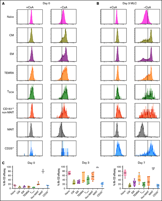
Multidrug resistance (MDR) transporters are cell-surface proteins that nonspecifically efflux cytoplasmic chemicals.24,25 Some MDR transporters are expressed on a subset of CD8+ T cells26 and are highly active in MAIT cells, conferring chemotherapy resistance to MAITs.20,21 Thus, we hypothesized that MDR activity contributes to CD8+ T-cell resistance to mafosfamide. To measure drug efflux capacity, we used an Rh-123 (fluorescent substrate of MDR) assay. Healthy donor CD8+ T cells had substantive functional efflux capacity only within MAITs (Figure 2A). However, at MLC day 3, all CD8+ T-cell subsets had increased efflux capacity, and this increase was most dramatically seen within naïve cells (Figure 2B). At MLC day 7, efflux capacity remained increased compared with day 0 but had declined in most subsets compared with day 3 (Figure 2C). By contrast, healthy donor CD4+ T-cell subsets had low percentages of Rh-123 effluxing cells, which were increased at day 3 or 7 of MLC only within a minority of naïve and memory conventional cells (supplemental Figure 5). CsA treatment before mafosfamide increased effluxing within central memory, CD161+ non-MAIT, and CD25+ CD8+ T-cell subsets at MLC day 7, while CsA treatment after mafosfamide reduced efflux capacity at day 7 across CD8+ T cells (supplemental Figure 6).
CD8+ T-cell subsets increase drug efflux capacity after stimulation in mixed lymphocyte culture. Human T cells were immunomagnetically isolated from fresh, healthy donor PBMCs and either assessed for Rh-123 effluxing immediately on day 0 or stimulated in MLC for 3 or 7 days before assessing for Rh-123 effluxing. Each sample was divided after Rh-123 loading, and half of the sample was treated with 600 ng/mL CsA, a multidrug resistance transporter inhibitor, during the effluxing to serve as a noneffluxing control for each sample. (A-B) Flow cytometry plots of Rh-123 are shown for a representative donor on day 0 (A) and day 3 of MLC (B). The vertical gray line indicates the threshold for effluxing determined based on the CsA control. (C) Percentages of different T-cell subsets that were Rh-123 effluxing are shown on day 0 (n = 3) and days 3 and 7 of MLC (n = 6). These percentages increased significantly for each subset between day 0 and day 3 except for the CD25+ subset (P = .15). These percentages overall somewhat declined between day 3 and day 7, but these differences were not significant except for the CM (P = .0086) and Tscm (P = .012) subsets.
CD8+ T-cell subsets increase drug efflux capacity after stimulation in mixed lymphocyte culture. Human T cells were immunomagnetically isolated from fresh, healthy donor PBMCs and either assessed for Rh-123 effluxing immediately on day 0 or stimulated in MLC for 3 or 7 days before assessing for Rh-123 effluxing. Each sample was divided after Rh-123 loading, and half of the sample was treated with 600 ng/mL CsA, a multidrug resistance transporter inhibitor, during the effluxing to serve as a noneffluxing control for each sample. (A-B) Flow cytometry plots of Rh-123 are shown for a representative donor on day 0 (A) and day 3 of MLC (B). The vertical gray line indicates the threshold for effluxing determined based on the CsA control. (C) Percentages of different T-cell subsets that were Rh-123 effluxing are shown on day 0 (n = 3) and days 3 and 7 of MLC (n = 6). These percentages increased significantly for each subset between day 0 and day 3 except for the CD25+ subset (P = .15). These percentages overall somewhat declined between day 3 and day 7, but these differences were not significant except for the CM (P = .0086) and Tscm (P = .012) subsets.
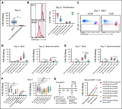
In exploring potential drivers of efflux capacity in MLC, we hypothesized that TCR signaling might modulate effluxing. Therefore, we cultured T cells either in MLC or with anti-CD3/CD28 stimulator beads. On day 3, bead-stimulated cells were nearly all noneffluxing in contrast with MLC-stimulated CD8+ T cells that had a substantial percentage of effluxing cells (Figure 3A). However, this stark difference appeared explainable not by the mitogen but instead by the proliferative state of the cell (Figure 3B-E). Indeed, whether bead-treated or MLC-stimulated, cells that had undergone multiple rounds of division (2+) at day 7 had lost the ability to efflux Rh-123, whereas a substantial percentage of cells that had not divided or only undergone 1 round of division had efflux capacity (Figure 3C-E). These results suggest that the proliferative state, rather than TCR signaling, is most closely associated with efflux capacity.
CD8+ T cells initially increase efflux capacity in antigen-stimulated conditions or when cultured alone in vitro and then lose efflux capacity as they proliferate. (A-F) Human T cells were stimulated in MLC as in Figure 1, stimulated with anti-CD3/CD28 beads, or cultured without allogeneic stimulators or beads. At 3 or 7 days of in vitro culture, Rh-123 effluxing was assessed as per Figure 2. The “+CsA” designation refers to culturing with CsA only during the effluxing to serve as a noneffluxing control, but these cells were not cultured with CsA during the 3 or 7 days of in vitro culture. In parts (A-E), T cells were labeled with CellTrace Violet before in vitro culture. Shown are combined results from 3 independent experiments for parts (A-B,D) and 2 independent experiments for parts (C,E). (A) The efflux capacity of CD8+ T cells in MLC was higher than with anti-CD3/CD28 bead stimulation. **P = .0075 on paired t test. (B) However, proliferation on day 3 was much less extensive after MLC, with negligible numbers of CD8+ T cells in MLC proliferating >1 generation. Example plots of proliferation as measured by CellTrace dilution are shown for T cells from a healthy donor treated either in MLC or with anti-CD3/CD28 beads. (C-E) On both day 3 (D) and day 7 (C,E) of in vitro culture, efflux capacity was increased in nonproliferating cells or those that had only divided a single generation regardless of MLC or anti-CD3/CD28 stimulation but decreased in CD8+ T cells that had proliferated more extensively. This divergence may explain the overall decreased effluxing seen in (A) after bead stimulation because of much more extensive proliferation with that treatment. On day 3 of MLC, effluxing was considered nonevaluable (NE) in the cells proliferating more than a single generation because of extremely low numbers of cells in that group. Representative flow cytometric plots are shown in (C). *P < .05, **P < .01, and ****P < .0001 on nonrepeated-measure one-way ANOVA followed by the Holm-Sidak post hoc test. (F) CD8+ T cells cultured with media alone increased effluxing capacity similarly compared with CD8+ T cells stimulated in MLC, and this was not augmented with treatment with common γ-chain cytokines. The combined results of 2 independent experiments are shown. *P < .05, **P < .01, and ns, not significantly different on repeated-measure one-way ANOVA followed by the Holm-Sidak post hoc test. (G) Human CD8+ T cells retrieved from the blood of patients (n = 5) on day 3 after allogeneic hematopoietic cell transplantation also had increased efflux capacity (P = .035 on paired t test) compared with effluxing seen in recipient or donor cells on day 0. These patients were not treated with any immunosuppression after transplant (PTCy or other) before sample collection on day 3.
CD8+ T cells initially increase efflux capacity in antigen-stimulated conditions or when cultured alone in vitro and then lose efflux capacity as they proliferate. (A-F) Human T cells were stimulated in MLC as in Figure 1, stimulated with anti-CD3/CD28 beads, or cultured without allogeneic stimulators or beads. At 3 or 7 days of in vitro culture, Rh-123 effluxing was assessed as per Figure 2. The “+CsA” designation refers to culturing with CsA only during the effluxing to serve as a noneffluxing control, but these cells were not cultured with CsA during the 3 or 7 days of in vitro culture. In parts (A-E), T cells were labeled with CellTrace Violet before in vitro culture. Shown are combined results from 3 independent experiments for parts (A-B,D) and 2 independent experiments for parts (C,E). (A) The efflux capacity of CD8+ T cells in MLC was higher than with anti-CD3/CD28 bead stimulation. **P = .0075 on paired t test. (B) However, proliferation on day 3 was much less extensive after MLC, with negligible numbers of CD8+ T cells in MLC proliferating >1 generation. Example plots of proliferation as measured by CellTrace dilution are shown for T cells from a healthy donor treated either in MLC or with anti-CD3/CD28 beads. (C-E) On both day 3 (D) and day 7 (C,E) of in vitro culture, efflux capacity was increased in nonproliferating cells or those that had only divided a single generation regardless of MLC or anti-CD3/CD28 stimulation but decreased in CD8+ T cells that had proliferated more extensively. This divergence may explain the overall decreased effluxing seen in (A) after bead stimulation because of much more extensive proliferation with that treatment. On day 3 of MLC, effluxing was considered nonevaluable (NE) in the cells proliferating more than a single generation because of extremely low numbers of cells in that group. Representative flow cytometric plots are shown in (C). *P < .05, **P < .01, and ****P < .0001 on nonrepeated-measure one-way ANOVA followed by the Holm-Sidak post hoc test. (F) CD8+ T cells cultured with media alone increased effluxing capacity similarly compared with CD8+ T cells stimulated in MLC, and this was not augmented with treatment with common γ-chain cytokines. The combined results of 2 independent experiments are shown. *P < .05, **P < .01, and ns, not significantly different on repeated-measure one-way ANOVA followed by the Holm-Sidak post hoc test. (G) Human CD8+ T cells retrieved from the blood of patients (n = 5) on day 3 after allogeneic hematopoietic cell transplantation also had increased efflux capacity (P = .035 on paired t test) compared with effluxing seen in recipient or donor cells on day 0. These patients were not treated with any immunosuppression after transplant (PTCy or other) before sample collection on day 3.
Given this unexpected finding, we hypothesized that cells undergoing lymphopenia-induced proliferation increased efflux capacity, and thus cytokines may modulate this pathway. Therefore, we cultured T cells either in media alone (not in MLC), with exogenous cytokines associated with lymphopenia, with stimulator beads, or with both cytokines and beads. Surprisingly, on day 3, T cells cultured alone had increased efflux capacity comparable with T cells cultured in MLC (Figure 3F). This efflux capacity was lost upon culture with serum-free media but maintained when cultured in the presence of human serum (supplemental Figure 7), suggesting this was not an artifact of culturing with FBS. IL-7 and IL-15 treatment significantly reduced the efflux capacity of CD8+ T cells, with IL-7 being the most potent modulator, while IL-2 had no appreciable effect (Figure 3F). Bead stimulation overshadowed any effect of exogenous cytokines, presumably because of cells losing efflux capacity after undergoing multiple rounds of cell division. Exogenous cytokine treatment without MLC did greatly alter the phenotype of the CD8+ T-cell compartment, with IL-7 and IL-15 treatment causing most phenotypically naïve cells to acquire CD95 expression (Tscm phenotype) despite lack of antigen exposure in these conditions and being presumably largely antigen-inexperienced (supplemental Figure 8). Among other common γ-chain cytokines, exogenous IL-4 treatment modestly reduced the efflux capacity of CD8+ T cells, while IL-9 and IL-21 had no effect (Figure 3F). Given that the highly effluxing MAITs are skewed toward Tc17 differentiation and that IL-6 has been associated with a multidrug-resistant phenotype in cancer cell lines,27 we tested the effects of the Tc17 pathway cytokines IL-6, IL-17, and IL-23, but found that these had no effect on efflux capacity (supplemental Figure 9). Overall, these findings suggest that drug efflux capacity is increased in CD8+ T cells when cultured in vitro and can be modulated by both the proliferative state of the cell and the cytokine milieu.
Drug efflux capacity is increased in CD8+ T cells in patients early after transplant
To assess the clinical applicability of these in vitro findings, we examined Rh-123 effluxing by CD8+ T cells from fresh patient samples obtained early after HCT. There was significantly increased efflux capacity on day 3 in recipients compared with that observed in donors or recipients on day 0 (Figure 3G), suggesting that this dynamic change also happens in CD8+ T cells after clinical HCT.
Aldehyde dehydrogenase 1A1 (ALDH1A1) expression is increased in MLC and by low-dose IL-2
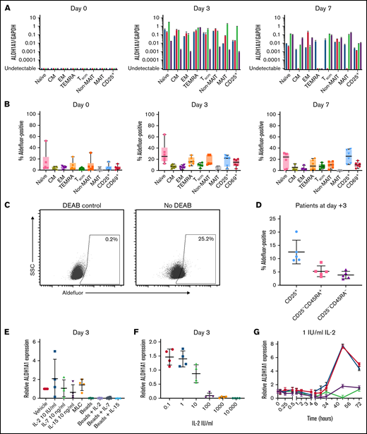
ALDH1A1 is the major enzyme involved in cyclophosphamide detoxification18 and contributes to human CD4+ Treg resistance to mafosfamide.7 Therefore, we hypothesized that ALDH1A1 expression is increased in MLC in CD8+ T cells, contributing to their resistance to mafosfamide. Before MLC, there were undetectable concentrations of ALDH1A1 in all CD8+ T-cell subsets; however, on day 3, all subsets had increased ALDH1A1 expression (Figure 4A). ALDH1A1 expression also was found on day 7 of MLC, albeit to a lesser extent than on day 3. Increased ALDH functional protein activity also was seen in MLC (Figure 4B-C), confirming the changes in ALDH1A1 expression. Furthermore, ALDH functional activity was seen on day +3 in patients after transplant (Figure 4D).
CD8+ T-cell subsets increase ALDH1A1 expression in mixed lymphocyte culture and after stimulation with low-dose IL-2. (A) T cells were isolated from healthy donor PBMCs and either taken directly for flow cytometric sorting as per supplemental Figure 1 or put in MLC as in Figure 1 and flow cytometrically sorted on day 3 or 7. Quantitative polymerase chain reaction (PCR) for ALDH1A1 or GAPDH was performed on RNA extracted from flow cytometrically-sorted CD8+ T-cell subsets, showing increased ALDH1A1 expression on day 3 that persisted but declined on day 7 of MLC. Each color shows a different healthy donor tested (n = 4). (B) ALDH functional activity was measured by Aldefluor positivity based on a DEAB negative control for each sample. There was overall increased Aldefluor positivity in CD8+ T-cell subsets on day 3 of MLC compared with freshly isolated healthy donor T cells (day 0); these differences were statistically significant on repeated-measure ANOVA for all but the CM, EM, and MAIT subsets. (C) A representative example of the positive shift in Aldefluor staining is shown for all CD8+ T cells on MLC day 3. The threshold of Aldefluor positivity is based on the sample-specific DEAB negative control. (D) Samples on day +3 after transplant from patients treated with myeloablative HLA-matched BM transplantation previously assessed for Aldefluor positivity within CD4+ T-cell subsets7 were assessed for Aldefluor positivity within CD4− T-cell subsets, also showing Aldefluor positivity within a subset of cells. (E) Human T cells were isolated and plated either in media alone; with 10 IU/mL IL-2, 10 ng/mL IL-7, or 10 ng/mL IL-15; in MLC; or with anti-CD3/CD28 stimulation beads ± IL-2, IL-7, or IL-15. T cells that were in MLC were immunomagnetically reisolated on day 3 to remove CD3− stimulator cells, after which ALDH1A1 and GAPDH expression were determined via quantitative PCR for all groups. The dose of IL-2 chosen (10 IU/mL) for the initial experiments resulted in a variable effect on ALDH1A1 expression, whereas bead stimulation decreased ALDH1A1 expression compared with cells cultured in media only. Relative expression is shown of ALDH1A1/GAPDH of the treatment divided by ALDH1A1/GAPDH of the vehicle-treated control group. (F) Therefore, a range of IL-2 doses was tested, showing increased ALDH1A1 expression at low doses and decreased ALDH1A1 expression at high doses. (G) A timecourse assessment showed that the peak increase in ALDH1A1 expression with low-dose IL-2 was at 48 hours in 3 of 4 healthy donors tested. Relative expression is shown of ALDH1A1/GAPDH of each time point divided by ALDH1A1/GAPDH of the pretreatment (time 0) sample. Combined results are shown from 2 independent experiments (n = 4) for (A,E-G) and from 3 independent experiments (n = 6) for (B). (D) n = 5.
CD8+ T-cell subsets increase ALDH1A1 expression in mixed lymphocyte culture and after stimulation with low-dose IL-2. (A) T cells were isolated from healthy donor PBMCs and either taken directly for flow cytometric sorting as per supplemental Figure 1 or put in MLC as in Figure 1 and flow cytometrically sorted on day 3 or 7. Quantitative polymerase chain reaction (PCR) for ALDH1A1 or GAPDH was performed on RNA extracted from flow cytometrically-sorted CD8+ T-cell subsets, showing increased ALDH1A1 expression on day 3 that persisted but declined on day 7 of MLC. Each color shows a different healthy donor tested (n = 4). (B) ALDH functional activity was measured by Aldefluor positivity based on a DEAB negative control for each sample. There was overall increased Aldefluor positivity in CD8+ T-cell subsets on day 3 of MLC compared with freshly isolated healthy donor T cells (day 0); these differences were statistically significant on repeated-measure ANOVA for all but the CM, EM, and MAIT subsets. (C) A representative example of the positive shift in Aldefluor staining is shown for all CD8+ T cells on MLC day 3. The threshold of Aldefluor positivity is based on the sample-specific DEAB negative control. (D) Samples on day +3 after transplant from patients treated with myeloablative HLA-matched BM transplantation previously assessed for Aldefluor positivity within CD4+ T-cell subsets7 were assessed for Aldefluor positivity within CD4− T-cell subsets, also showing Aldefluor positivity within a subset of cells. (E) Human T cells were isolated and plated either in media alone; with 10 IU/mL IL-2, 10 ng/mL IL-7, or 10 ng/mL IL-15; in MLC; or with anti-CD3/CD28 stimulation beads ± IL-2, IL-7, or IL-15. T cells that were in MLC were immunomagnetically reisolated on day 3 to remove CD3− stimulator cells, after which ALDH1A1 and GAPDH expression were determined via quantitative PCR for all groups. The dose of IL-2 chosen (10 IU/mL) for the initial experiments resulted in a variable effect on ALDH1A1 expression, whereas bead stimulation decreased ALDH1A1 expression compared with cells cultured in media only. Relative expression is shown of ALDH1A1/GAPDH of the treatment divided by ALDH1A1/GAPDH of the vehicle-treated control group. (F) Therefore, a range of IL-2 doses was tested, showing increased ALDH1A1 expression at low doses and decreased ALDH1A1 expression at high doses. (G) A timecourse assessment showed that the peak increase in ALDH1A1 expression with low-dose IL-2 was at 48 hours in 3 of 4 healthy donors tested. Relative expression is shown of ALDH1A1/GAPDH of each time point divided by ALDH1A1/GAPDH of the pretreatment (time 0) sample. Combined results are shown from 2 independent experiments (n = 4) for (A,E-G) and from 3 independent experiments (n = 6) for (B). (D) n = 5.
Exogenous cytokines alone modulated ALDH1A1 expression in vitro, whereas bead stimulation led to low ALDH1A1 expression regardless of cytokine administration (Figure 4E). Given that IL-2 appeared to have the greatest positive effect, we explored a range of IL-2 doses and found that low-dose IL-2 (0.1-1 IU/mL) increased ALDH expression, whereas high-dose IL-2 (100-10 000 IU/mL) actually reduced expression well below levels observed in T cells cultured in media alone (Figure 4F). The increased ALDH1A1 expression by low-dose IL-2 peaked at ∼48 hours of culture (Figure 4G).
Inhibition of MDR and ALDH activity sensitizes CD8+ T cells to mafosfamide, and this sensitivity can be modulated by low-dose IL-2
Given that both MDR activity and ALDH expression were increased in vitro, we examined their contributions to mafosfamide resistance by assessing cell viability after pharmacologically blocking ALDH activity with DEAB28 and/or blocking MDR activity with either CsA or PK11195 (broad ABC transporter inhibitor29,30 ) before mafosfamide treatment. Treatment with DEAB or PK11195 alone before mafosfamide did reduce CD8+ T-cell viability (Figure 5A). However, inhibition of MRD and ALDH together with PK11195 and DEAB before mafosfamide led to most CD8+ T cells being nonviable, suggesting an additive or perhaps even synergistic effect. To account for potential additive nonspecific drug toxicity, we halved the doses of PK11195 and DEAB and found that they continued to greatly sensitize CD8+ T cells to mafosfamide (Figure 5B), suggesting that the combination of MDR and ALDH appeared necessary for CD8+ T cells to survive mafosfamide in MLC.
ALDH1A1 expression and efflux capacity both appear to contribute to protection from mafosfamide-induced cytotoxicity. Human MLC was performed as per Figure 1, except that some groups received CsA (multidrug resistance [MDR] transporter inhibitor), PK11195 (broader MDR transporter inhibitor), or DEAB (ALDH inhibitor) from days 0 to 3. All drugs were washed off after the 1-hour treatment with vehicle or mafosfamide on day 3; CsA, PK11195, and DEAB were not added back to the cultures. (A-B) Blocking ALDH and/or MDR transporter activity sensitized CD8+ T cells to mafosfamide with the combination of PK11195 and DEAB having a maximal effect, suggesting that both pathways may contribute to CD8+ T-cell resistance to mafosfamide. (C-D) T cells were isolated from healthy donor PBMCs and plated either alone (C) or in MLC (D) as in Figure 1; some groups received exogenous IL-2 (1 IU/mL) or IL-7 (10 ng/mL) on day 0, and the IL-2 or IL-7 was not washed out until after mafosfamide (or vehicle) treatment on day 3. The addition of IL-2 or IL-7 before mafosfamide increased the percentage of viable CD8+ T cells on day 7. For IL-7 but not IL-2, this was in part because of significantly increased proliferation and apparent expansion of surviving T cells after IL-7 treatment. Combined results from 3 (A) or 2 (B-D) independent experiments are shown. *P < .05, **P < .01, ***P < .001, ****P < .0001, and ns, not significantly different on repeated-measure one-way ANOVA followed by the Holm-Sidak post hoc test. Comparisons for (B) were performed between all groups. For (C-D), mafosfamide-treated groups were only compared with each other, as were non-mafosfamide-treated groups, to assess the relative impact of IL-2 or IL-7 administration, the scientific question of interest.
ALDH1A1 expression and efflux capacity both appear to contribute to protection from mafosfamide-induced cytotoxicity. Human MLC was performed as per Figure 1, except that some groups received CsA (multidrug resistance [MDR] transporter inhibitor), PK11195 (broader MDR transporter inhibitor), or DEAB (ALDH inhibitor) from days 0 to 3. All drugs were washed off after the 1-hour treatment with vehicle or mafosfamide on day 3; CsA, PK11195, and DEAB were not added back to the cultures. (A-B) Blocking ALDH and/or MDR transporter activity sensitized CD8+ T cells to mafosfamide with the combination of PK11195 and DEAB having a maximal effect, suggesting that both pathways may contribute to CD8+ T-cell resistance to mafosfamide. (C-D) T cells were isolated from healthy donor PBMCs and plated either alone (C) or in MLC (D) as in Figure 1; some groups received exogenous IL-2 (1 IU/mL) or IL-7 (10 ng/mL) on day 0, and the IL-2 or IL-7 was not washed out until after mafosfamide (or vehicle) treatment on day 3. The addition of IL-2 or IL-7 before mafosfamide increased the percentage of viable CD8+ T cells on day 7. For IL-7 but not IL-2, this was in part because of significantly increased proliferation and apparent expansion of surviving T cells after IL-7 treatment. Combined results from 3 (A) or 2 (B-D) independent experiments are shown. *P < .05, **P < .01, ***P < .001, ****P < .0001, and ns, not significantly different on repeated-measure one-way ANOVA followed by the Holm-Sidak post hoc test. Comparisons for (B) were performed between all groups. For (C-D), mafosfamide-treated groups were only compared with each other, as were non-mafosfamide-treated groups, to assess the relative impact of IL-2 or IL-7 administration, the scientific question of interest.
Since low-dose IL-2 increased ALDH expression and IL-7 reduced efflux capacity, we hypothesized that cytokines could modulate CD8+ T-cell survival following mafosfamide treatment. Compared with mafosfamide alone, treatment with low-dose IL-2 before mafosfamide modestly but significantly decreased the percentages of CD8+ T cells that were nonviable on day 7 both when cultured without stimulator cells (Figure 5C) or in MLC (Figure 5D). Surprisingly, IL-7 pretreatment led to improved viability of CD8+ T cells after mafosfamide both in MLC and when T cells were cultured alone. Unlike with low-dose IL-2 treatment, IL-7–treated cells were more highly proliferative on day 7 (Figure 5D), suggesting that perhaps the better proliferation of surviving cells, rather than simply enhanced resistance, may have contributed to this higher viability.
Reconstitution of CD8+ T-cell subsets in patients treated with PTCy
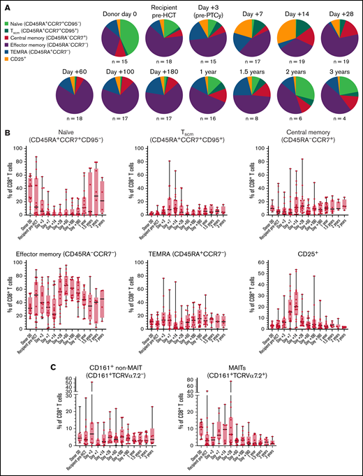
Since there may be a distinction between initial survival after mafosfamide/cyclophosphamide treatment and the proliferation and differentiation occurring during immune recovery, we studied immune reconstitution in patients treated in a prospective clinical study of reduced intensity BM transplantation using PTCy.19 The percentages of phenotypically naïve CD8+ T cells rapidly decreased; conversely, CD25+, Tscm, and MAIT cells were relatively increased early after PTCy, suggesting initial preferential survival and/or early reconstitution of these subsets. However, the increased percentages declined between days +14 and +28 and remained at lower percentages thereafter (Figure 6; supplemental Figure 10).
Activated and phenotypically memory, including T stem cell memory and MAIT, CD8+ T-cell subsets dominate early immune reconstitution in patients treated with PTCy. All 20 patients treated in a prospective study19 of reduced intensity conditioning HCT, as well as all 15 donors who consented for research, had immunophenotyping performed on fresh blood at serial time points to assess the relative composition of recovering CD8+ T-cell subsets. The 1 patient with primary graft failure was excluded from analysis. (A-B) CD8+ T cells were first gated as CD25+ or CD25−, and then the CD25− cells were subgated into the other subsets. (A) Median values at each time point are shown to provide the overall relative CD8+ T-cell composition. (B) For each CD8+ T-cell subset are shown the percentages data over the serial timepoints from all patients. (C) Markers to identify MAIT cells were on a separate panel that was run if sufficient cells were available. Therefore, recovery of CD161+TCR-Vα7.2+ MAIT and CD161+TCR-Vα7.2− non-MAIT cells are included separate from the other 6 subsets shown in (A).
Activated and phenotypically memory, including T stem cell memory and MAIT, CD8+ T-cell subsets dominate early immune reconstitution in patients treated with PTCy. All 20 patients treated in a prospective study19 of reduced intensity conditioning HCT, as well as all 15 donors who consented for research, had immunophenotyping performed on fresh blood at serial time points to assess the relative composition of recovering CD8+ T-cell subsets. The 1 patient with primary graft failure was excluded from analysis. (A-B) CD8+ T cells were first gated as CD25+ or CD25−, and then the CD25− cells were subgated into the other subsets. (A) Median values at each time point are shown to provide the overall relative CD8+ T-cell composition. (B) For each CD8+ T-cell subset are shown the percentages data over the serial timepoints from all patients. (C) Markers to identify MAIT cells were on a separate panel that was run if sufficient cells were available. Therefore, recovery of CD161+TCR-Vα7.2+ MAIT and CD161+TCR-Vα7.2− non-MAIT cells are included separate from the other 6 subsets shown in (A).
Naïve-derived but phenotypically memory T cells preferentially reconstitute after transplant in mice treated with or without PTCy
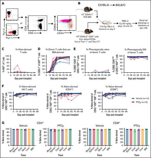
The relative increase in phenotypic memory T cells at the expense of phenotypic naïve T cells seemed to conflict with data suggesting that clinical T-cell reconstitution after PTCy preferentially derives from phenotypic Tscms originating from differentiating naïve T cells.9,10 Yet, other data had shown that after 2 to 3 months, PTCy-treated patients had a resurgence and dominance of CD8+ T cells derived from memory cells within the donor.12 To better understand the potential discrepancy between these data, we used an MHC-disparate murine HCT model in which we could track both the phenotype and the origin of naïve vs memory T cells (Figure 7). The T cells that were peripherally expanded (rather than BM-derived) were nearly exclusively phenotypically effector memory, similar to what we observed in patients. Yet, despite this memory phenotype, the vast majority of these T cells were derived from the donor-naïve compartment (Figure 7; supplemental Figure 11). These findings were similar within CD4+ and CD8+ T cells and in mice treated with or without PTCy. These data support that differentiated naïve T cells were indeed responsible for the bulk of early reconstitution after transplant, highlighting potential differences between initial survival and subsequent differentiation and reconstitution after PTCy.
The dominance of peripherally expanded phenotypically memory T cells during immune reconstitution after PTCy primarily derives from the differentiation of transplanted naïve donor T cells. (A) T cells from the spleens and cutaneous (cervical, brachial, axillary, and inguinal) lymph nodes (CLNs) from Thy1.1+ or Thy1.2+ 8- to 12-week-old female C57BL/6 mice (CD45.1−CD45.2+) were flow cytometrically sorted to isolate naïve (CD62L+CD44−) or central/effector memory (CD62L+CD44+ and CD62L−CD44+) T cells. (B) Either Thy1.1+ naïve and Thy1.2+ memory T cells or Thy1.1+ memory and Thy1.2+ naïve T cells were mixed together at the ratio at which they were sorted (approximately 5:1 naïve to memory). After 7.75 Gy irradiation in 2 divided fractions 8 hours apart, each recipient mouse received 106 total T cells and 107 CD45.1+ T-cell–depleted BM cells. Mice were treated with PBS or PTCy 33 mg/kg per day on days +3 and +4, and then (C-F) underwent serial tail bleeds or (G) were euthanized at day +28 for assessment of various organs. Nearly all T cells were donor-derived at all assessments (C), but substantial thymic-dependent T-cell recovery occurred within the first month after transplant (D), leading to a dominance of BM-derived T cells by day +28 to +42. (E) Nearly all of the recovered T cells that were originally transplanted and thus peripherally expanded (not BM-derived) displayed an effector memory (EM) phenotype. (F) Despite this memory phenotype, the vast majority of these cells were derived from naïve T cells that had now assumed an effector memory phenotype. This was true in CD4+ and CD8+ T cells and in those mice treated with PBS or PTCy. (G) This dominance of naïve-derived T cells was also true in various tissue compartments. MLN, mesenteric lymph nodes.
The dominance of peripherally expanded phenotypically memory T cells during immune reconstitution after PTCy primarily derives from the differentiation of transplanted naïve donor T cells. (A) T cells from the spleens and cutaneous (cervical, brachial, axillary, and inguinal) lymph nodes (CLNs) from Thy1.1+ or Thy1.2+ 8- to 12-week-old female C57BL/6 mice (CD45.1−CD45.2+) were flow cytometrically sorted to isolate naïve (CD62L+CD44−) or central/effector memory (CD62L+CD44+ and CD62L−CD44+) T cells. (B) Either Thy1.1+ naïve and Thy1.2+ memory T cells or Thy1.1+ memory and Thy1.2+ naïve T cells were mixed together at the ratio at which they were sorted (approximately 5:1 naïve to memory). After 7.75 Gy irradiation in 2 divided fractions 8 hours apart, each recipient mouse received 106 total T cells and 107 CD45.1+ T-cell–depleted BM cells. Mice were treated with PBS or PTCy 33 mg/kg per day on days +3 and +4, and then (C-F) underwent serial tail bleeds or (G) were euthanized at day +28 for assessment of various organs. Nearly all T cells were donor-derived at all assessments (C), but substantial thymic-dependent T-cell recovery occurred within the first month after transplant (D), leading to a dominance of BM-derived T cells by day +28 to +42. (E) Nearly all of the recovered T cells that were originally transplanted and thus peripherally expanded (not BM-derived) displayed an effector memory (EM) phenotype. (F) Despite this memory phenotype, the vast majority of these cells were derived from naïve T cells that had now assumed an effector memory phenotype. This was true in CD4+ and CD8+ T cells and in those mice treated with PBS or PTCy. (G) This dominance of naïve-derived T cells was also true in various tissue compartments. MLN, mesenteric lymph nodes.
Discussion
Herein, we explored the immunologic impact of PTCy on CD8+ T-cell survival and reconstitution, including what cellular pathways drive CD8+ T-cell resistance to PTCy. In human MLC, mafosfamide preserved a relatively normal distribution of phenotypically naïve and memory CD8+ T cells, but the percentages of MAIT and phenotypically Tscm subsets were increased. Some activated and proliferating CD8+ T cells persisted, albeit at lower levels, after mafosfamide treatment compared with vehicle. By contrast, CsA or rapamycin treatment preferentially maintained nonproliferating CD25− naïve cells. CD8+ T cells increased efflux capacity and ALDH activity in MLC, both of which appeared to contribute to CD8+ T-cell survival after mafosfamide. Efflux capacity and ALDH1A1 expression were modulated by cytokines and the proliferative state of the cell, suggesting a complex interaction between various survival pathways involved in PTCy resistance. Early immune reconstitution after transplant showed that phenotypically Tscm, MAIT, and CD25+ CD8+ T cells were increased in percentages also in PTCy-treated HCT patients. Yet, initial survival and PB reconstitution were distinct as some subsets that were relatively expanded early decreased in percentages with time. This change, in part, may be because of dynamics of CD8+ T-cell reconstitution and trafficking but also was related to in vivo differentiation and phenotypic changes of reconstituting T cells. Indeed, although most peripherally expanded donor T cells were phenotypically memory over the first 3 months after murine HCT, the vast majority of these T cells actually were derived from transplanted naïve T cells that had differentiated in the host.
T-cell survival after chemotherapy has long been of scientific and medical interest. Thirty years ago, functional ABCB1 activity was described within T cells, particularly CD8+ T cells,26,31 and this has been shown to be responsible for the transition from early-activated naïve to cytotoxic memory CD8+ T cells, in part through suppression of oxidative stress.32 Yet, ABCB1 activity also was lost in activated cells, allowing for the depletion of activated T cells unable to efflux photosensitizing agents, an effect that could be augmented via ABCB1 inhibition.33 The role played by MDR transporters, including ABCB1, in mediating resistance to cyclophosphamide is not well studied, although they have been implicated.34 Our data showing a broad increase in efflux capacity in naïve CD8+ T cells beginning to proliferate under cytokine or allogeneic stimulatory conditions followed by loss of efflux capacity in CD8+ T cells that had proliferated ≥2 generations may explain some of these prior findings. Even so, our data support that subsets of memory T cells maintain MDR activity, consistent with prior work.35 Our data also are consistent with prior data showing a lack of MDR activity in CD4+ Tregs.36
Although phenotypically naïve T cells are reduced after cyclophosphamide in our studies, consistent with prior findings in CD4+ T cells,7,12,37 naïve CD8+ T cells appear more resistant than naïve CD4+ T cells. This effect may be explained by higher efflux capacity and ALDH expression within naïve CD8+ T cells and may contribute to better CD8+ T-cell reconstitution after HCT. The contextual expression of ALDH within T cells has not been appreciated except in our prior work in CD4+ Tregs.7 Our findings that ALDH1A1 expression may be modulated by IL-2 is novel but may not be unique to IL-2 as a prior study had shown that tumor necrosis factor and IL-1 may modulate ALDH1 expression within other BM cells.38 Understanding these processes better is quite relevant clinically, particularly given the high risk for cytokine release syndrome early after HLA-haploidentical HCT at the time when PTCy is administered.39
The therapeutic efficacy of PTCy was previously attributed to the selective killing of alloreactive T cells that become activated and preferentially proliferate early after HCT.6 Although we have shown here and in prior studies that PTCy does broadly kill a substantial number of T cells,6,7,14,16,17 this effect does not appear selective for alloreactive T cells.6,14,16,17 Even so, the allure of the idea of cyclophosphamide selectively killing proliferating alloreactive T cells endures. We found that proliferating CD8+ T cells were reduced after PTCy, but a sizable population remained, distinct from that seen with CsA or rapamycin treatment. Furthermore, CD25+activated CD8+ T cells persisted at much higher levels after PTCy than after CsA or rapamycin and also were found early after HCT at high percentages in PTCy-treated patients. In HLA-mismatched MLC, these activated/proliferating T cells were derived both from naïve and memory precursors.
The chemotherapeutic action of cyclophosphamide is thought to rely on its alkylating properties, and thus cyclophosphamide is thought not to be a cell-cycle–dependent chemotherapeutic. Yet, inhibition of DNA damage repair does not modulate cyclophosphamide’s killing in vivo.40 Furthermore, at higher doses, mafosfamide induces transcriptional inhibition and cell-cycle–independent caspase-dependent apoptosis, confirming that the toxic effects of cyclophosphamide are not specific to proliferating cells.41 At the cellular level, both cyclophosphamide resistance and ALDH functional activity may be modulated by reactive oxygen species, cyclophosphamide metabolites, and thiols,42-44 suggesting that the complete picture is likely even more complex than we have previously hypothesized.45
Our data showing distinctions between initial survival and the subsequent differentiation and expansion occurring during immune reconstitution may in part explain the apparent discrepancies between previous datasets. We hypothesize that T-cell reconstitution within the first 2 to 3 months after PTCy stems largely from the expansion of infused naïve T cells that proliferate either because of lymphopenic conditions46 or antigenic stimulation (alloantigen- or pathogen-specific). Yet, this early ascendancy of expanded naïve-derived T cells is not maintained longterm in many patients who, after 2 to 3 months, have a strong resurgence of memory responses to immunodominant pathogens such as CMV.12 The lack of such a conversion back to memory-derived T cells in our mouse studies may stem from the specific pathogen-free housing conditions or more rapid and robust thymic recovery in mice. The enrichment of Tscms after PTCy9,10 may be an artifact of naïve T cells becoming CD95+ under homeostatic cytokine stimulation, consistent with prior data47 and our data here showing that IL-7 or IL-15 treatment alone without antigenic stimulation makes most naïve T cells adopt a Tscm phenotype (supplemental Figure 8), but phenotypic Tscms on MLC day 7 were uniformly CXCR3+ (supplemental Figure 12), and we cannot rule out that Tscms preferentially survive mafosfamide/cyclophosphamide.
There are several limitations of our study. Firstly, we do not know how our drug doses correspond to doses in patients; we focused on an intermediate/high dose of mafosfamide7 as shown to be optimal for PTCy14 and on high therapeutic doses of CsA and rapamycin, which had yielded similar results as low therapeutic doses in our prior studies.7 We also do not know whether the kinetics of T-cell activation and proliferation and the relative cytokine milieu differ between in vitro vs clinical allogeneic responses, with the potential for modulating drug effluxing or ALDH expression differently in patients than observed in MLC. Even so, we did show increases in both drug effluxing and ALDH functional activity in patient T cells on day +3, suggesting that these cellular processes are also active clinically and may play roles in T-cell resistance to PTCy. Furthermore, we do not know to what extent our in vitro findings apply clinically wherein both autologous and allogeneic antigen-presenting cells exist early after transplant or to patients conditioned with chemotherapy rather than radiation; nevertheless, the patients studied received only chemotherapy-based conditioning, so the similar findings between MLC and patients suggest potential broader applicability of our results. Lastly, our murine studies are limited by a lack of immunosuppression beyond PTCy as clinically PTCy is more frequently used in combination with other immunosuppression.1,48
Ultimately, this work provides mechanistic insight into how CD8+ T cells survive and reconstitute after cyclophosphamide. By better understanding how to modulate these survival pathways, it may be possible to enhance or reduce T-cell survival broadly or to preferentially increase or decrease particular T-cell subsets in specific clinical circumstances. Such applications may include improving host T-cell depletion before HCT or other adoptive cellular therapy or boosting antiviral T-cell immunity, such as to CMV or other herpesviruses, while not augmenting graft-versus-host responses. Yet, these findings are important not just for HCT or other cellular therapy but also for the ubiquitous use of cyclophosphamide across an array of other clinical applications, including as an anticancer cytotoxic chemotherapeutic and as an immunomodulatory or immunosuppressive agent for autoimmunity or solid organ transplantation.
Acknowledgments
The authors thank the Preclinical Development and Clinical Monitoring Facility, particularly Frances Hakim, Jeremy Rose, and Frank Flomerfelt, within the Experimental Transplantation and Immunotherapy Branch of the National Cancer Institute (NCI) for patient sample processing for flow cytometry; Allan Hess and Christopher Thoburn at Johns Hopkins for performing some of the polymerase chain reaction analyses; and Richard J. Jones (Johns Hopkins) for providing the mafosfamide.
This work was supported by the Intramural Research Program of the National Institutes of Health (NIH)/NCI and by the Lasker Foundation. This work also was supported by grants from the NIH/National Heart, Lung, and Blood Institute (NHLBI) (R01HL110907) and NIH/NCI (P01CA225618) to L.L.
Authorship
Contribution: C.G.K. designed the study; M.T.P. and L.L. contributed to the study design; M.T.P., N.S.N., L.P.W., A.P., and C.G.K. performed experiments; M.T.P., N.S.N., L.P.W., R.E.F., and C.G.K. analyzed data; D.D. and J.A.K. contributed patient samples; all authors interpreted the data; M.T.P., N.S.N., S.M.K., J.A.K., and C.G.K. prepared figures; M.T.P. and C.G.K. wrote the manuscript; and all authors revised the manuscript.
Conflict-of-interest disclosure: The authors declare no competing financial interests.
Correspondence: Christopher G. Kanakry, Building 10-CRC, Room 4-3142, 10 Center Drive, Bethesda, MD 20892; e-mail: christopher.kanakry@nih.gov.
References
Author notes
For original data, please contact christopher.kanakry@nih.gov.
The full-text version of this article contains a data supplement.

![ALDH1A1 expression and efflux capacity both appear to contribute to protection from mafosfamide-induced cytotoxicity. Human MLC was performed as per Figure 1, except that some groups received CsA (multidrug resistance [MDR] transporter inhibitor), PK11195 (broader MDR transporter inhibitor), or DEAB (ALDH inhibitor) from days 0 to 3. All drugs were washed off after the 1-hour treatment with vehicle or mafosfamide on day 3; CsA, PK11195, and DEAB were not added back to the cultures. (A-B) Blocking ALDH and/or MDR transporter activity sensitized CD8+ T cells to mafosfamide with the combination of PK11195 and DEAB having a maximal effect, suggesting that both pathways may contribute to CD8+ T-cell resistance to mafosfamide. (C-D) T cells were isolated from healthy donor PBMCs and plated either alone (C) or in MLC (D) as in Figure 1; some groups received exogenous IL-2 (1 IU/mL) or IL-7 (10 ng/mL) on day 0, and the IL-2 or IL-7 was not washed out until after mafosfamide (or vehicle) treatment on day 3. The addition of IL-2 or IL-7 before mafosfamide increased the percentage of viable CD8+ T cells on day 7. For IL-7 but not IL-2, this was in part because of significantly increased proliferation and apparent expansion of surviving T cells after IL-7 treatment. Combined results from 3 (A) or 2 (B-D) independent experiments are shown. *P < .05, **P < .01, ***P < .001, ****P < .0001, and ns, not significantly different on repeated-measure one-way ANOVA followed by the Holm-Sidak post hoc test. Comparisons for (B) were performed between all groups. For (C-D), mafosfamide-treated groups were only compared with each other, as were non-mafosfamide-treated groups, to assess the relative impact of IL-2 or IL-7 administration, the scientific question of interest.](https://ash.silverchair-cdn.com/ash/content_public/journal/bloodadvances/6/17/10.1182_bloodadvances.2022006961/1/m_advancesadv2022006961f5.png?Expires=1769105463&Signature=lwqDoWL~CoCvld1TU44sxQFq1IsgGPMw9~YJcJBColtocOAk9J110Jum0zZHGebYyVA~5Fn~NaV5x~LSHDriI9izxpbuQPo8w1Q9epEHR9MoGD-hRn3zPVZdtx6pE0sXErpvOOaO-OQbXUiDqxBBl0nA0T9UpUNu4kZtLl9nVzLq-5opAmrr-D3bYsXNUvduyoHcXw15WwE0oWGgfYda-O55Kmr~ICqeP7kC0KT0xGj761oU6yfnjcAKZkOoJRJIFgkxTsrusEFFhUWE8LVVaYa8fnav~vrSDTabUefKdBaCOBvzcz~RpFmoOaI3-PFQYz6-g7wm2kCBS6U8G5RCuw__&Key-Pair-Id=APKAIE5G5CRDK6RD3PGA)
