Key Points
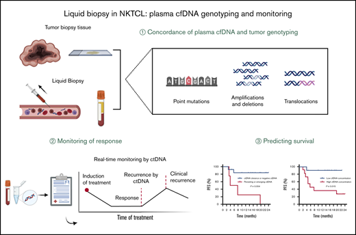
Plasma cfDNA mirrors tumor genomic DNA in detecting NKTCL-associated mutations and overcomes tumor spatial heterogeneity.
Plasma cfDNA could be used as a real-time noninvasive approach to track treatment response and survival in NKTCL.
Abstract
Satisfactory tumor material is often hard to obtain for molecular analysis in extranodal natural killer (NK)/T-cell lymphoma (NKTCL) at present. However, the accuracy and utility of circulating cell-free DNA (cfDNA) genotyping have not been adequately assessed in NKTCL. We therefore performed targeted next-generation sequencing on tumor tissues and a series of longitudinal plasma samples prospectively collected from a cohort of high-risk NKTCL patients. Concordance of genotyping results of paired baseline tumor and cfDNA and the predictive value of dynamic cfDNA monitoring were evaluated. At baseline, 59 somatic variants in 31 genes were identified in tumor and/or plasma cfDNA among 19 out of 24 high-risk NKTCL patients (79.2%). Plasma cfDNA had a sensitivity of 72.4% for detection of somatic variants identified in tumor biopsies before treatment. Plasma cfDNA also allowed the identification of mutations that were undetectable in tumor biopsies. These results were also verified in a validation cohort of an additional 23 high-risk NKTCL patients. Furthermore, longitudinal analysis showed that patients with rapid clearance of NKTCL-related mutations from plasma had higher complete remission rates (80.0% vs 0%; P = .004) and more favorable survival (1-year progression-free survival [PFS] rate, 79.0% vs 20.0%; P = .002) compared with those with persisting or emerging mutations in plasma. In addition, low cfDNA concentration before treatment was associated with favorable survival outcome for patients with NKTCL (1-year PFS, 90.0% vs 36.4%; P = .012). In conclusion, cfDNA mirrors tumor biopsy for detection of genetic alterations in NKTCL and noninvasive dynamic plasma cfDNA monitoring might be a promising approach for tracking response and survival outcome for patients with NKTCL.
Introduction
Extranodal natural killer (NK)/T-cell lymphoma (NKTCL) is a highly aggressive heterogeneous malignancy with a geographical predilection for Asian and Latin American populations.1 Remission from cytotoxic chemotherapy was not durable for advanced or relapsed disease, and prognosis is usually poor, with 3-year overall survival (OS) of 15% to 61%.2-4 Effective targeted therapies and predictive molecular biomarkers are urgently needed.
Recent work using comparative genomic hybridization, genomic microarray, and molecular genetics has provided novel insights into the pathogenesis and prognosis of NKTCL.5-9 Germ line single-nucleotide polymorphisms (SNPs), especially variation at HLA-DPB1, were reported to be strongly linked to individual NKTCL susceptibility.10 In addition, frequent somatic mutations (in JAK3, STAT3, DDX3X, TP53, EZH2, and KMT2D, among others) were identified in NKTCL, some of which might have the potential to be new molecular biomarkers with therapeutic or diagnostic implications for NKTCL.11-17
However, these studies were all performed using tumor tissues with or without the combination of NK cell lines, and in clinical practice, there are several barriers to tumor biopsy for genotyping in NKTCL. NKTCL tumors are usually largely necrotic, making the obtainability of sufficient high-quality material for molecular analysis extremely difficult. Another limitation of tumor biopsy is that it is usually performed at one time point with limited material from a heterogeneous lesion and therefore only reflects a small part of the whole story. Additionally, the invasive nature of the tumor biopsy procedure itself, putting patients at risk of harm and complications, also prevents it from being used as a real-time tool for monitoring tumor burden and evaluating treatment responses.
Circulating cell-free DNA (cfDNA) are small nucleotides shed into the bloodstream by cells undergoing apoptosis or necrosis. cfDNA harbors tumor-derived genetic alterations and reflects molecular heterogeneity across multiple disease sites.18 Currently, high-throughput next-generation sequencing (NGS) facilitates cfDNA genotyping with satisfactory sensibility and specificity in B-cell lymphomas, especially Hodgkin lymphoma (HL) and diffuse large B-cell lymphoma (DLBCL),19,20 and shows great promise in response monitoring and individualized medicine for these patients.21-24 However, to our knowledge, no study has evaluated cfDNA genotyping or assessed dynamic cfDNA monitoring in NKTCL.
Here we performed NGS on tumor tissues and a series of longitudinal plasma samples obtained from a cohort of high-risk NKTCL patients undergoing systemic chemotherapy. We analyzed the concordance of genotyping results of the tumor genome (gDNA) and plasma cfDNA among patients with paired tumor/plasma samples before treatment, as well as the predictive value of dynamic cfDNA monitoring in this study.
Methods
Patients
This study included high-risk NKTCL patients who were prospectively enrolled at the National Cancer Center/Cancer Hospital, Chinese Academy of Medical Sciences and Peking Union Medical College, between February 2018 and April 2019. Inclusion criteria were as follows: radiological and pathological diagnosis of NKTCL according to 2016 World Health Organization classification of lymphoma; male or female patient age ≥18 years; advanced (stage III/IV), relapsed, or newly diagnosed stage I/II disease with primary tumor invasion and/or regional lymph node involvement; sufficient tumor biopsy material for molecular analysis at enrollment; willing to offer peripheral blood samples before treatment, during treatment, and during follow-up; adequate organ function to undergo systemic treatment; presence of at least 1 measurable lesion; and evidence of signed consent. Patients who were diagnosed with bone marrow or peripheral blood involvement, other malignancies, or severe inflammatory disease were excluded. A total of 24 patients fulfilled the inclusion criteria and were prospectively recruited for the study as the training cohort. We also collected pretreatment blood samples and paired tumor biopsy tissues from an additional 23 patients with relapsed or advanced NKTCL at Peking University Cancer Hospital & Institute as the validation cohort.
All enrolled patients in the training cohort completed at least 1 cycle of chemotherapy according to National Comprehensive Cancer Network Clinical Practice Guidelines in T-cell lymphomas (version 1, 2018) and treatment advances in NKTCL. In the training cohort, most patients (22 [91.7%] of 24) received pegaspargase/gemcitabine-based chemotherapy: 13 received pagaspargase, gemcitabine, and oxaliplatin; 7 received gemcitabine, dexamethasone, and cisplatin; and 2 received etoposide, ifosphamide, dexamethasone, and pagaspargase. The remaining 2 patients who were heavily pretreated received chidamide orally in this study. Additional radiation treatment was administered in 8 cases. Response assessment followed the Lugano response criteria for non-Hodgkin lymphoma.25 All patients in the validation cohort also received pegaspargase/gemcitabine-based chemotherapy as first-line or salvage treatment during December 2019 to March 2020.
This study was approved by the Ethics Committee at the National Cancer Center/Cancer Hospital Chinese Academy of Medical Sciences and Peking Union Medical College (18-078/1656). The study was conducted in accordance with the Declaration of Helsinki.
Sample collection
The following biological materials were collected from each patient in the training cohort: tissue biopsies performed at diagnosis for newly diagnosed patients; tumor rebiopsies for those with relapsed disease at enrollment; and peripheral blood samples at enrollment, after every 2 to 3 cycles of chemotherapy, at the end of treatment, and at every follow-up visit in 3-month intervals. Tumor tissues were stored as formalin-fixed paraffin-embedded (FFPE) samples and analyzed within 3 to 4 weeks. Baseline peripheral blood samples were collected within 2 weeks before or after tumor biopsy and sent to the laboratory immediately at −4°C. Blood samples were processed immediately in the laboratory. Pretreatment blood samples as well as paired tumor biopsy tissues were collected retrospectively from patients in the validation cohort.
DNA extraction
Diagnosis of NKTCL was confirmed by 1 pathologist. Tissues with tumor proportions ≥10% and necrotic proportions ≤50% were selected for extraction of gDNA. Tumor gDNA and plasma cfDNA were extracted from tumor tissue and plasma using the QIAamp DNA FFPE Tissue Kit (Qiagen, Valencia, CA) and QIAamp Circulating Nucleic Acid Kit (Qiagen), respectively. Germ line gDNA was extracted from paired white blood cells using the QIAamp DNA Blood Mini Kit (Qiagen). DNA quantification was performed using a Qubit 2.0 fluorimeter (Thermo Fisher Scientific).
Targeted DNA sequencing and data analysis
Targeted sequencing was performed using Nextseq500 (Illumina, San Diego, CA) with paired-end reads. Profiling of DNA was performed using a capture-based NGS panel of 112 genes (supplemental Table 1), which covers genetic alterations of diagnostic use and/or therapeutic or prognostic value in lymphoid neoplasm.26 Single-nucleotide variants, copy-number variants (CNVs), and indels were detected using this panel in this study.
Sequencing data were mapped to the human genome (hg19) using a BWA aligner (version 0.7.10). Local alignment optimization, variant calling, and annotation were performed using GATK (version 3.2-2), MuTect (Broad Institute, Cambridge, MA), and VarScan (Genome Institute, Washington University, Washington, DC). DNA quality was assessed using high-sensitivity DNA assay. Different mutation calling thresholds were applied in samples with different DNA quality to avoid false-positive mutation calls resulting from DNA damage. Loci with a depth of <100 were filtered out by the VarScan filter pipeline. Variants with a frequency of >0.1% were categorized as SNPs and excluded from further analysis. Single-nucleotide variants and indels were annotated using the dbNSFP (v30a), COSMIC (v69), and dbSNP (snp138) databases. CNV analysis was performed by normalization and read counts from each target region, and the gene-level CNV was assessed by z test. The Integrative Genomics Viewer (Broad Institute) was used to visualize variants aligned against the reference genome to confirm the accuracy of variant calls by checking for possible strand biases and sequencing errors. Germ line variants were filtered out by analyzing genotyping results using gDNA extracted from paired white blood cells of each patient.
PD-L1 expression
FFPE tumor samples were stained with the programmed death-ligand 1 (PD-L1) antibody (clone 22C3) on the Dako automated staining platform following the manufacturer’s standard protocol (Dako 22C3 PharmDx Assay, Dako Autostainer Link 48; Agilent, Santa Clara, CA). PD-L1 expression was determined using the tumor proportion score in the training cohort. PD-L1 positivity was defined as demonstration of at least partial membrane staining with moderate or strong intensity in a minimum of 1% of tumor cells.
Statistical analysis
The sensitivity and specificity of plasma cfDNA genotyping were calculated in comparison with tumor gDNA (gold standard). Pretreatment plasma cfDNA concentrations were compared using the Mann-Whitney U test. Circulating tumor (ctDNA) was defined as the fraction of cfDNA carrying mutations. The correlation between baseline cfDNA concentration and ctDNA detection rate, as well as the correlation between allele frequencies (AFs) of mutations in cfDNA and tumor, was determined by Spearman correlation analysis. Progression-free survival (PFS) and OS were analyzed using Kaplan-Meier estimates. Comparisons of survival between groups were performed using the log-rank test. A P value of <.05 was considered statistically significant. All statistical analyses were performed using R software (version 3.6.1).
Results
Patient population description and treatment response of the training cohort
A series of 24 consecutive NKTCL patients were prospectively enrolled in the training cohort. Patients’ clinical features at enrollment are listed in Table 1. All enrolled patients represented a high-risk population. Relapsed or systemically advanced disease (stage III/IV) accounted for two-thirds of all patients. Most cases (91.7%) were diagnosed with primary tumor invasion, 54.2% had elevated serum lactate dehydrogenase, and 62.5% had elevated serum EBV DNA copy numbers. According to PINK and PINK plus EBV (PINKE) risk stratification, 62.5% and 58.4% of enrolled patients were at intermediate and high risk, respectively. A majority (87.5%) of patients had PD-L1+ expression, and half were diagnosed with a high level of PD-L1 expression.
Patient characteristics at enrollment in the training cohort
| Characteristic . | n . | % . |
|---|---|---|
| Age, y | ||
| ≤60 | 20 | 83.3 |
| >60 | 4 | 16.7 |
| Sex | ||
| Male | 15 | 62.5 |
| Female | 9 | 37.5 |
| ECOG PS | ||
| 0-1 | 22 | 91.7 |
| 2 | 2 | 8.3 |
| Ann Arbor stage | ||
| I-II | 13 | 54.2 |
| III-IV | 11 | 45.8 |
| Disease status | ||
| Relapsed | 11 | 45.8 |
| Newly diagnosed | 13 | 54.2 |
| Primary tumor location | ||
| UADT | 19 | 79.2 |
| Extra-UADT | 4 | 20.8 |
| Distant involvement | ||
| Skin | 4 | 16.7 |
| Lung | 2 | 8.3 |
| Bone | 3 | 12.5 |
| Distant lymph node | 3 | 12.5 |
| Adrenal gland | 1 | 4.2 |
| B symptoms | ||
| Yes | 14 | 58.3 |
| No | 20 | 41.7 |
| Primary tumor invasion | ||
| Yes | 22 | 91.7 |
| No | 2 | 8.3 |
| Elevated LDH | ||
| Yes | 13 | 54.2 |
| No | 11 | 45.8 |
| Serum EBV DNA | ||
| Positive | 15 | 62.5 |
| Negative | 7 | 29.2 |
| Not available | 2 | 8.3 |
| PD-L1 expression, % | ||
| 0 | 3 | 12.5 |
| ≥1 and <50 | 9 | 37.5 |
| ≥50 | 12 | 50.0 |
| IPI score | ||
| 0-1 | 12 | 50.0 |
| 2 | 7 | 29.2 |
| 3 | 5 | 20.8 |
| PINK score | ||
| 0 | 9 | 37.5 |
| ≥1 | 15 | 62.5 |
| PINKE score | ||
| ≤1 | 10 | 41.6 |
| >1 | 14 | 58.4 |
EBV, Epstein-Barr virus; ECOG PS, Eastern Cooperative Oncology Group performance score; IPI, International Prognostic Index; LDH, lactate dehydrogenase; PINK, prognostic index for NK cell lymphoma; PINKE, PINK plus EBV; UADT, upper aerodigestive tract.
Upon treatment, 16 patients (66.7%) achieved complete remission (CR), 3 (12.5%) achieved partial remission, 2 (8.3%) showed stable disease, and 3 (12.5%) had rapid progressive disease (PD). As of the last follow-up in March 2020, 7 patients had died as a result of lymphoma and 1 as a result of treatment-related toxicity, 4 were alive with lymphoma, and 12 showed no evidence of disease.
Plasma cfDNA mirrors tumor biopsy in identifying somatic mutations in NKTCL
A total of 24 tumor specimens and 102 plasma samples were collected from the enrolled training cohort, and each tumor specimen was pathologically confirmed. Paired baseline plasma samples were collected for all tumor biopsies, except in 3 patients who had undergone local irradiation or surgery before blood samples were collected. Therefore, there were 21 patients with intact measurable lesions who provided paired tumor/plasma samples before treatment.
The median sequencing depths were 1745× and 15490× in tumor gDNA and plasma cfDNA genotyping, respectively. The coverage of sequencing was 99.9%. Baseline plasma cfDNA concentration was 19.82 ± 15.03 ng/mL. Patients with elevated EBV DNA copy numbers had higher baseline cfDNA concentration. However, disease status, stage, age, B symptoms, lactate dehydrogenase level, Ki-67, and other clinical or pathological parameters were demonstrated to be unrelated to cfDNA concentration in this population (supplemental Figure 1). The detection rate of ctDNA before treatment was moderately associated with cfDNA concentration (R2 = 0.475; P = .026). Patients with elevated EBV DNA copy numbers (84.6% vs 0%; P = .001) or higher PINKE score (83.3% vs 22.2%; P = .025) had higher ctDNA detection rate in baseline plasma. It is suggested that the pathogenesis of NKTCL is complicated, and the inner relationship between EBV and somatic gene alternations in NKTCL may deserve further exploration.
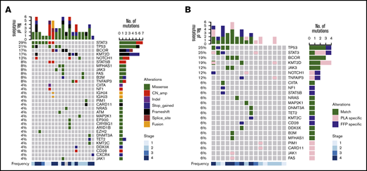
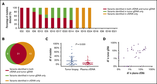
Baseline genotyping results of all patients are summarized in Figure 1A. Before treatment, somatic mutations were identified in 66.7% (16 of 24) of tumor biopsies and 57.1% (12 of 21) of cfDNA. Among the 21 paired baseline tumor/plasma samples, 5 cases harbored mutations in tumor biopsy but not plasma. We speculate that the lack of detection of any somatic variant from the plasma of these patients could be contributed to the limited ctDNA concentration. Therefore, to accurately assess cfDNA genotyping in NKTCL, we compared the cfDNA and tumor genotyping results among the remaining 16 patients (Figure 1B). The percentages of variants identified in tumor and/or cfDNA in each individual are presented in Figure 2A. A total of 39 somatic variants were identified, including 29 mutations detected by tumor gDNA and 31 by paired plasma cfDNA (Figure 2B). Although a majority of these mutations could be detected in both samples (n = 21), gene amplifications (n = 6) were detected only in the tumor biopsy, not the plasma. In addition, 10 somatic mutations that were not detectable in tumor biopsies were additionally identified in paired cfDNA. These mutations may be discordant with biopsy results because of intratumoral heterogeneity rather than false-positive results through rigorous bioinformatic analysis. Thus, plasma cfDNA achieved a sensitivity of 72.4% (21 of 29) in detecting variants verified in tumor specimens.
NGS of plasma cfDNA and tumor gDNA in NKTCL of the training cohort. (A) Overview of baseline mutational profiles in the entire training cohort. (B) Case-level genetic alterations of tumor gDNA and plasma cfDNA in 16 patients with paired plasma/tumor samples. Match indicates variants identified in both tumor gDNA and plasma cfDNA. Formalin-fixed paraffin-embedded (FFP) specific indicates variants identified in tumor gDNA only. Plasma (PLA) specific indicates variants identified in plasma cfDNA only. CN, copy number.
NGS of plasma cfDNA and tumor gDNA in NKTCL of the training cohort. (A) Overview of baseline mutational profiles in the entire training cohort. (B) Case-level genetic alterations of tumor gDNA and plasma cfDNA in 16 patients with paired plasma/tumor samples. Match indicates variants identified in both tumor gDNA and plasma cfDNA. Formalin-fixed paraffin-embedded (FFP) specific indicates variants identified in tumor gDNA only. Plasma (PLA) specific indicates variants identified in plasma cfDNA only. CN, copy number.
Concordance between plasma cfDNA and tumor gDNA genotyping in 16 patients with paired tumor/plasma patients in the training cohort. (A) The percentage of tumor biopsy–confirmed mutations that were detected in cfDNA. (B) The number of mutations discovered in baseline plasma cfDNA and/or tumor gDNA. (C) Comparison of AFs of mutations identified in baseline tumor gDNA and plasma cfDNA. (D) Scatter plot of mutation AF in cfDNA vs AF in tumor gDNA for each variant.
Concordance between plasma cfDNA and tumor gDNA genotyping in 16 patients with paired tumor/plasma patients in the training cohort. (A) The percentage of tumor biopsy–confirmed mutations that were detected in cfDNA. (B) The number of mutations discovered in baseline plasma cfDNA and/or tumor gDNA. (C) Comparison of AFs of mutations identified in baseline tumor gDNA and plasma cfDNA. (D) Scatter plot of mutation AF in cfDNA vs AF in tumor gDNA for each variant.
The mean AFs of mutations in tumor tissues and plasma cfDNA were 17.53% ± 22.28% and 7.31% ± 10.48% (P = .020; Figure 2C), respectively. In addition, AFs of mutations in tumor tissues and plasma cfDNA were moderately correlated (R2 = 0.547; P = .003; Figure 2D). Tumor-specific mutations that were not discovered in cfDNA were low-abundance variants in tissue genotyping (2 of 2). Likewise, most plasma cfDNA–specific mutations also had low abundance in cfDNA genotyping (9 of 11). AFs of cfDNA-specific and shared mutations were 1.71% ± 2.60% and 25.72% ± 22.09% in plasma, respectively (P = .006).
Overview of the genetic profile of NKTCL
We summarized the sequencing results of tumor gDNA and cfDNA at baseline to provide an overview of the genetic landscape of high-risk NKTCL (Figure 1A). Among the 24 patients, a total of 59 somatic alterations involving 31 genes were identified from either tumor tissue or plasma. The median number of variants was 2 (range, 0-6) per patient. Consistent with the typical spectrum of mutated genes in NKTCL reported in previous studies, STAT3 was the most frequent genetic alteration identified in this study, with somatic mutations or amplifications in 29.2% of cases (7 of 24), followed by TP53 in 20.8% (5 of 24), KMT2D and BCOR in 16.7% (4 of 24), and NOTCH1 in 12.5% (3 of 24). These recurrently affected genes are primarily involved in tumor-suppressing, JAK/STAT signaling, and epigenetic modulation pathways.
Validation of plasma cfDNA genotyping in NKTCL
To validate our approach of targeted sequencing, baseline peripheral blood samples (n = 23) and paired tumor biopsy tissues (n = 8) from another 23 patients with high-risk relapsed or advanced NKTCL were collected and sequenced. Patients’ characteristics are summarized in supplemental Table 2.
A total of 51 somatic variants were identified from 18 (78.3%) of 23 baseline plasma samples (supplemental Figure 2A). Consistent with genotyping results in the training cohort, BCOR (17.0%), JAK3 (17.0%), TP53 (17%), and STAT3 (13%) were among the most frequently mutated genes in patients with NKTCL in the validation cohort. Moreover, among the 8 patients with paired pretreatment tumor tissues and blood samples (supplemental Figure 2B), pairwise analysis showed plasma cfDNA had a sensitivity of 66.7% (14 of 21; supplemental Figure 3A-B) in detecting alternations identified in tumor gDNA genotyping, similar to the sensitivity in the training cohort.
In line with results of the training cohort, the mean AF of mutations identified from tumor sequencing was much higher than that of mutations in plasma cfDNA for patients with paired tumor and plasma samples (20.3% vs 2.06%; P < .01; supplemental Figure 3C). Similarly, AFs of mutations in tumor tissues and plasma cfDNA were also mildly correlated (R2 = 0.4832; P = .017).
Longitudinal monitoring of NKTCL genotyping by cfDNA in the training cohort
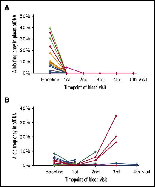
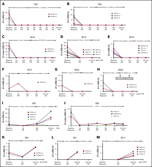
By the last follow-up, 20 patients in the training cohort had at least 1 plasma cfDNA monitoring beyond baseline, among whom 14 were still attending regular plasma and radiology visits. Longitudinal analysis showed a rapid clearance of ctDNA (no detection of any mutation in cfDNA within 2-4 cycles of chemotherapy) or persisting undetectable ctDNA in patients who achieved CR from treatment, whereas mutations persisted in the plasma of those who did not achieve CR (80% vs 0%; P = .004; Figure 3). After treatment, ctDNA became undetectable in the plasma of 8 patients, a majority (7 [87.5%] of 8) of whom achieved CR (Figure 4A-G); only 1 showed PD at response evaluation (Figure 4H). Meanwhile, 5 (71.4%) of 7 patients with persisting undetectable ctDNA at baseline and subsequent monitoring achieved CR, whereas the other 2 had PD. The remaining 5 patients who had persisting detectable or emerging ctDNA (JAK1/XPO1) in cfDNA monitoring either progressed or relapsed rapidly (Figure 4I-M). On the other hand, 2 patients achieving radiological remission at the end of treatment ultimately relapsed, in line with molecular progression/relapse through cfDNA monitoring (Figure 4K,M).
Longitudinal assessment of mutation abundance in cfDNA upon treatment. (A) NKTCL-related mutations disappeared during cfDNA monitoring among patients who achieved CR at the end of treatment. (B) NKTCL-related mutations persisted during cfDNA monitoring among patients who did not achieve CR at the end of treatment.
Longitudinal assessment of mutation abundance in cfDNA upon treatment. (A) NKTCL-related mutations disappeared during cfDNA monitoring among patients who achieved CR at the end of treatment. (B) NKTCL-related mutations persisted during cfDNA monitoring among patients who did not achieve CR at the end of treatment.
Noninvasive real-time monitoring of cfDNA genotyping and disease changes in patients with NKTCL. Patient identification (ID) numbers 2 (A), 5 (B), 10 (C), 12 (D), 15 (E), 17 (F), 24 (G), 20 (H), 3 (I), 6 (J), 4 (K), 1 (L), and 11 (M). PR, partial remission.
Noninvasive real-time monitoring of cfDNA genotyping and disease changes in patients with NKTCL. Patient identification (ID) numbers 2 (A), 5 (B), 10 (C), 12 (D), 15 (E), 17 (F), 24 (G), 20 (H), 3 (I), 6 (J), 4 (K), 1 (L), and 11 (M). PR, partial remission.
Of note, we suggested the 4 patients with molecular residual disease (MRD) receive subsequent intensive or consolidation treatment. One male patient with stage IV disease had PD-L1 expression of 80% and was shown to harbor EZH2 and STAT3 mutations at his peripheral blood visit. He received subsequent PD-1 blockade treatment of MRD, and as of last follow-up, he was alive, with a median OS of 24.6 months. We recommended oral chidamide for a relapsed 72-year-old patient whose mutations were related to epigenetic genes (DNMT2A and TET2). He was in durable partial remission, with a median OS of 18.9 months, as of his last visit. However, the remaining 2 patients who refused further treatment progressed and died as a result of lymphoma (median OS, 7.6 and 9.8 months, respectively). Although the number of informative cases in our study was quite small, these results provided a hint that dynamic cfDNA might be a potential tool in disease monitoring and individual medicine in NKTCL.
Survival outcome and plasma cfDNA genotyping in the training cohort
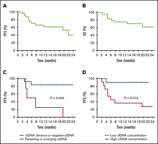
By the last visit in March 2020, the median follow-up period was 15.5 (range, 2.3-24.6) months. The 1-year PFS and OS rates for this cohort of patients were 57.0% and 78.4%, respectively (Figure 5A-B). Patients who had plasma ctDNA clearance or consistently undetectable ctDNA in plasma had a much more favorable PFS than those with persisting detectable or emerging ctDNA during therapy and surveillance (1-year PFS, 83.9% vs 28.6%; P = .004; Figure 5C). Besides, patients with high cfDNA concentration before treatment had a 1-year PFS of 36.4%, which was inferior in comparison with those with low cfDNA concentration (90.0%; P = .015; Figure 5D). However, baseline EBV DNA level was found to be unrelated to survival in NKTCL patients in this current study. Patient heterogeneity, small sample size, and relatively short follow-up period of our study might account for this result. Therefore, further more explorative research is warranted to compare or combine plasma cfDNA and EBV DNA in predicting survival in NKTCL patients.
Survival curves of NKTCL patients included in this study. PFS (A) and OS (B) of the entire cohort. (C) Comparison of PFS between patients with a clearance of ctDNA and those with persisting detectable ctDNA during monitoring. (D) Comparison of PFS between patients with high and low baseline plasma cfDNA concentration.
Survival curves of NKTCL patients included in this study. PFS (A) and OS (B) of the entire cohort. (C) Comparison of PFS between patients with a clearance of ctDNA and those with persisting detectable ctDNA during monitoring. (D) Comparison of PFS between patients with high and low baseline plasma cfDNA concentration.
Discussion
Tumor tissue samples are notoriously difficult to obtain in patients with NKTCL. However, in this study, we successfully collected pathologically confirmed tumor biopsies from 24 NKTCL patients along with a series of peripheral blood samples. An additional 23 patients with relapsed or advanced NKTCL were sequenced to validate these results. To our knowledge, this is the first and largest prospective study to ascertain the feasibility of plasma cfDNA genotyping among NKTCL patients compared with tumor sequencing and explore dynamic cfDNA monitoring during treatment. First, we demonstrated plasma cfDNA mirrors tumor gDNA sequencing in detecting NKTCL-associated mutations. Second, we showed plasma cfDNA overcomes tumor spatial heterogeneity in NKTCL and allows the identification of genetic alterations that are undetectable in tumor biopsies. Finally, we provided evidence that plasma cfDNA might be used as a real-time, noninvasive approach to track treatment response and survival in NKTCL.
Most studies on cfDNA assessment in lymphoma to date have centered on B-cell lymphomas,20-24,27,28 and there are few available data on the role of cfDNA genotyping in NKTCL. In fact, detecting cfDNA in NKTCL compared with DLBCL may be more challenging. It was reported that patients with DLBCL harbored a significantly higher number of mutations in cfDNA (7.27 ± 5.38) compared with those with other lymphoma subtypes, including NKTCL (0.65 ± 0.79; P = .026).29 Although there was a higher level of cfDNA concentration in NKTCL than DLBCL (19.60 ± 26.04 vs 11.70 ± 12.05 ng/mL; P = .027), the abundance of somatic mutations detected per sample was indeed significantly lower in NKTCL (AF >10%; 12.5% vs 75%; P = .003).30 Detecting mutations in cfDNA in NKTCL cases may be more difficult because of the common pathological necrosis and less mutation-driven nature of this disease.
Ultrasensitive panel-directed NGS is 1 method of cfDNA assessment with satisfactory sensibility and specificity in lymphomas.30 Unlike sequencing of immunoglobins or T-cell receptors, panel-directed NGS captures the complex landscape of somatic variation in lymphomas and is independent of the availability of tumor sequencing results. In DLBCL, targeted NGS has been shown to detect somatic mutations in 93% to 100% of tumor tissues and 64% to 98% of pretreated plasmas.23,24,28,31,32 In this study, we designed a panel targeting 112 lymphoma-related genes. By performing NGS with this panel in baseline plasma and tumor samples, 79.2% of patients were identified as harboring at least 1 genetic alteration: 66.7% were identified in tumors and 57.1% in plasmas. In addition, the genetic profile of the NKTCL mutations revealed in this study was in line with the known signature in published reports.5,8,33 Results in the validation cohort in our study were also consistent with those of the training cohort. On the basis of these data, our panel is rational and informative enough to track the genetic features of NKTCL, and panel-directed NGS can reliably provide molecular information about NKTCL mutations.
The concordance of cfDNA and tumor genotyping has been beyond satisfactory in B-cell lymphomas. In a study of 50 patients with previously untreated DLBCL, pretreatment plasma cfDNA correctly discovered tumor-confirmed mutations with >90% sensitivity and ∼100% specificity.24 Similarly, we found plasma cfDNA genotyping achieved satisfactory sensitivity (72.4%) in detecting variants verified in tumors in NKTCL patients. We also showed the AFs of mutations in plasma and tumors were mildly correlated (R2 = 0.547; P = .001). Taken together, our data indicate cfDNA can mirror tumor biopsy for providing valuable molecular information about NKTCL, especially when tumor biopsy is not accessible.
We propose cfDNA is an important complementary source of the tumor genome, but not a thorough substitute for tumor biopsy for NKTCL genotyping at present. In this study, cfDNA allowed the identification of somatic mutations that were undetectable in tumor biopsies because of tumor spatial heterogeneity, although they presented in plasma with a comparably low abundance. On the other hand, amplifications were only detected from tumor but not plasma in our study, suggesting that small fragmental cfDNA might be unsuitable for detecting certain alterations. Furthermore, in its current modality, cfDNA genotyping cannot accurately distinguish metastatic tumors from primary lesions.28
Standard methods for response assessment in lymphomas are positron emission tomography and computed tomography scans, which are costly, pose health risks, and are limited in detecting the presence of MRD. Plasma cfDNA has been reported to be a promising biomarker in monitoring MRD ahead of radiological imaging in DLBCL and classical HL.19,20,24,34 In addition, cfDNA genotyping can be easily repeated at multiple time points, offering a complete picture of clonal evolution over the course of the disease. Indeed, in a retrospective analysis of 126 patients with DLBCL who received standard first-line treatment, interim ctDNA monitoring showed positive and negative predictive values of 63% and 80%, respectively, in patients with and without detectable ctDNA; the corresponding 5-year time-to-progression rates were 41.7% and 80.2%, respectively (P < .0001).32 Consistently, longitudinal cfDNA monitoring in our study showed that the clearance of ctDNA might be a predictor of CR after treatment and favorable survival for patients with high-risk NKTCL. We hold that both radiological and cfDNA techniques have their strengths and weaknesses and will likely to be used together rather than alone for MRD diagnosis in the future.
Our study is limited by its small sample size and relatively short follow-up period. Therefore, it was difficult to determine the lead time until ctDNA positivity before overt progression. We did not quantify ctDNA concentration, and the definition of ctDNA clearance was also nonquantitative. The true value of plasma cfDNA in NKTCL should be explored in a large, prospective clinical trial in the future.
Send data sharing requests to corresponding authors, Xiaoli Feng (fengxl@hotmail.com) or Mei Dong (dongmei030224@163.com).
Acknowledgments
The authors thank all physicians and nurses who participated in this study as well as all enrolled patients at the Cancer Hospital, Chinese Academy of Medical Sciences and Peking Union Medical College, and Burning Rock Biotech.
This study was supported by the Beijing Hope Run Project (LC2018L04) and Chinese Geriatric Oncology Society Scientific Research Fund (CGOS-06-2014-1-1-01600).
Authorship
Contribution: M.D. and X.F. designed the study; J.L., Y.W., J.Y., H.L., H.H.-Z., and X.M. contributed to the acquisition of data; F.Q., B.C., Z.C., and Y.C. analyzed and interpreted the data; F.Q. and Z.C. contributed to the writing of the manuscript; and all authors revised the manuscript and gave final approval.
Conflict-of-interest disclosure: The authors declare no competing financial interests.
Correspondence: Mei Dong, Department of Medical Oncology, National Cancer Center/National Clinical Research Center for Cancer/Cancer Hospital, Chinese Academy of Medical Sciences and Peking Union Medical College, No. 17 Panjiayuan Nanli, Chaoyang District, Beijing 100021, China; e-mail: dongmei030224@163.com; and Xiaoli Feng, Department of Pathology, National Cancer Center/National Clinical Research Center for Cancer/Cancer Hospital, Chinese Academy of Medical Sciences and Peking Union Medical College, No. 17 Panjiayuan Nanli, Chaoyang District, Beijing 100021, China; e-mail: fengxl@hotmail.com.
References
Author notes
F.Q. and Z.C. contributed equally to this work as first authors.
The full-text version of this article contains a data supplement.

