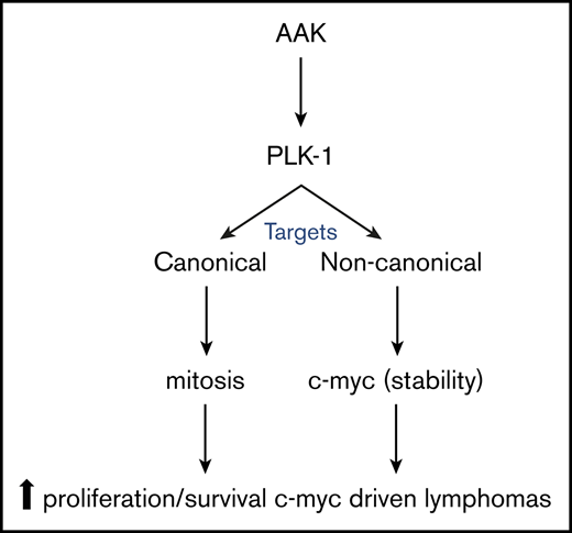
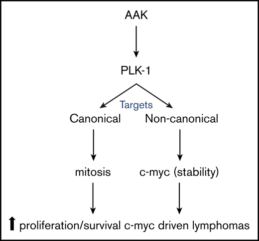
High-risk lymphomas (HRLs) are associated with dismal outcomes and remain a therapeutic challenge. Recurrent genetic and molecular alterations, including c-myc expression and aurora A kinase (AAK) and polo-like kinase-1 (PLK1) activation, promote cell proliferation and contribute to the highly aggressive natural history associated with these lymphoproliferative disorders. In addition to its canonical targets regulating mitosis, the AAK/PLK1 axis directly regulates noncanonical targets, including c-myc. Recent studies demonstrate that HRLs, including T-cell lymphomas and many highly aggressive B-cell lymphomas, are dependent upon the AAK/PLK1 axis. Therefore, the AAK/PLK1 axis has emerged as an attractive therapeutic target in these lymphomas. In addition to reviewing these recent findings, we summarize the rationale for targeting AAK/PLK1 in high-risk and c-myc–driven lymphoproliferative disorders.

Elucidation of the “cell of origin,” by gene expression profiling or immunohistochemistry-based algorithms, has led to the identification of clinically and molecularly distinct subsets among aggressive non-Hodgkin lymphomas, including diffuse large B-cell1,2  and peripheral T-cell lymphomas.3,4  Integrated genomic analyses have since unveiled the considerable genetic heterogeneity within each of these subsets,5-7  including the identification of those that are “high risk,” as demonstrated by resistance to traditional anthracycline-based regimens (eg, cyclophosphamide, doxorubicin, vincristine, and prednisone [CHOP]) and inferior survival.8,9  Despite the histopathologic diversity observed among these high-risk lymphomas (HRLs), many of these HRLs share common transcriptional programs, particularly those that regulate cell-cycle progression and proliferation, and harbor common genetic/epigenetic alterations, including those that culminate in the overexpression of c-myc and the loss of p53.7,8,10  Therefore, novel therapeutic strategies targeting cell-cycle regulators, including the oncogenes and tumor suppressors associated with these HRLs, are needed. Herein, we review recent data suggesting that aurora and polo-like kinases are attractive therapeutic targets in these HRLs.

Disruption of the “polo” and “aurora” genes in Drosophila was observed, almost 3 decades ago, to impair centrosome separation, mitotic spindle formation, and the proper spatiotemporal separation of chromosomes during mitosis.11,12  The homologs of these highly conserved genes were discovered in humans,13-16  and subsequent work has demonstrated their central role in regulating mitotic entry and progression.17,18  The 3 aurora kinases (aurora A [AAK], B, and C) and 5 polo-like kinases (PLK1-5) in humans are serine-threonine kinases that, with cyclin B/cyclin-dependent kinase 1 (CDK1) and various adaptor/scaffold proteins, collaborate in various spatiotemporal contexts to regulate mitosis and cytokinesis. PLKs are characterized by C-terminal polo box domains, which regulate their spatiotemporal localization by binding phosphorylated motifs, largely generated by CDK1, on PLK-associated proteins. Polo box domain binding induces a conformational change, exposing the N-terminal kinase domain, including a threonine residue (T210 on PLK1), phosphorylation of which is required for full kinase activation.19,20  The coordinated expression and spatiotemporal localization of AAK and PLK1 during the G2/M phase of the cell cycle have indirectly linked these kinases since their initial discovery, but this association was bolstered upon recognition that the PLK1 activation loop (T210) is directly phosphorylated by AAK.21,22  Both AAK and PLK1, in complex with multiple cofactors, each of which is tightly regulated, choreograph cell entry into, and smooth progression through, mitosis. Therefore, given its central role in mitosis, the AAK/PLK1 axis is a significant cancer dependency.23 

Importantly, nuclear expression of AAK has been demonstrated in different subsets of HRLs, with predominant expression in peripheral T-cell lymphoma (PTCL) cases.24  Similarly, increased expression of AAK and PLK1 has been demonstrated in cutaneous T-cell lymphoma, and other subtypes of HRLs.24-28  The AAK/PLK1 axis and its role in mitosis have been recently reviewed.17,18  In contrast, more recent findings demonstrate that the AAK/PLK1 axis promotes cancer cell growth and survival independently from its well-established role in mitosis by phosphorylating noncanonical substrates, many of which are of significant interest in hematologic malignancies, including “high-risk” lymphoproliferative disorders.

c-myc–driven lymphoproliferative disorders

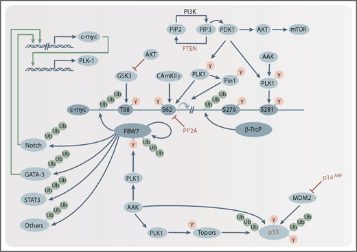
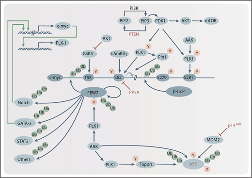
In many aggressive lymphoproliferative disorders, c-myc is highly expressed secondary to increased transcription (eg, in B-cell lymphomas harboring translocations placing the c-myc locus under the control of alternative enhancer elements), and/or alterations that increase c-myc stability (eg, hotspot mutations that prevent phosphorylation-dependent ubiquitination and proteasomal degradation). Importantly, increased c-myc expression in specific B- and T- lymphoma subsets is associated with poor outcomes with conventional anthracycline-based regimens. Burkitt lymphomas, the overwhelming majority of which are highly curable with current therapies, are a notable exception.29  In stark contrast, a subset of diffuse large B-cell lymphomas (DLBCLs) and high-grade B-cell lymphomas harbor c-myc translocations, often in conjunction with bcl-2 and/or bcl-6 translocations (ie, “double/triple hits”), and highly express c-myc (reviewed in Rosenthal and Younes30 ). A subset of aggressive peripheral T-cell lymphomas, which are similarly resistant to standard chemotherapy regimens,9  was recently described and shown to highly express c-myc.3,4,7  At the very least, these lymphomas, “double/triple hits” and a recently described PTCL, not other specified subset, may be fairly described as HRLs, as they are associated with relatively high rates of primary refractory disease and poor outcomes in the setting of relapsed/refractory disease. The expression of c-myc in these HRLs, while transcriptionally regulated, is also a consequence of several mechanisms operating simultaneously to increase c-myc stability (reviewed in Figure 1). Multiple phosphorylation-dependent ubiquitin ligases have opposing roles in tagging c-myc for proteasomal degradation. PLK1 or calcium/calmodulin-dependent kinase II γ (CAMKIIγ) phosphorylate c-myc at serine 62 (S62),31,32  priming c-myc for binding by glycogen synthase kinase 3 (GSK3), culminating in GSK3-mediated phosphorylation of threonine 58 (T58).33-35  Phosphorylation at T58 and the subsequent dephosphorylation at S62 mediated by protein phosphatase 2A (PP2A)36-38  are required for optimal c-myc ubiquitination by a Skp1/Cul1/F-box protein (SCF), SCFFBW7 (Welcker et al33  and Hao et al39 ; reviewed in Welcker and Clurman40 ). However, PP2A is a trans-specific phosphatase, which requires cis/trans isomerization of the S62 and proline 63 peptide bond prior to S62 dephosphorylation. Isomerization is accomplished by peptidylprolyl cis/trans isomerase, NIMA-interacting 1 (Pin1).36,37  Pin1 regulates other relevant transcription factors, including NF-κB and Notch,41,42  and also promotes c-myc recruitment to its target genes,43  and is thus required for B-cell proliferation in c-myc–driven lymphomas.44  Importantly, PLK1 directly phosphorylates and stabilizes Pin1 by impairing Pin1 ubiquitination and proteasomal degradation.45  Ubiquitination mediated by F-box and WD repeat domain-containing 7 (FBW7), or alternative ubiquitin ligases,46  targets c-myc for proteasome-mediated degradation. In short, the sequential and interdependent phosphorylation, isomerization, and dephosphorylation of the highly conserved T58/S62 residues regulates c-myc ubiquitination and proteasomal degradation.

Figure 1.

The AAK/PLK1 axis in HRLs. AAK and PLK1 directly, or indirectly, regulate the stability of oncogenes (eg, c-myc, Notch) and tumor suppressors (eg, p53) implicated in the pathogenesis of most HRLs. Schematic representations of posttranslational phosphorylation (Y) and ubiquitination (Ub) modifications are indicated (blue arrows), as are inhibitory modifications (red bars). Tumor suppressors that are recurrently deleted in many HRLs are indicated in red. Transcription factors promoting c-myc or PLK1 expression are shown (green arrows). β-Trcp, β-transducin repeat-containing protein; CAMKIIγ, calcium/calmodulin-dependent kinase II γ; GSK3, glycogen synthase kinase 3; Mdm2, mouse double minute 2 homolog; mTOR, mammalian target of rapamycin; p14ARF, p14 alternative reading frame; PDK1, phosphoinositide-dependent kinase 1; PI3K, phosphatidylinositol 3-kinase; Pin1, peptidylprolyl cis/trans isomerase, NIMA-interacting 1; PIP2, phosphatidylinositol 4,5-bisphosphate; PIP3, phosphatidylinositol (3,4,5)-trisphosphate; PP2A, protein phosphatase 2A; PTEN, phosphatase and tensin homolog.

Close modal

In contrast to T58/S62, a pair of serine residues (S279/S281) is required for β-transducin repeat-containing protein (β-Trcp)–mediated ubiquitination events that not only impair c-myc proteasomal degradation, but also are likely required for c-myc–dependent cell transformation and growth.47,48  Phosphorylation of S279 primes c-myc for PLK1-dependent phosphorylation of S281,48,49  which is required for binding of β-Trcp, an E3 ubiquitin ligase that ubiquitinates and stabilizes c-myc.48  In addition to its role in phosphorylating the β-Trcp–binding motif, PLK1 also phosphorylates FBW7, promoting its autoubiquitination and degradation.50  Similarly, AAK directly, or indirectly, promotes c-myc stability,51,52  further implicating the AAK/PLK1 axis in c-myc expression. Furthermore, AAK and PLK1 likely collaborate with the genetic landscape (eg, phosphatase and tensin homolog [PTEN] deletions) and dysregulated signaling pathways (eg, phosphatidylinositol 3-kinase [PI3K]/AKT) to regulate c-myc expression in aggressive lymphomas.7,32,53-55  The available evidence not only highlights the fundamental role of the AAK/PLK1 axis in regulating c-myc, but also has significant therapeutic implications in c-myc–driven lymphoproliferative disorders.

Recently, PLK1 was identified in a short hairpin RNA–based “Achilles heel” screen in T-cell lymphomas.26  In both patient-derived cell lines and primary T-cell lymphoma specimens, PLK1 inhibition with volasertib induced apoptosis, modestly impaired bromodomain containing 4 (BRD4) binding to the c-myc enhancer, and decreased c-myc transcription.26,56  In addition, an apparent decrease in c-myc phosphorylation at S62 was also observed, and was associated with loss of detectable c-myc expression in volasertib-treated cell lines and tumor xenografts.26  These observations are consistent with previous studies demonstrating that PLK1-dependent phosphorylation impairs proteasome-mediated degradation of c-myc. In addition to its role in conferring c-myc stability, a “feed-forward” mechanism has also been described in which c-myc increases PLK1 transcription upon binding the PLK1 promoter.55  Therefore, it may not be surprising that PLK1 and c-myc expression were strongly correlated in DLBCLs and high-grade lymphomas, including “double hits.”26,28,55  Furthermore, PLK1 inhibition with volasertib induced apoptosis and impaired c-myc expression in DLBCL cell lines.26  The association between PLK1 and c-myc was subsequently validated, and, not surprisingly, PLK1 expression was associated with inferior survival in a cohort of DLBCL patients following rituximab plus CHOP.55  Collectively, the available data suggest that AAK and PLK1 are attractive therapeutic targets in c-myc–driven lymphoproliferative disorders.

p53-deficient lymphoproliferative disorders

In isolation, overexpression of c-myc may induce an endogenous apoptotic program that is dependent upon the tumor suppressor p53.57-59  Therefore, lymphoproliferative disorders that highly express and are driven by c-myc are frequently characterized by genetic aberrations that directly, or indirectly, inactivate p53.7,29  For example, a high-risk subset of GATA-3+ PTCL that highly expresses c-myc also harbors highly recurrent deletions in cyclin-dependent kinase inhibitor 2A (CDKN2A) and/or p53.7  CDKN2A deletions lead to the loss of the p14 alternative reading frame (p14ARF), which normally binds mouse double minute 2 homolog (Mdm2) and impairs its E3 ubiquitin ligase activity, culminating in Mdm2-dependent p53 destabilization. This cooperativity between c-myc and alterations that culminate in p53 inactivation suggests that therapeutic strategies targeting c-myc may be effective in p53-deficient lymphomas.58  This principle was elegantly demonstrated in a mouse model of Nras-driven (and p53-deficient) liver cancer.60  Consistent with earlier studies demonstrating that AAK directly binds and stabilizes N-Myc,52,61,62  Dauch et al demonstrated a direct interaction between AAK and c-myc in p53-deficient cells, and further demonstrated that AAK preferentially bound and stabilized doubly phosphorylated (T58/S62) c-myc.60  However, this interaction was prevented by a subset of AAK inhibitors, including alisertib, that induce a conformational change in AAK.61,62  Therefore, treatment with AAK inhibitors in this model culminated in c-myc degradation and apoptosis in p53-deficient tumors.

In addition to its role in c-myc–driven and p53-deficient tumors, the AAK/PLK1 axis also regulates the function of p53 and its family members. The N-terminal domain of PLK1 is required for binding to the p53 DNA-binding domain, and PLK1 binding impairs p53, establishing PLK1 as a negative regulator of p53.63  Although PLK1-mediated inhibition of p53 was dependent upon PLK1 kinase activity, more recent evidence suggests that PLK1-dependent phosphorylation regulates the activity of Topors, a p53-binding protein that has a dual function, mediating sumoylation or ubiquitination of client proteins.64,65  Whereas Topors-mediated ubiquitination predictably leads to p53 degradation, Topors-mediated sumoylation promotes p53 expression.66  More importantly, PLK1 phosphorylates Topors, leading to enhanced Topors-mediated p53 ubiquitination, impaired sumoylation, and ultimately p53 destabilization.65  PLK1 also negatively regulates other members of the p53 family.67,68  Similarly, p53 is also an AAK substrate, with AAK-dependent phosphorylation culminating in p53 inactivation and destabilization.69-71  Not surprisingly then, the extent to which AAK/PLK1 inhibition culminates in apoptotic cell death in many c-myc–independent tumors is dependent upon p53 or p53 family members.71-76  Therefore, AAK/PLK1 inhibitors may be rationally combined with therapies, including Mdm2 inhibitors, which further bolster p53 expression and function.77 

Current therapeutic approaches in lymphoproliferative neoplasms

Multiple aurora kinase inhibitors have been developed, many of which are pan-selective and inhibit AAK and aurora B and/or aurora C kinases (reviewed in Yan et al78 ). In contrast, MLN8054 is an adenosine triphosphate–competitive, reversible, and selective inhibitor of aurora A that impaired G2/M progression and induced apoptosis in preclinical studies at submicromolar concentrations.79  In phase 1 studies, the dose-limiting toxicity in patients with advanced solid tumors was somnolence and was attributed to γ-aminobutyric acid A (GABAA) binding in the central nervous system.80  Efforts to increase the therapeutic window and decrease GABAA binding led to the development of a second-generation compound, MLN8237 (alisertib; reviewed in Sells et al81 ). Alisertib is an orally bioavailable and highly selective AAK inhibitor,82,83  and is the first aurora kinase inhibitor to be examined in a phase 3 clinical trial. Based on significant activity in T-cell lymphoproliferative disorders in preclinical studies and early-phase clinical trials,84-89  alisertib was compared with a “dealer’s choice” (including pralatrexate, romidepsin, or gemcitabine) in a randomized phase 3 trial in patients with relapsed/refractory PTCL. Although a significant improvement in response rate was not observed relative to the comparators, an overall response rate comparable with earlier studies (≈30%) was observed.90  Although failing to achieve its primary end point, it is notable that the overall response rate with alisertib is comparable to those observed with US Food and Drug Administration (FDA)–approved agents.

As the AAK/PLK1 axis is an attractive therapeutic target, multiple PLK1 inhibitors have been developed, many of which also inhibit PLK2 and PLK3, among other targets (reviewed in Talati et al91  and Strebhardt92 ). However, BI2536 was one of the first adenosine triphosphate–competitive PLK1 inhibitors developed and was shown to inhibit PLK1/PLK2/PLK3 in the low nanomolar range, leading to G2/M arrest and the induction of apoptosis in multiple leukemia cell lines.93-95  BI6727 (volasertib), a derivative of BI2536 and second-generation PLK1 inhibitor, is a potent PLK1 (and PLK2/PLK3) inhibitor with significant activity in leukemia and lymphoma cell lines.26,96-98  In addition to its PLK1-inhibitory activity at low nanomolar concentrations, a recent screen for dual kinase-bromodomain inhibitors identified volasertib as a potent BRD4 inhibitor.56  Therefore, it has been suggested that volasertib is best classified as a dual kinase-bromodomain inhibitor.26  This is significant, as volasertib was shown to inhibit BRD4 binding to the c-myc locus, impairing c-myc transcription and expression in lymphoma cell lines.26  The phase 1/2 experience with either single-agent volasertib or in combinations with low-dose cytosine arabinoside (LDAC) in patients with acute myeloid leukemia (AML) informed a randomized phase 3 trial (POLO-AML-2 trial) comparing volasertib/LDAC with placebo/LDAC in AML patients 65 years and older who were not suitable candidates for standard induction regimens.99,100  A nonsignificant increase in response rate was observed in the volasertib/LDAC arm, but a significant increase in severe adverse events, including fatal infectious complications, was observed in this arm and contributed to a nonsignificant, but notable, decrease in overall survival. Given its dual mechanism of action, future studies investigating its role in highly proliferative (and c-myc–driven) lymphoproliferative disorders, including “double hit” B-cell lymphomas and PTCL subsets, seem warranted.7,26,28,55,101 

Optimizing AAK/PLK1 inhibitors with novel and rational combinations

F-box protein FBW7 (Fbxw7), a well-characterized SCF-type E3 ubiquitin ligase and tumor suppressor, is mutated in many human cancers, including ≈33% of T-cell acute lymphoblastic leukemia,102,103  where it is associated with poor survival.104  FBW7 stability is regulated in a phosphorylation- and PLK1-dependent manner, disruption of which leads to the accumulation of oncogenic FBW7 substrates. For example, PLK1 phosphorylates FBW7 at serine and threonine sites (S58/T284), which culminates in FBW7 self-ubiquitination and proteasomal degradation. Thus, AAK/PLK1 inhibition and FBW7 stabilization promotes the degradation of its substrates, many of which are oncogenic, and therapeutically targetable, in high-risk hematologic malignancies. For example, AAK itself is a FBW7 substrate.105  GATA-3, a zinc-finger transcription factor that identifies an aggressive subset of PTCL,3,4,7  upon phosphorylation by CDK2 is a target for FBW7-dependent ubiquitination and proteasomal degradation.106  It is noteworthy that these GATA-3–expressing T-cell lymphomas highly express c-myc, which as previously noted, is also a FBW7 substrate. A number of FBW7 substrates, or the pathways they regulate, are therapeutically targetable, including Mcl-1,107,108  mammalian target of rapamycin,109  Notch1/Notch2,110-112  NF-κB,113-115  and STAT3.116  Therefore, AAK/PLK1 inhibition may be a rational strategy in combination with agents targeting these oncogenic pathways that are regulated by FBW7. For example, PLK1 inhibition led to Mcl-1 degradation and was synergistic with the Bcl-2 inhibitor venetoclax in venetoclax-resistant, and Mcl-1 expressing, B-cell lymphoma lines.55  AAK/PLK1 inhibition is potentially synergistic with PI3K inhibition,117  BRD4 (and hence c-myc) inhibition,118  and JAK/STAT inhibition.119  Despite their pleiotropic mechanism of action, which remain poorly defined, histone deacetylase inhibitors lead to the inhibition of FBW7 substrates,120-123  and are synergistic with alisertib in T-cell lymphoma cells.124  Proteasome inhibitors have been exploited as a strategy to inhibit both NF-κB and GATA-3 in T-cell lymphomas,125  and may be safely combined with alisertib.126  However, as AAK/PLK1 inhibition acts, at least in part, by inducing the proteasomal degradation of FBW7 substrates that are oncogenic, a potential antagonism between AAK/PLK1 and proteasomal inhibition may be anticipated, and is supported by preclinical studies performed in B-cell and T-cell lymphoma cell lines.124  In stark contrast, proteasome and PLK1 inhibition were effectively combined in AML, including primary AML cells.127  Despite the mitotic arrest imposed by AAK/PLK1 inhibition, malignant cells slowly degrade mitotic cyclins (eg, cyclin B) and inappropriately escape mitotic arrest (“mitotic slippage”). However, proteasome inhibition impaired the proteasomal inhibition of cyclin B in AML cells and prolonged the mitotic delay observed with PLK1 inhibition in AML cells, thus enhancing volasertib’s efficacy.127 

The ubiquitin-proteasome pathway plays a critically important role in regulating the stability of c-myc and other oncogenic FBW7 substrates. Therefore, upstream signaling pathways that promote c-myc phosphorylation, priming it for ubiquitination and degradation, are attractive therapeutic targets. The PI3K/AKT pathway may be chief among them. Loss-of-function mutations and/or deletion in the tumor-suppressor PTEN lead to unopposed PI3K activity and are highly recurrent in many tumors, including many lymphoproliferative disorders. PTEN deletions, for example, are observed in a substantial minority of T-cell lymphoproliferative disorders.7,128-139  In addition, alternative mechanisms antagonize PTEN. For example, the Notch target gene Hes-1 is a potent transcriptional repressor that suppresses PTEN transcription,140  whereas c-myc–induced microRNA (eg, miR-17-92 cluster) target PTEN messenger RNA.141-143  Unopposed PI3K activity leads to phosphoinositide-dependent kinase 1 and AKT activation and phosphorylation of their downstream substrates, including PLK1 and GSK3, respectively. Consequently, PI3K/AKT activation indirectly promotes the stability of c-myc and other FBW7 substrates.32,144,145  In addition to c-myc, AKT-dependent phosphorylation and inhibition of GSK3 impairs the downstream phosphorylation and proteasomal degradation of alternative, and therapeutically relevant, GSK3 and FBW7 substrates (reviewed in Hermida et al54 ). The apparent convergence of PI3K/AKT and AAK/PLK1 at FBW7 and its substrates suggests that combined inhibition of both pathways may be synergistic, and is supported in preclinical models.32,74,117,146  Enthusiasm for this combination is further bolstered by the significant single-agent activity observed with PI3K inhibitors in many lymphoproliferative disorders.

Novel targeted and immuno-oncology therapies are needed for patients with HRLs, as few durable remissions are achieved with many conventional chemotherapy regimens. The AAK/PLK1 axis has emerged as a promising target in these HRLs and future clinical studies are warranted.

This work was supported in part by the National Cancer Institute of the National Institutes of Health under award numbers K08-CA218460 (C.M.-Z.) and R01CA217994 (R.A.W.).

Contribution: C.M.-Z., K.V.I., and R.A.W. wrote the manuscript.

Conflict-of-interest disclosure: The authors declare no competing financial interests.

Correspondence: Ryan A. Wilcox, University of Michigan, 4431 Rogel Cancer Center, 1400 E Medical Center Dr, Ann Arbor, MI 48104; e-mail: rywilcox@med.umich.edu; or Carlos Murga-Zamalloa, University of Michigan, 4431 Rogel Cancer Center, 1400 E Medical Center Dr, Ann Arbor, MI 48104; e-mail: carlosmu@med.umich.edu.

1.
Alizadeh
AA
,
Eisen
MB
,
Davis
RE
, et al
.
Distinct types of diffuse large B-cell lymphoma identified by gene expression profiling
.
Nature
.
2000
;
403
(
6769
):
503
-
511
.
2.
Hans
CP
,
Weisenburger
DD
,
Greiner
TC
, et al
.
Confirmation of the molecular classification of diffuse large B-cell lymphoma by immunohistochemistry using a tissue microarray
.
Blood
.
2004
;
103
(
1
):
275
-
282
.
3.
Iqbal
J
,
Wright
G
,
Wang
C
, et al;
Lymphoma Leukemia Molecular Profiling Project and the International Peripheral T-cell Lymphoma Project
.
Gene expression signatures delineate biological and prognostic subgroups in peripheral T-cell lymphoma
.
Blood
.
2014
;
123
(
19
):
2915
-
2923
.
4.
Wang
T
,
Feldman
AL
,
Wada
DA
, et al
.
GATA-3 expression identifies a high-risk subset of PTCL, NOS with distinct molecular and clinical features
.
Blood
.
2014
;
123
(
19
):
3007
-
3015
.
5.
Chapuy
B
,
Stewart
C
,
Dunford
AJ
, et al
.
Molecular subtypes of diffuse large B cell lymphoma are associated with distinct pathogenic mechanisms and outcomes [published corrections appear in Nat Med. 2018;24(8):1292 and Nat Med. 2018;24(8):1290-1291]
.
Nat Med
.
2018
;
24
(
5
):
679
-
690
.
6.
Schmitz
R
,
Wright
GW
,
Huang
DW
, et al
.
Genetics and pathogenesis of diffuse large B-cell lymphoma
.
N Engl J Med
.
2018
;
378
(
15
):
1396
-
1407
.
7.
Heavican
TB
,
Bouska
A
,
Yu
J
, et al
.
Genetic drivers of oncogenic pathways in molecular subgroups of peripheral T-cell lymphoma
.
Blood
.
2019
;
133
(
15
):
1664
-
1676
.
8.
Sha
C
,
Barrans
S
,
Cucco
F
, et al
.
Molecular high-grade B-cell lymphoma: defining a poor-risk group that requires different approaches to therapy
.
J Clin Oncol
.
2019
;
37
(
3
):
202
-
212
.
9.
Wang
T
,
Lu
Y
,
Polk
A
, et al
.
T-cell receptor signaling activates an ITK/NF-κB/GATA-3 axis in T-cell lymphomas facilitating resistance to chemotherapy [published correction appears in Clin Cancer Res. 2017;23(19):5995]
.
Clin Cancer Res
.
2017
;
23
(
10
):
2506
-
2515
.
10.
Pasqualucci
L
,
Khiabanian
H
,
Fangazio
M
, et al
.
Genetics of follicular lymphoma transformation
.
Cell Reports
.
2014
;
6
(
1
):
130
-
140
.
11.
Llamazares
S
,
Moreira
A
,
Tavares
A
, et al
.
polo encodes a protein kinase homolog required for mitosis in Drosophila
.
Genes Dev
.
1991
;
5
(
12A
):
2153
-
2165
.
12.
Glover
DM
,
Leibowitz
MH
,
McLean
DA
,
Parry
H
.
Mutations in aurora prevent centrosome separation leading to the formation of monopolar spindles
.
Cell
.
1995
;
81
(
1
):
95
-
105
.
13.
Golsteyn
RM
,
Schultz
SJ
,
Bartek
J
,
Ziemiecki
A
,
Ried
T
,
Nigg
EA
.
Cell cycle analysis and chromosomal localization of human Plk1, a putative homologue of the mitotic kinases Drosophila polo and Saccharomyces cerevisiae Cdc5
.
J Cell Sci
.
1994
;
107
(
Pt 6
):
1509
-
1517
.
14.
Holtrich
U
,
Wolf
G
,
Bräuninger
A
, et al
.
Induction and down-regulation of PLK, a human serine/threonine kinase expressed in proliferating cells and tumors
.
Proc Natl Acad Sci USA
.
1994
;
91
(
5
):
1736
-
1740
.
15.
Sen
S
,
Zhou
H
,
White
RA
.
A putative serine/threonine kinase encoding gene BTAK on chromosome 20q13 is amplified and overexpressed in human breast cancer cell lines
.
Oncogene
.
1997
;
14
(
18
):
2195
-
2200
.
16.
Bischoff
JR
,
Anderson
L
,
Zhu
Y
, et al
.
A homologue of Drosophila aurora kinase is oncogenic and amplified in human colorectal cancers
.
EMBO J
.
1998
;
17
(
11
):
3052
-
3065
.
17.
Asteriti
IA
,
De Mattia
F
,
Guarguaglini
G
.
Cross-talk between AURKA and Plk1 in mitotic entry and spindle assembly
.
Front Oncol
.
2015
;
5
:
283
.
18.
Joukov
V
,
De Nicolo
A
.
Aurora-PLK1 cascades as key signaling modules in the regulation of mitosis
.
Sci Signal
.
2018
;
11
(
543
):
19.
Elia
AE
,
Rellos
P
,
Haire
LF
, et al
.
The molecular basis for phosphodependent substrate targeting and regulation of Plks by the Polo-box domain
.
Cell
.
2003
;
115
(
1
):
83
-
95
.
20.
Elia
AE
,
Cantley
LC
,
Yaffe
MB
.
Proteomic screen finds pSer/pThr-binding domain localizing Plk1 to mitotic substrates
.
Science
.
2003
;
299
(
5610
):
1228
-
1231
.
21.
Macůrek
L
,
Lindqvist
A
,
Lim
D
, et al
.
Polo-like kinase-1 is activated by aurora A to promote checkpoint recovery
.
Nature
.
2008
;
455
(
7209
):
119
-
123
.
22.
Seki
A
,
Coppinger
JA
,
Jang
CY
,
Yates
JR
,
Fang
G
.
Bora and the kinase Aurora a cooperatively activate the kinase Plk1 and control mitotic entry
.
Science
.
2008
;
320
(
5883
):
1655
-
1658
.
23.
Liu
X
,
Erikson
RL
.
Polo-like kinase (Plk)1 depletion induces apoptosis in cancer cells
.
Proc Natl Acad Sci USA
.
2003
;
100
(
10
):
5789
-
5794
.
24.
Kanagal-Shamanna
R
,
Lehman
NL
,
O’Donnell
JP
, et al
.
Differential expression of aurora-A kinase in T-cell lymphomas
.
Mod Pathol
.
2013
;
26
(
5
):
640
-
647
.
25.
Humme
D
,
Haider
A
,
Möbs
M
, et al
.
Aurora Kinase A Is Upregulated in Cutaneous T-Cell Lymphoma and Represents a Potential Therapeutic Target
.
J Invest Dermatol
.
2015
;
135
(
9
):
2292
-
2300
.
26.
Murga-Zamalloa
C
,
Polk
A
,
Hanel
W
, et al
.
Polo-like-kinase 1 (PLK-1) and c-myc inhibition with the dual kinase-bromodomain inhibitor volasertib in aggressive lymphomas
.
Oncotarget
.
2017
;
8
(
70
):
114474
-
114480
.
27.
Hassan
QN
II
,
Alinari
L
,
Byrd
JC
.
PLK1: a promising and previously unexplored target in double-hit lymphoma
.
J Clin Invest
.
2018
;
128
(
12
):
5206
-
5208
.
28.
Feng
Y
,
Lin
J
,
Liu
Y
,
Tang
Y
,
Zhou
Y
,
Zhong
M
.
Investigation of expressions of PDK1, PLK1 and c-Myc in diffuse large B-cell lymphoma
.
Int J Exp Pathol
.
2019
;
100
(
1
):
32
-
40
.
29.
Schmitz
R
,
Young
RM
,
Ceribelli
M
, et al
.
Burkitt lymphoma pathogenesis and therapeutic targets from structural and functional genomics
.
Nature
.
2012
;
490
(
7418
):
116
-
120
.
30.
Rosenthal
A
,
Younes
A
.
High grade B-cell lymphoma with rearrangements of MYC and BCL2 and/or BCL6: double hit and triple hit lymphomas and double expressing lymphoma
.
Blood Rev
.
2017
;
31
(
2
):
37
-
42
.
31.
Gu
Y
,
Zhang
J
,
Ma
X
, et al
.
Stabilization of the c-Myc protein by CAMKIIgamma promotes T cell lymphoma
.
Cancer Cell
.
2017
;
32
(
1
):
115
-
128.e7
.
32.
Tan
J
,
Li
Z
,
Lee
PL
, et al
.
PDK1 signaling toward PLK1-MYC activation confers oncogenic transformation, tumor-initiating cell activation, and resistance to mTOR-targeted therapy
.
Cancer Discov
.
2013
;
3
(
10
):
1156
-
1171
.
33.
Welcker
M
,
Orian
A
,
Jin
J
, et al
.
The Fbw7 tumor suppressor regulates glycogen synthase kinase 3 phosphorylation-dependent c-Myc protein degradation [published correction appears in Proc Natl Acad Sci USA. 2006;103(2):504]
.
Proc Natl Acad Sci USA
.
2004
;
101
(
24
):
9085
-
9090
.
34.
Yada
M
,
Hatakeyama
S
,
Kamura
T
, et al
.
Phosphorylation-dependent degradation of c-Myc is mediated by the F-box protein Fbw7
.
EMBO J
.
2004
;
23
(
10
):
2116
-
2125
.
35.
Sears
R
,
Nuckolls
F
,
Haura
E
,
Taya
Y
,
Tamai
K
,
Nevins
JR
.
Multiple Ras-dependent phosphorylation pathways regulate Myc protein stability
.
Genes Dev
.
2000
;
14
(
19
):
2501
-
2514
.
36.
Yeh
E
,
Cunningham
M
,
Arnold
H
, et al
.
A signalling pathway controlling c-Myc degradation that impacts oncogenic transformation of human cells
.
Nat Cell Biol
.
2004
;
6
(
4
):
308
-
318
.
37.
Arnold
HK
,
Sears
RC
.
Protein phosphatase 2A regulatory subunit B56alpha associates with c-myc and negatively regulates c-myc accumulation
.
Mol Cell Biol
.
2006
;
26
(
7
):
2832
-
2844
.
38.
Arnold
HK
,
Sears
RC
.
A tumor suppressor role for PP2A-B56alpha through negative regulation of c-Myc and other key oncoproteins
.
Cancer Metastasis Rev
.
2008
;
27
(
2
):
147
-
158
.
39.
Hao
B
,
Oehlmann
S
,
Sowa
ME
,
Harper
JW
,
Pavletich
NP
.
Structure of a Fbw7-Skp1-cyclin E complex: multisite-phosphorylated substrate recognition by SCF ubiquitin ligases
.
Mol Cell
.
2007
;
26
(
1
):
131
-
143
.
40.
Welcker
M
,
Clurman
BE
.
FBW7 ubiquitin ligase: a tumour suppressor at the crossroads of cell division, growth and differentiation
.
Nat Rev Cancer
.
2008
;
8
(
2
):
83
-
93
.
41.
Ryo
A
,
Suizu
F
,
Yoshida
Y
, et al
.
Regulation of NF-kappaB signaling by Pin1-dependent prolyl isomerization and ubiquitin-mediated proteolysis of p65/RelA
.
Mol Cell
.
2003
;
12
(
6
):
1413
-
1426
.
42.
Franciosa
G
,
Diluvio
G
,
Gaudio
FD
, et al
.
Prolyl-isomerase Pin1 controls Notch3 protein expression and regulates T-ALL progression
.
Oncogene
.
2016
;
35
(
36
):
4741
-
4751
.
43.
Farrell
AS
,
Pelz
C
,
Wang
X
, et al
.
Pin1 regulates the dynamics of c-Myc DNA binding to facilitate target gene regulation and oncogenesis
.
Mol Cell Biol
.
2013
;
33
(
15
):
2930
-
2949
.
44.
D’Artista
L
,
Bisso
A
,
Piontini
A
, et al
.
Pin1 is required for sustained B cell proliferation upon oncogenic activation of Myc
.
Oncotarget
.
2016
;
7
(
16
):
21786
-
21798
.
45.
Eckerdt
F
,
Yuan
J
,
Saxena
K
, et al
.
Polo-like kinase 1-mediated phosphorylation stabilizes Pin1 by inhibiting its ubiquitination in human cells
.
J Biol Chem
.
2005
;
280
(
44
):
36575
-
36583
.
46.
Huber
AL
,
Papp
SJ
,
Chan
AB
, et al
.
CRY2 and FBXL3 cooperatively degrade c-MYC
.
Mol Cell
.
2016
;
64
(
4
):
774
-
789
.
47.
Adhikary
S
,
Marinoni
F
,
Hock
A
, et al
.
The ubiquitin ligase HectH9 regulates transcriptional activation by Myc and is essential for tumor cell proliferation
.
Cell
.
2005
;
123
(
3
):
409
-
421
.
48.
Popov
N
,
Schülein
C
,
Jaenicke
LA
,
Eilers
M
.
Ubiquitylation of the amino terminus of Myc by SCF(β-TrCP) antagonizes SCF(Fbw7)-mediated turnover
.
Nat Cell Biol
.
2010
;
12
(
10
):
973
-
981
.
49.
Padmanabhan
A
,
Li
X
,
Bieberich
CJ
.
Protein kinase A regulates MYC protein through transcriptional and post-translational mechanisms in a catalytic subunit isoform-specific manner
.
J Biol Chem
.
2013
;
288
(
20
):
14158
-
14169
.
50.
Xiao
D
,
Yue
M
,
Su
H
, et al
.
Polo-like kinase-1 regulates myc stabilization and activates a feedforward circuit promoting tumor cell survival
.
Mol Cell
.
2016
;
64
(
3
):
493
-
506
.
51.
Richards
MW
,
Burgess
SG
,
Poon
E
, et al
.
Structural basis of N-Myc binding by Aurora-A and its destabilization by kinase inhibitors
.
Proc Natl Acad Sci USA
.
2016
;
113
(
48
):
13726
-
13731
.
52.
Otto
T
,
Horn
S
,
Brockmann
M
, et al
.
Stabilization of N-Myc is a critical function of Aurora A in human neuroblastoma
.
Cancer Cell
.
2009
;
15
(
1
):
67
-
78
.
53.
Zhang
HH
,
Lipovsky
AI
,
Dibble
CC
,
Sahin
M
,
Manning
BD
.
S6K1 regulates GSK3 under conditions of mTOR-dependent feedback inhibition of Akt
.
Mol Cell
.
2006
;
24
(
2
):
185
-
197
.
54.
Hermida
MA
,
Dinesh Kumar
J
,
Leslie
NR
.
GSK3 and its interactions with the PI3K/AKT/mTOR signalling network
.
Adv Biol Regul
.
2017
;
65
:
5
-
15
.
55.
Ren
Y
,
Bi
C
,
Zhao
X
, et al
.
PLK1 stabilizes a MYC-dependent kinase network in aggressive B cell lymphomas
.
J Clin Invest
.
2018
;
128
(
12
):
5517
-
5530
.
56.
Ciceri
P
,
Müller
S
,
O’Mahony
A
, et al
.
Dual kinase-bromodomain inhibitors for rationally designed polypharmacology [published correction appears in Nat Chem Biol. 2014;10(8):692]
.
Nat Chem Biol
.
2014
;
10
(
4
):
305
-
312
.
57.
Evan
GI
,
Wyllie
AH
,
Gilbert
CS
, et al
.
Induction of apoptosis in fibroblasts by c-myc protein
.
Cell
.
1992
;
69
(
1
):
119
-
128
.
58.
Meng
X
,
Carlson
NR
,
Dong
J
,
Zhang
Y
.
Oncogenic c-Myc-induced lymphomagenesis is inhibited non-redundantly by the p19Arf-Mdm2-p53 and RP-Mdm2-p53 pathways
.
Oncogene
.
2015
;
34
(
46
):
5709
-
5717
.
59.
Huser
CA
,
Gilroy
KL
,
de Ridder
J
, et al
.
Insertional mutagenesis and deep profiling reveals gene hierarchies and a Myc/p53-dependent bottleneck in lymphomagenesis
.
PLoS Genet
.
2014
;
10
(
2
):
e1004167
.
60.
Dauch
D
,
Rudalska
R
,
Cossa
G
, et al
.
A MYC-aurora kinase A protein complex represents an actionable drug target in p53-altered liver cancer
.
Nat Med
.
2016
;
22
(
7
):
744
-
753
.
61.
Brockmann
M
,
Poon
E
,
Berry
T
, et al
.
Small molecule inhibitors of aurora-a induce proteasomal degradation of N-myc in childhood neuroblastoma [published correction appears in Cancer Cell. 2016;30(2):357-358]
.
Cancer Cell
.
2013
;
24
(
1
):
75
-
89
.
62.
Gustafson
WC
,
Meyerowitz
JG
,
Nekritz
EA
, et al
.
Drugging MYCN through an allosteric transition in Aurora kinase A
.
Cancer Cell
.
2014
;
26
(
3
):
414
-
427
.
63.
Ando
K
,
Ozaki
T
,
Yamamoto
H
, et al
.
Polo-like kinase 1 (Plk1) inhibits p53 function by physical interaction and phosphorylation
.
J Biol Chem
.
2004
;
279
(
24
):
25549
-
25561
.
64.
Zhou
R
,
Wen
H
,
Ao
SZ
.
Identification of a novel gene encoding a p53-associated protein
.
Gene
.
1999
;
235
(
1-2
):
93
-
101
.
65.
Yang
X
,
Li
H
,
Zhou
Z
, et al
.
Plk1-mediated phosphorylation of Topors regulates p53 stability
.
J Biol Chem
.
2009
;
284
(
28
):
18588
-
18592
.
66.
Weger
S
,
Hammer
E
,
Heilbronn
R
.
Topors acts as a SUMO-1 E3 ligase for p53 in vitro and in vivo
.
FEBS Lett
.
2005
;
579
(
22
):
5007
-
5012
.
67.
Koida
N
,
Ozaki
T
,
Yamamoto
H
, et al
.
Inhibitory role of Plk1 in the regulation of p73-dependent apoptosis through physical interaction and phosphorylation
.
J Biol Chem
.
2008
;
283
(
13
):
8555
-
8563
.
68.
Soond
SM
,
Barry
SP
,
Melino
G
,
Knight
RA
,
Latchman
DS
,
Stephanou
A
.
p73-mediated transcriptional activity is negatively regulated by polo-like kinase 1
.
Cell Cycle
.
2008
;
7
(
9
):
1214
-
1223
.
69.
Liu
Q
,
Kaneko
S
,
Yang
L
, et al
.
Aurora-A abrogation of p53 DNA binding and transactivation activity by phosphorylation of serine 215
.
J Biol Chem
.
2004
;
279
(
50
):
52175
-
52182
.
70.
Katayama
H
,
Sasai
K
,
Kawai
H
, et al
.
Phosphorylation by aurora kinase A induces Mdm2-mediated destabilization and inhibition of p53
.
Nat Genet
.
2004
;
36
(
1
):
55
-
62
.
71.
Iqbal
J
,
Weisenburger
DD
,
Chowdhury
A
, et al;
International Peripheral T-cell Lymphoma Project
.
Natural killer cell lymphoma shares strikingly similar molecular features with a group of non-hepatosplenic γδ T-cell lymphoma and is highly sensitive to a novel aurora kinase A inhibitor in vitro [published correction appears in Leukemia. 2011;25(8):1377]
.
Leukemia
.
2011
;
25
(
2
):
348
-
358
.
72.
Dar
AA
,
Belkhiri
A
,
Ecsedy
J
,
Zaika
A
,
El-Rifai
W
.
Aurora kinase A inhibition leads to p73-dependent apoptosis in p53-deficient cancer cells
.
Cancer Res
.
2008
;
68
(
21
):
8998
-
9004
.
73.
Nair
JS
,
Ho
AL
,
Schwartz
GK
.
The induction of polyploidy or apoptosis by the Aurora A kinase inhibitor MK8745 is p53-dependent
.
Cell Cycle
.
2012
;
11
(
4
):
807
-
817
.
74.
Shionome
Y
,
Yan
L
,
Liu
S
,
Saeki
T
,
Ouchi
T
.
Integrity of p53 associated pathways determines induction of apoptosis of tumor cells resistant to Aurora-A kinase inhibitors
.
PLoS One
.
2013
;
8
(
1
):
e55457
.
75.
Tentler
JJ
,
Ionkina
AA
,
Tan
AC
, et al
.
p53 family members regulate phenotypic response to aurora kinase a inhibition in triple-negative breast cancer
.
Mol Cancer Ther
.
2015
;
14
(
5
):
1117
-
1129
.
76.
Marxer
M
,
Ma
HT
,
Man
WY
,
Poon
RY
.
p53 deficiency enhances mitotic arrest and slippage induced by pharmacological inhibition of Aurora kinases
.
Oncogene
.
2014
;
33
(
27
):
3550
-
3560
.
77.
Vilgelm
AE
,
Pawlikowski
JS
,
Liu
Y
, et al
.
Mdm2 and aurora kinase a inhibitors synergize to block melanoma growth by driving apoptosis and immune clearance of tumor cells
.
Cancer Res
.
2015
;
75
(
1
):
181
-
193
.
78.
Yan
M
,
Wang
C
,
He
B
, et al
.
Aurora-A kinase: a potent oncogene and target for cancer therapy
.
Med Res Rev
.
2016
;
36
(
6
):
1036
-
1079
.
79.
Manfredi
MG
,
Ecsedy
JA
,
Meetze
KA
, et al
.
Antitumor activity of MLN8054, an orally active small-molecule inhibitor of Aurora A kinase
.
Proc Natl Acad Sci USA
.
2007
;
104
(
10
):
4106
-
4111
.
80.
Macarulla
T
,
Cervantes
A
,
Elez
E
, et al
.
Phase I study of the selective Aurora A kinase inhibitor MLN8054 in patients with advanced solid tumors: safety, pharmacokinetics, and pharmacodynamics
.
Mol Cancer Ther
.
2010
;
9
(
10
):
2844
-
2852
.
81.
Sells
TB
,
Chau
R
,
Ecsedy
JA
, et al
.
MLN8054 and Alisertib (MLN8237): discovery of selective oral aurora a inhibitors
.
ACS Med Chem Lett
.
2015
;
6
(
6
):
630
-
634
.
82.
Dees
EC
,
Cohen
RB
,
von Mehren
M
, et al
.
Phase I study of aurora A kinase inhibitor MLN8237 in advanced solid tumors: safety, pharmacokinetics, pharmacodynamics, and bioavailability of two oral formulations
.
Clin Cancer Res
.
2012
;
18
(
17
):
4775
-
4784
.
83.
Manfredi
MG
,
Ecsedy
JA
,
Chakravarty
A
, et al
.
Characterization of Alisertib (MLN8237), an investigational small-molecule inhibitor of aurora A kinase using novel in vivo pharmacodynamic assays
.
Clin Cancer Res
.
2011
;
17
(
24
):
7614
-
7624
.
84.
Maris
JM
,
Morton
CL
,
Gorlick
R
, et al
.
Initial testing of the aurora kinase A inhibitor MLN8237 by the Pediatric Preclinical Testing Program (PPTP)
.
Pediatr Blood Cancer
.
2010
;
55
(
1
):
26
-
34
.
85.
Tomita
M
,
Mori
N
.
Aurora A selective inhibitor MLN8237 suppresses the growth and survival of HTLV-1-infected T-cells in vitro
.
Cancer Sci
.
2010
;
101
(
5
):
1204
-
1211
.
86.
Qi
W
,
Spier
C
,
Liu
X
, et al
.
Alisertib (MLN8237) an investigational agent suppresses Aurora A and B activity, inhibits proliferation, promotes endo-reduplication and induces apoptosis in T-NHL cell lines supporting its importance in PTCL treatment
.
Leuk Res
.
2013
;
37
(
4
):
434
-
439
.
87.
Cervantes
A
,
Elez
E
,
Roda
D
, et al
.
Phase I pharmacokinetic/pharmacodynamic study of MLN8237, an investigational, oral, selective aurora a kinase inhibitor, in patients with advanced solid tumors
.
Clin Cancer Res
.
2012
;
18
(
17
):
4764
-
4774
.
88.
Barr
PM
,
Li
H
,
Spier
C
, et al
.
Phase II intergroup trial of alisertib in relapsed and refractory peripheral T-cell lymphoma and transformed mycosis fungoides: SWOG 1108
.
J Clin Oncol
.
2015
;
33
(
21
):
2399
-
2404
.
89.
Friedberg
JW
,
Mahadevan
D
,
Cebula
E
, et al
.
Phase II study of alisertib, a selective Aurora A kinase inhibitor, in relapsed and refractory aggressive B- and T-cell non-Hodgkin lymphomas
.
J Clin Oncol
.
2014
;
32
(
1
):
44
-
50
.
90.
O’Connor
OA
,
Özcan
M
,
Jacobsen
ED
, et al;
Lumiere Study Investigators
.
Randomized phase III study of alisertib or investigator’s choice (selected single agent) in patients with relapsed or refractory peripheral T-cell lymphoma
.
J Clin Oncol
.
2019
;
37
(
8
):
613
-
623
.
91.
Talati
C
,
Griffiths
EA
,
Wetzler
M
,
Wang
ES
.
Polo-like kinase inhibitors in hematologic malignancies
.
Crit Rev Oncol Hematol
.
2016
;
98
:
200
-
210
.
92.
Strebhardt
K
.
Multifaceted polo-like kinases: drug targets and antitargets for cancer therapy
.
Nat Rev Drug Discov
.
2010
;
9
(
8
):
643
-
660
.
93.
Steegmaier
M
,
Hoffmann
M
,
Baum
A
, et al
.
BI 2536, a potent and selective inhibitor of polo-like kinase 1, inhibits tumor growth in vivo
.
Curr Biol
.
2007
;
17
(
4
):
316
-
322
.
94.
Oliveira
JC
,
Pezuk
JA
,
Brassesco
MS
, et al
.
PLK1 expression and BI 2536 effects in childhood acute lymphoblastic leukemia
.
Pediatr Blood Cancer
.
2014
;
61
(
7
):
1227
-
1231
.
95.
Münch
C
,
Dragoi
D
,
Frey
AV
, et al
.
Therapeutic polo-like kinase 1 inhibition results in mitotic arrest and subsequent cell death of blasts in the bone marrow of AML patients and has similar effects in non-neoplastic cell lines
.
Leuk Res
.
2015
;
39
(
4
):
462
-
470
.
96.
Rudolph
D
,
Steegmaier
M
,
Hoffmann
M
, et al
.
BI 6727, a Polo-like kinase inhibitor with improved pharmacokinetic profile and broad antitumor activity
.
Clin Cancer Res
.
2009
;
15
(
9
):
3094
-
3102
.
97.
Abbou
S
,
Lanvers-Kaminsky
C
,
Daudigeos-Dubus
E
, et al;
within the ITCC Biology and Preclinical Evaluation Committee
.
Polo-like kinase inhibitor volasertib exhibits antitumor activity and synergy with vincristine in pediatric malignancies
.
Anticancer Res
.
2016
;
36
(
2
):
599
-
609
.
98.
Gorlick
R
,
Kolb
EA
,
Keir
ST
, et al
.
Initial testing (stage 1) of the Polo-like kinase inhibitor volasertib (BI 6727), by the Pediatric Preclinical Testing Program
.
Pediatr Blood Cancer
.
2014
;
61
(
1
):
158
-
164
.
99.
Döhner
H
,
Lübbert
M
,
Fiedler
W
, et al
.
Randomized, phase 2 trial of low-dose cytarabine with or without volasertib in AML patients not suitable for induction therapy
.
Blood
.
2014
;
124
(
9
):
1426
-
1433
.
100.
Ottmann
OG
,
Müller-Tidow
C
,
Krämer
A
, et al
.
Phase I dose-escalation trial investigating volasertib as monotherapy or in combination with cytarabine in patients with relapsed/refractory acute myeloid leukaemia
.
Br J Haematol
.
2019
;
184
(
6
):
1018
-
1021
.
101.
Manso
R
,
Bellas
C
,
Martín-Acosta
P
, et al
.
C-MYC is related to GATA3 expression and associated with poor prognosis in nodal peripheral T-cell lymphomas
.
Haematologica
.
2016
;
101
(
8
):
e336
-
e338
.
102.
Malyukova
A
,
Dohda
T
,
von der Lehr
N
, et al
.
The tumor suppressor gene hCDC4 is frequently mutated in human T-cell acute lymphoblastic leukemia with functional consequences for Notch signaling [published correction appears in Cancer Res. 2008;68(6):2051]
.
Cancer Res
.
2007
;
67
(
12
):
5611
-
5616
.
103.
Close
V
,
Close
W
,
Kugler
SJ
, et al
.
FBXW7 mutations reduce binding of NOTCH1, leading to cleaved NOTCH1 accumulation and target gene activation in CLL
.
Blood
.
2019
;
133
(
8
):
830
-
839
.
104.
Yuan
L
,
Lu
L
,
Yang
Y
, et al
.
Genetic mutational profiling analysis of T cell acute lymphoblastic leukemia reveal mutant FBXW7 as a prognostic indicator for inferior survival
.
Ann Hematol
.
2015
;
94
(
11
):
1817
-
1828
.
105.
Kwon
YW
,
Kim
IJ
,
Wu
D
, et al
.
Pten regulates Aurora-A and cooperates with Fbxw7 in modulating radiation-induced tumor development
.
Mol Cancer Res
.
2012
;
10
(
6
):
834
-
844
.
106.
Kitagawa
K
,
Shibata
K
,
Matsumoto
A
, et al
.
Fbw7 targets GATA3 through cyclin-dependent kinase 2-dependent proteolysis and contributes to regulation of T-cell development
.
Mol Cell Biol
.
2014
;
34
(
14
):
2732
-
2744
.
107.
Inuzuka
H
,
Shaik
S
,
Onoyama
I
, et al
.
SCF(FBW7) regulates cellular apoptosis by targeting MCL1 for ubiquitylation and destruction
.
Nature
.
2011
;
471
(
7336
):
104
-
109
.
108.
Wertz
IE
,
Kusam
S
,
Lam
C
, et al
.
Sensitivity to antitubulin chemotherapeutics is regulated by MCL1 and FBW7 [published correction appears in Nature. 2011;475(7354):122]
.
Nature
.
2011
;
471
(
7336
):
110
-
114
.
109.
Mao
JH
,
Kim
IJ
,
Wu
D
, et al
.
FBXW7 targets mTOR for degradation and cooperates with PTEN in tumor suppression
.
Science
.
2008
;
321
(
5895
):
1499
-
1502
.
110.
Thompson
BJ
,
Buonamici
S
,
Sulis
ML
, et al
.
The SCFFBW7 ubiquitin ligase complex as a tumor suppressor in T cell leukemia
.
J Exp Med
.
2007
;
204
(
8
):
1825
-
1835
.
111.
Fukushima
H
,
Shimizu
K
,
Watahiki
A
, et al
.
NOTCH2 Hajdu-Cheney mutations escape SCF(FBW7)-dependent proteolysis to promote osteoporosis
.
Mol Cell
.
2017
;
68
(
4
):
645
-
658.e5
.
112.
Fryer
CJ
,
White
JB
,
Jones
KA
.
Mastermind recruits CycC:CDK8 to phosphorylate the Notch ICD and coordinate activation with turnover
.
Mol Cell
.
2004
;
16
(
4
):
509
-
520
.
113.
Busino
L
,
Millman
SE
,
Scotto
L
, et al
.
Fbxw7α- and GSK3-mediated degradation of p100 is a pro-survival mechanism in multiple myeloma
.
Nat Cell Biol
.
2012
;
14
(
4
):
375
-
385
.
114.
Fukushima
H
,
Matsumoto
A
,
Inuzuka
H
, et al
.
SCF(Fbw7) modulates the NFkB signaling pathway by targeting NFkB2 for ubiquitination and destruction
.
Cell Reports
.
2012
;
1
(
5
):
434
-
443
.
115.
Arabi
A
,
Ullah
K
,
Branca
RM
, et al
.
Proteomic screen reveals Fbw7 as a modulator of the NF-κB pathway
.
Nat Commun
.
2012
;
3
:
976
.
116.
Yao
S
,
Xu
F
,
Chen
Y
, et al
.
Fbw7 regulates apoptosis in activated B-cell like diffuse large B-cell lymphoma by targeting Stat3 for ubiquitylation and degradation
.
J Exp Clin Cancer Res
.
2017
;
36
(
1
):
10
.
117.
Islam
S
,
Vick
E
,
Huber
B
, et al
.
Co-targeting aurora kinase with PD-L1 and PI3K abrogates immune checkpoint mediated proliferation in peripheral T-cell lymphoma: a novel therapeutic strategy
.
Oncotarget
.
2017
;
8
(
59
):
100326
-
100338
.
118.
Felgenhauer
J
,
Tomino
L
,
Selich-Anderson
J
,
Bopp
E
,
Shah
N
.
Dual BRD4 and AURKA inhibition is synergistic against MYCN-amplified and nonamplified neuroblastoma
.
Neoplasia
.
2018
;
20
(
10
):
965
-
974
.
119.
Yang
H
,
Lawrence
HR
,
Kazi
A
, et al
.
Dual Aurora A and JAK2 kinase blockade effectively suppresses malignant transformation
.
Oncotarget
.
2014
;
5
(
10
):
2947
-
2961
.
120.
Valdez
BC
,
Brammer
JE
,
Li
Y
, et al
.
Romidepsin targets multiple survival signaling pathways in malignant T cells
.
Blood Cancer J
.
2015
;
5
:
e357
.
121.
Wozniak
MB
,
Villuendas
R
,
Bischoff
JR
, et al
.
Vorinostat interferes with the signaling transduction pathway of T-cell receptor and synergizes with phosphoinositide-3 kinase inhibitors in cutaneous T-cell lymphoma
.
Haematologica
.
2010
;
95
(
4
):
613
-
621
.
122.
Imre
G
,
Gekeler
V
,
Leja
A
,
Beckers
T
,
Boehm
M
.
Histone deacetylase inhibitors suppress the inducibility of nuclear factor-kappaB by tumor necrosis factor-alpha receptor-1 down-regulation
.
Cancer Res
.
2006
;
66
(
10
):
5409
-
5418
.
123.
Shin
DY
,
Kim
A
,
Kang
HJ
,
Park
S
,
Kim
DW
,
Lee
SS
.
Histone deacetylase inhibitor romidepsin induces efficient tumor cell lysis via selective down-regulation of LMP1 and c-myc expression in EBV-positive diffuse large B-cell lymphoma
.
Cancer Lett
.
2015
;
364
(
2
):
89
-
97
.
124.
Zullo
KM
,
Guo
Y
,
Cooke
L
, et al
.
Aurora A kinase inhibition selectively synergizes with histone deacetylase inhibitor through cytokinesis failure in T-cell lymphoma
.
Clin Cancer Res
.
2015
;
21
(
18
):
4097
-
4109
.
125.
Boonstra
PS
,
Polk
A
,
Brown
N
, et al
.
A single center phase II study of ixazomib in patients with relapsed or refractory cutaneous or peripheral T-cell lymphomas
.
Am J Hematol
.
2017
;
92
(
12
):
1287
-
1294
.
126.
Rosenthal
A
,
Kumar
S
,
Hofmeister
C
, et al
.
A Phase Ib study of the combination of the aurora kinase inhibitor alisertib (MLN8237) and bortezomib in relapsed multiple myeloma
.
Br J Haematol
.
2016
;
174
(
2
):
323
-
325
.
127.
Schnerch
D
,
Schüler
J
,
Follo
M
, et al
.
Proteasome inhibition enhances the efficacy of volasertib-induced mitotic arrest in AML in vitro and prolongs survival in vivo
.
Oncotarget
.
2017
;
8
(
13
):
21153
-
21166
.
128.
Bonnet
M
,
Loosveld
M
,
Montpellier
B
, et al
.
Posttranscriptional deregulation of MYC via PTEN constitutes a major alternative pathway of MYC activation in T-cell acute lymphoblastic leukemia
.
Blood
.
2011
;
117
(
24
):
6650
-
6659
.
129.
Gutierrez
A
,
Sanda
T
,
Grebliunaite
R
, et al
.
High frequency of PTEN, PI3K, and AKT abnormalities in T-cell acute lymphoblastic leukemia
.
Blood
.
2009
;
114
(
3
):
647
-
650
.
130.
Hales
EC
,
Orr
SM
,
Larson Gedman
A
,
Taub
JW
,
Matherly
LH
.
Notch1 receptor regulates AKT protein activation loop (Thr308) dephosphorylation through modulation of the PP2A phosphatase in phosphatase and tensin homolog (PTEN)-null T-cell acute lymphoblastic leukemia cells
.
J Biol Chem
.
2013
;
288
(
31
):
22836
-
22848
.
131.
Jenkinson
S
,
Kirkwood
AA
,
Goulden
N
,
Vora
A
,
Linch
DC
,
Gale
RE
.
Impact of PTEN abnormalities on outcome in pediatric patients with T-cell acute lymphoblastic leukemia treated on the MRC UKALL2003 trial
.
Leukemia
.
2016
;
30
(
1
):
39
-
47
.
132.
Larson Gedman
A
,
Chen
Q
,
Kugel Desmoulin
S
, et al
.
The impact of NOTCH1, FBW7 and PTEN mutations on prognosis and downstream signaling in pediatric T-cell acute lymphoblastic leukemia: a report from the Children’s Oncology Group
.
Leukemia
.
2009
;
23
(
8
):
1417
-
1425
.
133.
Mendes
RD
,
Canté-Barrett
K
,
Pieters
R
,
Meijerink
JP
.
The relevance of PTEN-AKT in relation to NOTCH1-directed treatment strategies in T-cell acute lymphoblastic leukemia
.
Haematologica
.
2016
;
101
(
9
):
1010
-
1017
.
134.
Mendes
RD
,
Sarmento
LM
,
Canté-Barrett
K
, et al
.
PTEN microdeletions in T-cell acute lymphoblastic leukemia are caused by illegitimate RAG-mediated recombination events
.
Blood
.
2014
;
124
(
4
):
567
-
578
.
135.
Mirantes
C
,
Dosil
MA
,
Hills
D
, et al
.
Deletion of Pten in CD45-expressing cells leads to development of T-cell lymphoblastic lymphoma but not myeloid malignancies
.
Blood
.
2016
;
127
(
15
):
1907
-
1911
.
136.
Palomero
T
,
Dominguez
M
,
Ferrando
AA
.
The role of the PTEN/AKT pathway in NOTCH1-induced leukemia
.
Cell Cycle
.
2008
;
7
(
8
):
965
-
970
.
137.
Tesio
M
,
Trinquand
A
,
Macintyre
E
,
Asnafi
V
.
Oncogenic PTEN functions and models in T-cell malignancies
.
Oncogene
.
2016
;
35
(
30
):
3887
-
3896
.
138.
Yuan
T
,
Yang
Y
,
Chen
J
, et al
.
Regulation of PI3K signaling in T-cell acute lymphoblastic leukemia: a novel PTEN/Ikaros/miR-26b mechanism reveals a critical targetable role for PIK3CD
.
Leukemia
.
2017
;
31
(
11
):
2355
-
2364
.
139.
Zuurbier
L
,
Petricoin
EF
III
,
Vuerhard
MJ
, et al
.
The significance of PTEN and AKT aberrations in pediatric T-cell acute lymphoblastic leukemia
.
Haematologica
.
2012
;
97
(
9
):
1405
-
1413
.
140.
Palomero
T
,
Sulis
ML
,
Cortina
M
, et al
.
Mutational loss of PTEN induces resistance to NOTCH1 inhibition in T-cell leukemia
.
Nat Med
.
2007
;
13
(
10
):
1203
-
1210
.
141.
Mu
P
,
Han
YC
,
Betel
D
, et al
.
Genetic dissection of the miR-17∼92 cluster of microRNAs in Myc-induced B-cell lymphomas
.
Genes Dev
.
2009
;
23
(
24
):
2806
-
2811
.
142.
Mavrakis
KJ
,
Wolfe
AL
,
Oricchio
E
, et al
.
Genome-wide RNA-mediated interference screen identifies miR-19 targets in Notch-induced T-cell acute lymphoblastic leukaemia
.
Nat Cell Biol
.
2010
;
12
(
4
):
372
-
379
.
143.
Olive
V
,
Bennett
MJ
,
Walker
JC
, et al
.
miR-19 is a key oncogenic component of mir-17-92
.
Genes Dev
.
2009
;
23
(
24
):
2839
-
2849
.
144.
Cross
DA
,
Alessi
DR
,
Cohen
P
,
Andjelkovich
M
,
Hemmings
BA
.
Inhibition of glycogen synthase kinase-3 by insulin mediated by protein kinase B
.
Nature
.
1995
;
378
(
6559
):
785
-
789
.
145.
Vartanian
R
,
Masri
J
,
Martin
J
, et al
.
AP-1 regulates cyclin D1 and c-MYC transcription in an AKT-dependent manner in response to mTOR inhibition: role of AIP4/Itch-mediated JUNB degradation
.
Mol Cancer Res
.
2011
;
9
(
1
):
115
-
130
.
146.
De Martino
D
,
Yilmaz
E
,
Orlacchio
A
,
Ranieri
M
,
Zhao
K
,
Di Cristofano
A
.
PI3K blockage synergizes with PLK1 inhibition preventing endoreduplication and enhancing apoptosis in anaplastic thyroid cancer
.
Cancer Lett
.
2018
;
439
:
56
-
65
.