Abstract
Between May 1988 and June 2000, 698 children were treated in the Medical Research Council acute myeloid leukemia 10 and 12 trials. The presenting features and outcomes of therapy in these children were compared by age. Although there was no single cutoff in age, younger children were more likely to have intermediate risk and less likely to have favorable cytogenetics (P < .001), and they had a higher incidence of translocations involving chromosome 11q23 (P < .001). The distribution of French-American-British (FAB) types also varied with age; FAB types M5 (P < .001) and M7 (P < .001) were more common in early childhood, whereas older children were more likely to have FAB types M0 (P = .03), M1 (P = .04), M2 (P = .005), and M3 (P < .001). Involvement of the central nervous system at diagnosis was also more common in the youngest children (P = .01). Younger children had more severe diarrhea (P = .002), whereas older children had worse nausea and vomiting (P = .01) after chemotherapy. When adjusted for other important factors, complete remission rates were similar (P = .5) and although there was less resistant disease in younger children (P = .003), this was partially balanced by a slight increase in deaths during induction therapy in younger patients (P = .06). On multivariate analysis overall survival (P = .02), event-free survival (P = .02), and disease-free survival were better (P = .06) in younger children due to a lower relapse rate (P = .02) especially in the bone marrow (P = .02).
Introduction
The outcome for children with acute myeloid leukemia (AML) has improved markedly over the last 3 decades, so that half of affected individuals remain disease free at 5 years from diagnosis and are probably cured.1-4 Age is a recognized prognostic factor, with poorer survival for older adults than children,5 but less attention has been given to the effects of age on prognosis within childhood. In particular, very young children have been described as having both a similar2,6-8and a poorer outlook5 9 than older children, and there are concerns regarding the toxicity of treatment in this age group. Dose reductions for infants under 1 year are standard in chemotherapy protocols, but these are generally arbitrary rather than guided by known differences in pharmacokinetics or therapeutic ratio. Accordingly, our experience with children in the Medical Research Council (MRC) AML 10 and AML 12 trials has been analyzed to address these issues.
Patients and methods
Between May 1988 and April 1995, children with AML and refractory anemia with excess blasts (RAEB) or in transformation (RAEBt) and aged under 15 years at diagnosis were eligible for registration in the pediatric part of the MRC AML 10 trial. Bone marrow morphology and cytogenetics were reviewed centrally. In summary, all children were scheduled to receive 4 courses of intensive chemotherapy with an anthracycline- and cytarabine-based induction.4There was an initial randomization between etoposide and thioguanine as the third induction agent. Children with a matched sibling donor were eligible for bone marrow transplantation (BMT) in first remission after completion of chemotherapy, whereas remaining children were eligible for a randomization between an autograft and no further therapy. All chemotherapy doses were reduced by 25% for children under 1 year of age at diagnosis. Since May 1995, AML 10 was replaced by AML 12,10 which remains open. This trial adopted risk-directed therapy based on 3 risk groups—good, standard, and poor—derived from cytogenetics and response to induction chemotherapy.11Good risk patients have translocations between chromosomes 15 and 17, 8 and 21, inversion of chromosome 16, or French-American-British (FAB) type M3. Poor risk patients have monosomy of chromosome 5, monosomy of chromosome 7, deletions of the long arm of chromosome 5, abnormality of the long arm of chromosome 3, complex cytogenetics with 5 or more separate abnormalities, or resistant disease defined as more than 15% bone marrow blasts on recovery from the first course of chemotherapy. All other children are standard risk. The main trial randomization was between 4 and 5 courses of treatment in total. Good risk children were ineligible for BMT in first remission, whereas remaining children with a matched sibling donor were eligible for first remission allogeneic transplantation as their final course of therapy.
Autografting in first remission was discontinued. Induction chemotherapy remained based on an anthracycline with cytarabine and now etoposide, and there was an initial randomization between daunorubicin and mitoxantrone. Central nervous system (CNS)-directed therapy comprised either 4 (AML 10) or 3 (AML 12) intrathecal injections of methotrexate, cytarabine, and hydrocortisone (triple therapy). CNS disease was defined as the presence of 5 × 10⁶ blast cells or more per liter of cerebrospinal fluid or isolated cranial nerve palsy. Children with CNS disease received twice weekly triple intrathecal therapy for 6 doses, then monthly until the end of therapy. At this time, children aged over 2 years with CNS disease who were not scheduled for BMT received 24-Gy cranial radiation. Physical condition at diagnosis was assessed using the play score in children under age 10 years,12 and by the World Health Organization (WHO) performance score above this age. Toxicities were recorded using the WHO toxicity score (scale 0 = no toxicity to 4 = severe toxicity).
Definitions of end points
Complete remission (CR) was defined as a normocellular bone marrow aspirate containing less than 5% blast cells and showing evidence of normal maturation of other marrow elements. Although blood count recovery was not mandatory for this definition, over 96% of children achieved a neutrophil count more than 1 × 10⁹/L and a platelet count more than 100 × 109/L. Remission failures were classified by the referring clinician as due either to induction death, that is, related to treatment or hypoplasia or both, or as resistant disease, that is, related to the failure of therapy to eliminate the disease (including partial remission with 5%-15% blasts). Where the clinician's evaluation was not available, deaths within 30 days of entry were classified as induction deaths and deaths at more than 30 days as resistant disease.
Overall survival was defined as the time from diagnosis until death and event-free survival as the time from diagnosis until first event (failure to achieve CR, death in remission or relapse), with nonremitters counted as having an event on day 1. For patients who achieved remission, disease-free survival was the time from CR to death in first remission or relapse and risk of relapse was the cumulative risk of relapse from remission, censoring at death in remission.
Statistical methods
To investigate changes in the pattern of disease and outcome of treatment, children were subdivided into 5 age groups: infants aged under 1 year at diagnosis, 1 year olds, and 2 to 4, 5 to 9, and 10 to 15 year olds. Clinical features at diagnosis, the percentage of children with grades 3 or 4 nausea/vomiting, oral toxicity, and diarrhea (ie, moderate/severe gastrointestinal toxicity), remission, reasons for remission failure, and first remission treatment were compared between age groups by the Mantel-Haenszel test for trend. Differences between the age groups in terms of time to hematologic recovery from course one were compared using the log-rank test.
Kaplan-Meier life tables of outcome were compared using the log-rank test, with surviving patients censored on May 1, 1999 (AML 10) and June 1, 2000 (AML 12), when follow-up was complete for more than 99% of patients (the small number of patients lost to follow-up are censored at the date they were last known to be alive). Median follow-up was 4 years 2 months (range, 2 days to 10 years 10 months). Logistic and Cox regression analyses were used to determine the most important predictors of outcome in children in multivariate analysis. AllP values quoted are 2-tailed.
The exclusion of the small number of children with Down syndrome and those with secondary AML made no material difference to the results.
Results
Between May 1988 and June 1, 2000, 698 eligible children were registered at diagnosis in the pediatric part of AML 10 (n = 341) and AML 12 (n = 357). Features at presentation are given in Table1. There were 384 boys and 314 girls, 653 with de novo AML, 35 with secondary AML, and 10 with myelodysplastic syndrome (MDS). Comparison of presenting features showed that the incidence of FAB type varied with age; FAB types M5 (P < .001) and M7 (P < .001) were more common in very young children, whereas FAB types M0 (P = .03), M1 (P = .04), M2 (P = .005), and M3 (P < .001) were more frequent in older children. Details of the distribution of FAB types in the first 6 years of life showed that there was no clear age cutoff. Younger children were more likely to have an intermediate cytogenetic risk and less likely to have favorable cytogenetic features (P < .001), and they had a high incidence of translocations involving chromosome 11 with breakpoints at 11q23 (P < .001). Despite the increased incidence of AML M7 in the first year of life, no child in this age group had Down syndrome, and Down-associated AML was most common between 1 and 4 years of age. There was no age-related trend in presenting white cell count (WCC), but involvement of the CNS at diagnosis was more common in younger children (P = .01). Younger children had significantly poorer play scores (P < .001), suggesting greater debility at diagnosis.
Features at presentation
| Parameter . | No. of patients (n = 698) . | Age (y) . | P value for trend . | ||||
|---|---|---|---|---|---|---|---|
| 0 (n = 57) . | 1 (n = 95) . | 2-4 (n = 140) . | 5-9 (n = 178) . | 10-15 (n = 228) . | |||
| Gender | |||||||
| Male | 384 | 60 | 55 | 49 | 60 | 54 | .9 |
| Female | 314 | 40 | 45 | 51 | 40 | 46 | |
| Type of disease | |||||||
| De novo | 653 | 91 | 96 | 89 | 95 | 95 | .1 |
| Secondary | 35 | 5 | 3 | 10 | 3 | 4 | .2 |
| MDS | 10 | 4 | 1 | 1 | 2 | 1 | .4 |
| Initial WCC × 109/L (n = 681) | |||||||
| Below 10 | 279 | 26 | 31 | 46 | 44 | 41 | .6 |
| 10-99 | 298 | 47 | 53 | 44 | 38 | 41 | |
| 100 or above | 104 | 25 | 14 | 7 | 16 | 17 | |
| FAB type (n = 673) | |||||||
| M0 | 22 | 0 | 2 | 3 | 2 | 5 | .03 |
| M1 | 83 | 4 | 13 | 11 | 10 | 16 | .04 |
| M2 | 203 | 16 | 13 | 25 | 44 | 30 | .005 |
| M3 | 53 | 0 | 2 | 6 | 10 | 11 | < .001 |
| M4 | 108 | 12 | 11 | 14 | 18 | 18 | .1 |
| M5 | 112 | 42 | 26 | 19 | 6 | 11 | < .001 |
| M6 | 12 | 0 | 3 | 2 | 1 | 2 | .9 |
| M7 | 43 | 14 | 18 | 10 | 2 | < 1 | < .001 |
| RAEBt | 37 | 9 | 9 | 6 | 3 | 4 | .06 |
| Performance status | |||||||
| 0 | 155 | 21 | 19 | 18 | 21 | 27 | < .001 |
| 1 | 297 | 37 | 29 | 44 | 47 | 45 | |
| 2 | 177 | 30 | 32 | 27 | 25 | 21 | |
| 3 | 56 | 12 | 17 | 9 | 4 | 6 | |
| 4 | 13 | 0 | 3 | 2 | 3 | 0 | |
| CNS involvement (n = 673) | |||||||
| Yes | 44 | 12 | 15 | 5 | 3 | 5 | .01 |
| No | 629 | 84 | 80 | 92 | 93 | 92 | |
| Down syndrome (n = 688) | |||||||
| Yes | 35 | 0 | 17 | 11 | 1 | < 1 | < .001 |
| No | 653 | 98 | 82 | 86 | 98 | 98 | |
| Abnormal 11q23 (n = 564) | |||||||
| Yes | 61 | 25 | 18 | 9 | 6 | 3 | < .001 |
| No | 503 | 54 | 63 | 70 | 78 | 77 | |
| Cytogenetic risk group (n = 564) | |||||||
| Favorable | 137 | 2 | 6 | 18 | 28 | 25 | < .001 |
| Intermediate | 354 | 68 | 60 | 46 | 47 | 48 | |
| Adverse | 73 | 9 | 15 | 15 | 9 | 7 | |
| Remission status after course 1 (n = 617) | |||||||
| CR | 421 | 67 | 62 | 58 | 62 | 58 | .004 |
| PR | 117 | 14 | 23 | 20 | 15 | 14 | |
| RD | 79 | 0 | 5 | 9 | 13 | 17 | |
| Risk group (n = 585) | |||||||
| Good | 164 | 2 | 6 | 21 | 32 | 31 | .03 |
| Standard | 301 | 72 | 57 | 45 | 37 | 34 | |
| Poor | 120 | 4 | 18 | 17 | 17 | 21 | |
Figures for each age group are column percentages.
MDS indicates myelodysplastic syndrome; WCC, white cell count; RAEBt, refractory anemia with excess blasts in transformation; CNS, central nervous system; CR, complete remission; PR, partial remission; RD, resistant disease.
There was no significant trend in remission rate or deaths in induction with increasing age (Table 2;P = .5, P = .1, respectively), but resistant disease was less common in the youngest children (P = .02).
Outcome
| . | Overall (n = 698) . | Age (y) . | P value for trend . | |||||
|---|---|---|---|---|---|---|---|---|
| 0 (n = 57) . | 1 (n = 95) . | 2-4 (n = 140) . | 5-9 (n = 178) . | 10-15 (n = 228) . | Unadjusted . | Adjusted* . | ||
| Complete remission and reasons for failure (%) | ||||||||
| Complete remission | 92 | 89 | 94 | 94 | 90 | 91 | .5 | .5 |
| Induction death | 4 | 11 | 5 | 4 | 3 | 4 | .1 | .06 |
| Resistant disease | 4 | 0 | 1 | 1 | 7 | 5 | .02 | .003 |
| Outcome after remission (actuarial percentage at 5 y) | ||||||||
| Death in remission | 9 | 8 | 8 | 10 | 5 | 11 | 1.0 | .7 |
| Relapse risk | ||||||||
| Any | 37 | 25 | 26 | 41 | 39 | 41 | .02 | .02 |
| BM | 35 | 21 | 22 | 39 | 38 | 39 | .008 | .02 |
| CNS | 2 | 2 | 6 | 3 | 2 | 1 | .05 | .2 |
| Disease-free survival | 58 | 69 | 68 | 53 | 58 | 52 | .04 | .06 |
| Overall outcome (actuarial percentage at 5 y) | ||||||||
| Overall survival | 61 | 65 | 66 | 64 | 60 | 56 | .2 | .02 |
| Event-free survival | 52 | 59 | 63 | 48 | 53 | 47 | .09 | .02 |
BM indicates bone marrow; CNS, central nervous system.
P values adjust for other prognostic factors.
There were no substantial differences in time to recovery of neutrophil count to 1.0 × 10⁹/L (P = .9) or platelet recovery to 100 × 109/L (P = .7). There were no differences in severity of oral toxicity (P = .8), but diarrhea was more severe in younger children (P = .002; Table 3). In contrast, nausea and vomiting were most common in children aged 10 to 15 years (P = .01).
Gastrointestinal toxicity during induction chemotherapy
| Toxicity . | Overall (n = 698) . | Age (y) . | P value for trend . | ||||
|---|---|---|---|---|---|---|---|
| 0 (n = 57) . | 1 (n = 95) . | 2-4 (n = 140) . | 5-9 (n = 178) . | 10-15 (n = 228) . | |||
| Nausea/vomiting | 25 | 25 | 24 | 20 | 19 | 33 | .01 |
| Oral toxicity | 23 | 24 | 27 | 28 | 15 | 25 | .8 |
| Diarrhea | 19 | 38 | 34 | 15 | 12 | 16 | .002 |
Percent of patients with WHO scores of 3 or 4 in each age group (higher score indicates worse toxicity). Score is available in at least 90% of each group (except oral toxicity in infants where it is available in 89% of cases).
A matched sibling donor was available for 134 of 641 children who achieved CR. Eighty-eight of these children (66%) received a bone marrow allograft (Table 4). BMT was more common in older children (P = .02). Known reasons for the decision not to proceed with BMT in the remaining children with a matched family donor were unfit (n = 13), relapse/death (n = 7), favorable cytogenetics (n = 6), parental refusal (n = 5), and physician decision (n = 5). Thirteen children received a matched unrelated donor BMT in first remission, even though this was not part of the treatment protocol.
Treatment received in first remission
| Treatment in first remission . | Overall (n = 631) . | Age (y) . | P value for trend4-150 . | ||||
|---|---|---|---|---|---|---|---|
| 0 (n = 49) . | 1 (n = 88) . | 2-4 (n = 128) . | 5-9 (n = 160) . | 10-15 (n = 206) . | |||
| Chemotherapy only | 466 | 35 | 69 | 1034-151 | 114 | 145‡ | |
| Any BMT | 165 | 14 | 19 | 25 | 46 | 61 | .09 |
| MFD BMT | 88 | 5 | 9 | 11 | 28 | 35 | .02 |
| UD BMT | 13 | 2 | 0 | 2 | 3 | 6 | .3 |
| Autologous BMT | 60 | 5 | 10 | 11 | 14 | 20 | .8 |
| Other BMT | 4 | 2 | 0 | 1 | 1 | 0 | .09 |
MFD indicates matched family donor; UD, unrelated donor; BMT, bone marrow transplant.
Compares BMT with chemotherapy only.
Includes 2 patients who had MFD BMT before remission.
Includes patients who received transplants before remission: one MFD BMT, one UD BMT, and one mismatched BMT.
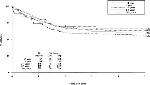
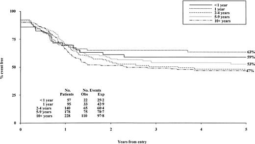
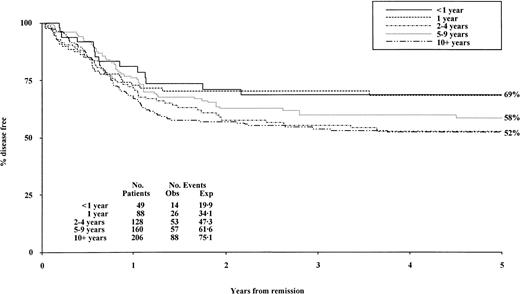
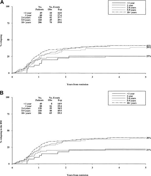
Reasons for treatment failure are shown in Table 2. There was no significant difference in overall survival (Figure1, P = .2) or event-free survival (Figure 2,P = .09), but disease-free survival was superior in younger children (Figure 3,P = .04) due to fewer relapses (P = .02, Figure 4A). The risk of bone marrow relapse was lower in younger children (P = .008, Figure4B) although this age group had slightly more CNS relapses (P = .05). However, CNS relapse was rare, only affecting 11 children overall. There was no difference in the incidence of deaths in first remission (8% overall, P = 1.0). Subdivision by age within the first year of life showed similar outcomes between groups.
Overall survival by age at diagnosis.
Obs indicates observed; Exp, expected.
Event-free survival by age at diagnosis.
Obs indicates observed; Exp, expected.
Disease-free survival by age at diagnosis.
Obs indicates observed; Exp, expected.
Relapse risk by age at diagnosis.
(A) Relapse risk by age at diagnosis. (B) Bone marrow relapse by age at diagnosis. Obs indicates observed; Exp, expected.
Relapse risk by age at diagnosis.
(A) Relapse risk by age at diagnosis. (B) Bone marrow relapse by age at diagnosis. Obs indicates observed; Exp, expected.
Multivariate analysis of prognostic factors (age, gender, initial WCC, FAB type and cytogenetic risk, with bone marrow status after course one of induction and MRC risk group also used in analyses of relapse risk, risk of death in remission, and disease-free survival) indicated that poor survival was related to increasing age (P = .02), higher presenting WCC (as a continuous variableP < .001), poor risk cytogenetics (P < .001), and FAB type other than M5 (P = .02). CR rates were lower in patients with adverse cytogenetic risk group (P = .002), lower initial WCC (P = .001), and FAB type M4 (P = .01), whereas induction deaths were more frequent with higher initial WCC (P = .02) and resistant disease was more common with adverse cytogenetic risk group (P < .001), FAB types M4 (P = .03) and M6 (P = .01), older age (P = .003), and higher initial WCC (P = .001). Significant adverse risk factors for relapse were increasing age (P = .02), male sex (P = .03), FAB type other than M5 (P = .04), higher WCC (P < .001), poor risk cytogenetics (P < .001), more than 15% blasts in the bone marrow following recovery from the first course of chemotherapy (P < .001), and MRC risk group (P < .001), whereas the only significant risk factor for death in remission was more than 15% blasts in the bone marrow following course one (P = .007). The only significant factor for CNS relapse was poor cytogenetic risk group (P = .001), whereas the risk factors for bone marrow relapse were identical to those for all relapses and achieved similar levels of significance. Disease-free survival was better with lower WCC (P < .001), good MRC risk group (P < .001), FAB type M5 (P = .03), and with 15% or fewer blasts in the marrow after the first course of induction (P < .001). Event-free survival was better with lower WCC (P < 0.001), favorable cytogenetic risk group (P < .001), M5 FAB type (P = .01), and younger age (P = 0.02). In multivariate analysis, age was not a significant factor for risk of CNS relapse (P = .2), risk of death in remission (P = .7), or CR rate (P = .5) and was not quite statistically significant for disease-free survival (worse prognosis with increasing ageP = .06) and induction death (better prognosis with increasing age P = .06). These findings are in line with those reported for the AML 10 trial with adult and pediatric data combined.12
Discussion
Although major collaborative groups have demonstrated an improved prognosis for AML,1-4 few publications have concentrated on the specific features of the disease at different ages in childhood.2,5-7 Two studies presented data for children aged under 2 years compared with older children7 8 and reported a similar pattern of AML between the first and second year of life with distinctive features when compared to older children. These very young children had high presenting WCCs and an increased incidence of monocytic disease. Analysis of cytogenetics indicated a high frequency of abnormalities of chromosome 11 with breakpoints at 11q23. A high incidence of CNS disease of 20% at diagnosis was noted, and the CNS was a common site of relapse. The children had an increased incidence of AML M7, which was not associated with Down syndrome.
We also found that FAB type varied with age with a high incidence of AML M5 and M7 in very young children, whereas the incidence of M0, M1, M2, and M3 increased with age. It was not possible to identify a precise age cutoff for these trends.
Cytogenetics have consistently been shown to carry major prognostic significance in AML,11,13-15 and there were clear differences in the distribution of cytogenetic risk groups in our series, with a high incidence of translocations involving chromosome 11 with breakpoints at 11q23 in young children, a change strongly associated with AML M5.15 Cytogenetic results were available for more than 80% of the children and represent the most complete data set published. The low incidence of favorable cytogenetics in the youngest children was reflected by the relative infrequency of the associated FAB types—AML M2 with t(8;21) and M3 with t(15;17).
There has been disagreement regarding the effect of age in childhood on prognosis with both similar2,6,7 and poorer outcome5,8,9 reported compared to older children. In the series of Vormoor and colleagues,8 survival was worse in children under age 2 years due to resistant disease and relapse, but this was ascribed to the pattern of FAB types because the younger children had an excess of FAB types M5 and M7 identified as carrying poor risk in children treated on Berlin-Frankfurt-Munster (BFM) group protocols.1 However, in AML 10 and AML 12, FAB type is not included in risk group assignment.11 An analysis of factors affecting response in AML 1011 showed that poor performance status was a risk factor for induction death. Adverse cytogenetics, non-M3 FAB type, and high presenting WCC were risk factors for resistant disease, but the effects of FAB type and WCC were relatively weak compared with cytogenetics. In our series there was no difference in remission rates but resistant disease was less common in younger patients partly counterbalanced by an increase in induction deaths. Following successful induction therapy, relapse was also less common in younger children and there were no differences in deaths in remission, resulting in better disease-free and overall survival.
Multivariate analysis of prognostic factors for children confirmed the importance of cytogenetics, response to the first course of chemotherapy, overall MRC risk group (a combination of cytogenetics and response to course one of chemotherapy), and the effects of high presenting WCCs, and demonstrated that age within childhood affected survival, relapse rate, and disease-free survival. Our experience suggests that better control of AML is achieved in these children using treatment of the intensity prescribed in MRC trials, irrespective of FAB types.
The increased incidence of CNS disease at diagnosis in younger children and the marginal increase in CNS relapse concurs with other reports.7 8 The low CNS relapse rate (only 11 overall) may relate to overall dose intensity of therapy because AML 10 and 12 are among the most intensive of regimens reported for the treatment of AML in children, and better systemic control may result in less CNS relapse. Intrathecal treatment in our patients was limited to triple therapy and cranial radiation was not routinely administered. It appears clear from MRC data that provided overall treatment intensity is appropriate cranial radiation is an unnecessary source of potential CNS toxicity.
A major concern is the appropriateness of current dose adjustments made in chemotherapy protocols for infants (generally based on prescription as milligram per kilogram or in our series on drug reductions by 25%) occasioned by the relatively high surface area for body weight and differences in pharmacokinetics.16 There is variation between the protocols of different collaborative groups both in drug dosages and scheduling and in the methods of dose reduction applied to young children that may have important effects on treatment intensity.
This concern is especially important given the increased severity of diarrhea in young children in our series and the reported increased risk of bacteremia associated with gastrointestinal toxicity of chemotherapy.17 However, the better disease-free survival rate in children under age 2 years is encouraging. Together these findings suggest that younger children may have received relatively more intensive therapy, and increased treatment intensity is associated with improved disease-free survival in childhood AML.3 The lack of good pharmacokinetic data to accurately determine dosages indicates the strong need for appropriate studies in this area to guide improvements to the therapeutic ratio. There was more severe diarrhea in younger children and a marginally statistically significant excess in deaths during induction, but less resistant disease and relapse. Accordingly, further dose reductions are inappropriate at this time, but attention to the supportive care needs of very young children must be emphasized.
Membership of the MRC Childhood Leukaemia working party during the AML 10 and AML 12 trials: C. Bailey, M. Caswell, J. Chessells, P. Darbyshire, S. Dempsey, J. Durrant, O. Eden, K. Forman, B. Gibson, A. Goodman, I. Hann, C. Harrison, C. Haworth, F. Hill, M. Jenney, D. King, S. Kinsey, J. Lilleyman, M. Madden, S. Meller, C. Mitchell, A. Oakhill, R. Pinkerton, M. Radford, M. Reid, S. Richards, V. Saha, J. Simpson, O. Smith, R. Stevens, A. Thomas, F. Vargha-Khadem, A. Vora, D. Webb, K. Wheatley, A. Will, and K. Windebank.
The publication costs of this article were defrayed in part by page charge payment. Therefore, and solely to indicate this fact, this article is hereby marked “advertisement” in accordance with 18 U.S.C. section 1734.
References
Author notes
David K. H. Webb, Haematology Department, Great Ormond Street Hospital, London WC1N 3JH, United Kingdom; e-mail:david.webb@gosh-tr.nthames.nhs.uk.


This feature is available to Subscribers Only
Sign In or Create an Account Close Modal