Abstract
Human telomerase reverse transcriptase (hTERT) is considered a potential target for cancer immunotherapy because it is preferentially expressed in malignant cells. hTERT-derived peptides carrying motifs for HLA-A24 (HLA-A*2402), the most common allele among Japanese and also frequently present in persons of European descent, were examined for their capacity to elicit antileukemia cytotoxic T lymphocytes (CTLs). Two of the 5 peptides tested, VYAETKHFL and VYGFVRACL, appeared capable of generating hTERT peptide-specific and HLA-A24–restricted CTLs. The CD8+ CTL clones specific for these hTERT peptides exerted cytotoxicity against leukemia cells in an HLA-A24–restricted manner. This cytotoxicity was inhibited by the addition of hTERT peptide-loaded autologous cells, suggesting that hTERT is naturally processed in leukemia cells and that hTERT-derived peptides are expressed on these cells and are recognized by CTLs in the context of HLA-A24. Taken together with the currently identified HLA-A2–restricted CTL epitopes derived from hTERT, identification of new CTL epitopes presented by HLA-A24 increases the feasibility of immunotherapy for leukemia using hTERT-derived peptides.
Introduction
Telomerase is a ribonucleoprotein enzyme that plays a key role in determining telomere length and cellular replicative life span.1,2 Three components of human telomerase, human telomerase RNA component (hTERC),3 human telomerase protein 1 (hTEP1),4,5 and human telomerase reverse transcriptase (hTERT),6,7 have been identified recently. Among them, mRNA expression of hTERT, the catalytic subunit of human telomerase, has been highly detected in telomerase-positive primary tumors and cancer cell lines but not in telomerase-negative cells. Induction of the hTERT gene in telomerase-negative cells resulted in expression levels comparable to those in immortal telomerase-positive cells.8 The evidence that telomerase is activated in more than 85% of cancer cells, including hematopoietic malignancies, but not in normal cells9-14has led to studies of the usefulness of telomerase for cancer diagnostics and therapeutics.
Cytotoxic T lymphocytes (CTLs) undoubtedly play a crucial role in resistance to cancer. Although various proteins have been identified as tumor-associated antigens for melanoma-specific and solid tumor–specific CTLs,15 the number of potential target antigens of CTLs directed against leukemia is still limited.16-24 Because hTERT is expressed in most types of leukemia but not in normal tissues, immunotherapy using CTLs directed against hTERT seems potentially efficacious. Indeed, it was reported recently that HLA-A2 (HLA-A*0201)-restricted hTERT peptide-specific CTLs can lyse various malignant cells but not normal cells.25 In the present study, we identified the peptide sequences derived from hTERT, which can elicit hTERT peptide-specific CTLs restricted by HLA-A24 (HLA-A*2402), the most common allele in Japanese (more than 60%) and also present in persons of European descent (nearly 20%). The CD8+ CTL clones specific for these hTERT peptides appear to be cytotoxic against HLA-A24+ leukemia cells but not against HLA-A24− leukemia cells or HLA-A24+ normal cells. The findings of this study support the feasibility of developing effective immunotherapy against hematopoietic malignancies using hTERT-derived peptides.
Study design
Cell lines
B-lymphoblastoid cell lines (B-LCLs) were established by the transformation of peripheral blood B lymphocytes using Epstein-Barr virus. LCLs and C1R cell line were cultured in RPMI 1640 medium supplemented with 10% fetal calf serum (FCS). TheHLA-A*2402 gene–transfected C1R cell line (C1R-A*2402)26 was cultured in RPMI 1640 medium supplemented with 10% FCS and 500 μg/mL hygromycin B (Sigma, St Louis, MO).
Synthetic peptides
According to peptide motifs for HLA molecules,27the sequence of hTERT was reviewed, and five 9-mer peptides consisting of binding motifs for HLA-A*2402 (the most common HLA class I type in the Japanese population) were synthesized. Amino acid sequences of the 5 synthetic peptides—designated TEL324, TEL385, TEL461, TEL845, and TEL1088—are as follows (underlined amino acids indicate binding motifs for HLA-A*2402): TEL324, VYAETKHFL (residues 324-332); TEL385, RYWQMRPLF (residues 385-393); TEL461, VYGFVRACL (residues 461-469); TEL845, CYGDMENKL (residues 845-853); and TEL1088, TYVPLLGSL (residues 1088-1096). Peptides derived from MAGE-3, IMPKAGLLI (residues 195-203)28 and human immunodeficiency virus type 1 (HIV-1) gp41, RYLKDQQLL (residues 583-591)29 were also synthesized and were used as positive controls because they can bind to HLA-A24 molecules and elicit the generation of peptide-specific and HLA-A24–restricted CTLs. Peptides were synthesized to a minimum purity of 90% using an automated peptide synthesizer (model 432A Synergy; Applied Biosystems, Foster City, CA) with the 9-fluorenyl-methoxycarbonyl procedure.
Generation of hTERT peptide-specific CTL clones
Peripheral blood monocyte-derived dendritic cells (DCs) were generated as described previously.22 Briefly, monocytes were isolated from the peripheral blood mononuclear cells (PBMCs) of HLA-A24+ healthy individuals. Plastic adherent cells were cultured in RPMI 1640 medium supplemented with 10% FCS, 500 U/mL recombinant human interleukin-4 (IL-4) (Genzyme, Boston, MA), and 800 U/mL recombinant human granulocyte-macrophage colony-stimulating factor (GM-CSF) (Kirin Brewery, Tokyo, Japan). On day 3 of incubation, fresh medium supplemented with IL-4 and GM-CSF was added, and on day 5, that medium was exchanged for fresh medium supplemented with IL-4, GM-CSF, and 100 U/mL recombinant human tumor necrosis factor-α (Dainippon Pharmaceutical, Osaka, Japan). On day 8 or 9, the cells were harvested and used as monocyte-derived DCs for antigen presentation. Generated cells appeared to express DC-associated antigens, such as CD1a, CD80, CD83, CD86, and HLA class I and class II.
The CD8+ T lymphocytes were isolated from peripheral blood lymphocytes from the same donors using polystyrene beads coated with an anti-CD8 monoclonal antibody (mAb) (DYNAL, Oslo, Norway). One million CD8+ T lymphocytes were cultured with 1 × 10⁵ autologous DCs treated with mitomycin C (MMC) (Kyowa Hakko, Tokyo, Japan) in RPMI 1640 medium supplemented with 10% human AB-type serum and 5 ng/mL recombinant human IL-7 (Genzyme), together with an hTERT synthetic peptide at a concentration of 10 μM in a 16-mm well. After culture for 7 days, half the medium was exchanged for fresh culture medium, and the cells were stimulated again by adding 1 × 10⁵ autologous DCs and 10 μM of an hTERT peptide. After culture for 7 additional days, the cells were stimulated a third time in the same manner. After culture for 3 more days (day 17 of culture), 10 U/mL recombinant human IL-2 (Boehringer Mannheim, Mannheim, Germany) was added to each well. The cytotoxicity of the growing cells was examined, and the bulk of the cells that were cytotoxic to an hTERT peptide–loaded autologous B-LCL were cloned.
To establish the T-cell clones, we performed a limiting dilution method as described previously.30 Bulk line cells were seeded at a concentration of 1 cell/well in round-bottomed microtiter wells containing 0.2 mL culture medium with IL-2 (10 U/mL) and 1 × 10⁵ MMC-treated autologous PBMC. After 2 weeks of culture, the cells were transferred into 16-mm wells and were expanded in the presence of IL-2 (10 U/mL).
HLA typing
HLA serotyping was performed using a microlymphocyte cytotoxicity assay with local qualified antisera. According to the serologic typing results, HLA class I alleles were amplified by the polymerase chain reaction using group-specific primers, then typed at the nucleotide sequence level. For example, HLA-A9 group alleles, including HLA-A*2402, were amplified using HLA-A9–specific primers and then analyzed using 9 probes used to distinguish 6 alleles (A*2301, A*2402-A*2406), as described previously.31 HLA-A24 expression on some leukemia cells was examined by flow cytometry using a fluorescein isothiocyanate (FITC)-conjugated anti–HLA-A24 mAb (One Lambda, Canoga Park, CA) and FITC-conjugated mouse IgG as the control.
Measurement of telomerase activity
Telomerase activity was measured by the telomeric repeat amplification protocol (TRAP) using the TRAPEZE assay kit (Intergen, Gaithersburg, MD) in accordance with the manufacturer's instructions.
Cytotoxicity assays
Cytotoxicity was examined by standard chromium 51 (51Cr) release assays, as described previously.32 Briefly, 1 × 10⁴ 51Cr (Na251CrO4) (New England Nuclear, Boston, MA)-labeled target cells suspended in 100 μL RPMI 1640 medium supplemented with 10% FCS (assay medium) were seeded into round-bottomed microtiter wells and incubated with or without a synthetic peptide for 2 hours. In some experiments, target cells were incubated with an anti-HLA class I framework mAb, w6/32 (ATCC, Rockville, MD), or an anti-HLA-DR mAb, L243 (ATCC), at an optimal concentration (10 μg/mL) for 30 minutes to determine whether cytotoxicity was restricted by HLA class I. Various numbers of effector cells suspended in 100 μL assay medium were added to the well and incubated for 4 hours, and 100 μL supernatant was collected from each well. The percentage of specific lysis was calculated as follows: (experimental release cpm − spontaneous release cpm)/(maximal release cpm − spontaneous release cpm).
Cold target inhibition assays
To examine whether hTERT-specific CTLs lyse leukemia cells through the recognition of hTERT peptides, which are naturally processed in leukemia cells in the context of HLA-A24, cold target inhibition assays were performed as follows. Autologous LCL cells were incubated with an hTERT-derived peptide at a concentration of 10 μM for 2 hours. After extensive washing, peptide-loaded cells were used as “cold” target cells. Various numbers of cold target cells were incubated with 1 × 10⁵ cytotoxic effector cells for 1 hour, and then 1 × 10451Cr-labeled leukemia cells were added to the wells. Cytotoxicity assays were performed as described above.
Results and discussion
Generation of hTERT peptide–specific CD8+ CTL clones
Two CD8+ CTL clones that lysed an hTERT peptide–loaded autologous LCL were generated from 2 healthy individuals, KYO and HAS. HLA class I types of the donors were as follows: KYO, HLA-A24/26 (*2402/*2601), B62/−, Cw9/w4; HAS, HLA-A24/33 (*2402/*3303), B62/54, Cw1/w9. CTL clones, which exerted cytotoxicity against hTERT peptide TEL324- and TEL461-loaded autologous LCL, were designated as KYO-1 and HAS-1, respectively. Antigen specificity and HLA restriction of cytotoxicity mediated by KYO-1 and HAS-1 are summarized in Table 1. KYO-1 lysed autologous LCL cells that were loaded with the TEL324, but was not cytotoxic to unloaded or TEL385-, TEL461-, TEL845-, or TEL1088-loaded autologous LCL. On the other hand, HAS-1 lysed only TEL461-loaded autologous LCL. Additionally, neither KYO-1 nor HAS-1 lysed autologous LCL loaded with peptides derived from either MAGE-3 or HIV-1 gp41 consisting of the binding motifs for HLA-A24. Cytotoxicity mediated by KYO-1 and HAS-1 seemed to be restricted by HLA-A24 because only hTERT peptide–loaded HLA-A24+ LCLs were lysed by these CTL clones. To further confirm the HLA-A24 restriction of these clones, we examined their cytotoxicity against the hTERT peptide–loaded HLA-A*2402 transfectant cell line, C1R-A*2402. In the presence of each peptide, KYO-1 and HAS-1 were cytotoxic to C1R-A*2402 but not to its HLA− parent cell line, C1R. Pretreatment of peptide-loaded autologous LCLs with an anti-HLA class I mAb inhibited the cytotoxicity of KYO-1 and HAS-1 (data not shown).
HLA-A24–restricted and human telomerase reverse transcriptase peptide–specific cytotoxicity of KYO-1 and HAS-1
| Target cells . | HLA class I alleles (HLA-A*alleles) . | Peptide . | % Specific lysis* . | |||||
|---|---|---|---|---|---|---|---|---|
| KYO-1 . | HAS-1 . | |||||||
| 10:1 . | 5:1 . | 2.5:1 . | 10:1 . | 5:1 . | 2.5:1 . | |||
| Autologous LCL | KYO: A24/26, B62/−, Cw9/w4 | None | 0 | 0 | 0 | 0 | 0 | 0 |
| (*2402/*2601) | TEL324 | 71.0 | 66.8 | 58.0 | 1.3 | 0 | 0 | |
| HAS: A24/33, B62/54, Cw1/w9 | TEL385 | 0 | 0 | 0 | 0 | 0 | 0 | |
| (*2402/*3303) | TEL461 | 0 | 0 | 0 | 65.4 | 52.8 | 47.4 | |
| TEL845 | 0 | 0 | 0 | 0.8 | 0 | 0 | ||
| TEL1088 | 0 | 0 | 0 | 0 | 0 | 0 | ||
| MAGE-3 | 0 | 0 | 0 | 0.8 | 0.4 | 0 | ||
| HIV-1 gp41 | 0 | 0 | 0 | 0.6 | 0.6 | 0 | ||
| Allogeneic LCL 1 | A24/−, B52/54, Cw1/w− | None | 0 | 0 | 0 | 1.2 | 0.6 | 0 |
| (*2402/*2402) | TEL324 | 69.4 | 56.2 | 47.8 | ND | ND | ND | |
| TEL461 | ND | ND | ND | 84.6 | 77.4 | 69.3 | ||
| Allogeneic LCL 2 | A24/2, B52/55, Cw1/w− | None | 0 | 0 | 0 | 0 | 0 | 0 |
| (*2402/*0201) | TEL324 | 78.2 | 68.0 | 56.2 | ND | ND | ND | |
| TEL461 | ND | ND | ND | 85.2 | 76.4 | 65.2 | ||
| Allogeneic LCL 3 | A26/31, B61/62, Cw3/− | None | 1.1 | 1.4 | 0.8 | 0.8 | 1.2 | 0.5 |
| (*2601/*3101) | TEL324 | 2.1 | 1.8 | 0.6 | ND | ND | ND | |
| TEL461 | ND | ND | ND | 0.8 | 0.8 | 0.6 | ||
| Allogeneic LCL 4 | A26/33, B44/51, Cw−/− | None | 0 | 0 | 0 | 0 | 0 | 0 |
| (*2601/*3303) | TEL324 | 0 | 0 | 0 | ND | ND | ND | |
| TEL461 | ND | ND | ND | 1.5 | 0 | 0 | ||
| C1R | Negative | None | 0 | 0 | 0 | 0 | 0 | 0 |
| TEL324 | 0 | 0 | 0 | ND | ND | ND | ||
| TEL461 | ND | ND | ND | 0 | 0 | 0 | ||
| C1R-A*2402 | A24 | None | 0 | 0 | 0 | 0.4 | 0 | 0 |
| (*2402) | TEL324 | 71.7 | 59.2 | 46.0 | ND | ND | ND | |
| TEL461 | ND | ND | ND | 55.2 | 43.2 | 37.6 | ||
LCL indicates lymphoblastoid cell line; ND, not determined.
The cytotoxicity of KYO-1 and HAS-1 to various LCLs loaded or not loaded with the hTERT peptide was determined by 4-hour 51Cr release assays at effector:target ratios of 10:1, 5:1, and 2.5:1.
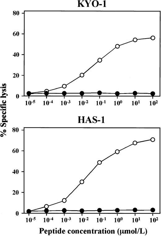
As shown in Figure 1, both CTL clones showed cytotoxicity to peptide-loaded autologous LCLs, depending on the peptide concentration. KYO-1 and HAS-1 showed cytotoxicity at a peptide concentration range of 1 nM to 100 μM. The optimal peptide concentration range of these clones seemed to be 1 μM to 100 μM.
hTERT peptide concentration–dependent cytotoxicity of KYO-1 and HAS-1.
The cytotoxicity of KYO-1 and HAS-1 to autologous (○) and HLA-A24− allogeneic (●) LCLs loaded with various concentrations of an hTERT peptide, TEL324 or TEL461, was examined. The cytotoxicity of KYO-1 and HAS-1 was determined by 4-hour51Cr release assays at an effector-target ratio of 5:1.
Lysis of leukemia cell lines by hTERT peptide–specific CTL clones
The cytotoxicity of KYO-1 and HAS-1 against normal cells and leukemia cell lines is shown in Table 2. All leukemia cell lines examined showed high telomerase activity, whereas telomerase activity in normal cells was low, in accordance with previous reports.9-14 Both KYO-1 and HAS-1 showed cytotoxicity against HLA-A24+ leukemia cell lines, whereas no cytotoxicity against HLA-A24− leukemia cell lines was detected. On the other hand, normal PBMCs and foreskin fibroblasts were not lysed by these CTL clones, regardless of HLA type.
Cytotoxicity of human telomerase reverse transcriptase peptide–specific cytotoxic T-lymphocyte clones to leukemia cell lines and normal cells
| Target cells . | HLA-A24 . | Telomerase activity* . | % Specific lysis† . | |||||
|---|---|---|---|---|---|---|---|---|
| KYO-1 . | HAS-1 . | |||||||
| 10:1 . | 5:1 . | 2.5:1 . | 10:1 . | 5:1 . | 2.5:1 . | |||
| Leukemia cell lines | ||||||||
| KH88 | + | 1.996 | 15.2 | 12.8 | 9.8 | 11.2 | 8.6 | 4.4 |
| MEG01 | + | 2.009 | 24.2 | 18.6 | 12.0 | 27.2 | 22.2 | 19.6 |
| OUN-1 | + | 1.894 | 19.7 | 17.9 | 14.1 | 13.6 | 11.2 | 8.5 |
| KT-1 | − | 1.522 | 0 | 0 | 0 | 0 | 0 | 0 |
| HL60 | − | 1.892 | 0 | 0 | 0 | 0 | 0 | 0 |
| K562 | − | 1.524 | 0 | 0 | 0 | 1.2 | 0.6 | 1.0 |
| Normal PBMCs | ||||||||
| Autologous (KYO) | + | 0.335 | 0 | 0 | 0 | ND | ND | ND |
| Autologous (HAS) | + | 0.271 | ND | ND | ND | 0 | 0 | 0 |
| Allogeneic 1 | + | 0.315 | 0 | 0 | 0 | 0 | 0 | 0 |
| Allogeneic 2 | − | 0.362 | 0 | 0 | 0 | 0 | 0 | 0 |
| Normal foreskin fibroblasts | ||||||||
| Allogeneic 1 | + | 0.024 | 0 | 0 | 0 | 0 | 0 | 0 |
| Allogeneic 2 | − | 0.023 | 0 | 0 | 0 | 0 | 0 | 0 |
PBMC indicates peripheral blood mononuclear cell; ND, not determined.
Telomerase activity was measured by TRAP assay.
Cytotoxicity of KYO-1 and HAS-1 to various leukemia cell lines, normal PBMCs, and normal foreskin fibroblasts in the absence of the hTERT peptide was determined by 4-hour 51Cr release assays at effector:target ratios of 10:1, 5:1, and 2.5:1.
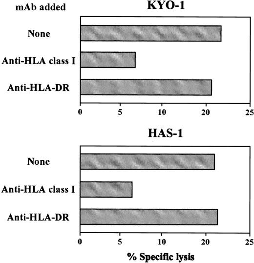
As shown in Figure 2, cytotoxicity of KYO-1 and HAS-1 against HLA-A24+ leukemia cell lines was inhibited by an anti-HLA class I mAb but not by an anti–HLA-DR mAb. These data suggest that KYO-1 and HAS-1 recognize hTERT as a target antigen and exert cytotoxicity against leukemia cells in an HLA-A24–restricted manner.
Effect of an anti-HLA class I mAb on cytotoxicity of KYO-1 and HAS-1 against leukemia cells.
The cytotoxicity of KYO-1 and HAS-1 against HLA A24+leukemia cell line, MEG01, preincubated with or without an anti-HLA class I mAb or an anti-HLA class II mAb was determined by 4-hour51Cr release assays at an effector-target ratio of 10:1.
Cold target inhibition assays
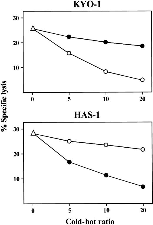
To further confirm that the cytotoxicity of hTERT peptide–specific CTL clones against leukemia cells was mediated by the specific recognition of endogenously processed hTERT, we performed cold target inhibition experiments. As shown in Figure3, the addition of TEL324-loaded autologous LCL resulted in decreased cytotoxicity of KYO-1 against MEG01, whereas the addition of TEL461-loaded autologous LCL showed no effect on cytotoxicity. On the other hand, cytotoxicity of HAS-1 was inhibited by adding TEL461-loaded, but not TEL324-loaded, autologous LCL. The same data were obtained from experiments using the KH88 leukemia cell line and the peptide-loaded autologous LCL as51Cr-labeled target and cold target cells, respectively (data not shown). These data strongly suggest that hTERT is naturally processed in leukemia cells and recognized by hTERT-specific CD8+ CTLs in the context of HLA-A24.
Cold target inhibition assays.
51Cr-labeled MEG01 cell line (1 × 10⁴ cells) was mixed with various numbers of unlabeled autologous LCL that had been loaded with TEL324 peptide (○) or TEL461 peptide (●). The cytotoxicity of KYO-1 and HAS-1 against the mixture of51Cr-labeled and unlabeled targets was determined by 4-hour51Cr release assays at an effector:51Cr-labeled target ratio of 10:1.
Although evidence that CTLs can lyse human leukemia cells through the specific recognition of tumor-associated antigens has been accumulating, the number of identified antigens recognized by antileukemia T lymphocytes is limited. So far, BCR-ABL, ETV6-AML1, proteinase-3, WT1, and cyclophilin B have been reported as the target antigens recognized by antileukemia human CTLs.16-24 Among them, fusion proteins such as BCR-ABL and ETV6-AML1 are expressed in leukemia cells exclusively; thus, these antigens are considered to be the potential targets for cancer immunotherapy. However, because the expression of fusion proteins is limited in certain kinds of leukemia, identification of leukemia-associated antigens expressed broadly in various subtypes of leukemia, so called universal tumor antigens, has been expected. According to recent findings that hTERT is expressed in more than 85% of malignancies, including various types of hematopoietic malignancy, and that its expression level in normal tissues is significantly low or undetectable,9-14 we attempted to establish hTERT-specific CTLs and to investigate their cytotoxic activity against leukemia cells.
In the present study, we have succeeded in establishing 2 hTERT peptide–specific HLA-A24–restricted CD8+ CTL clones, namely KYO-1 and HAS-1, from 2 healthy individuals. Both CTL clones lysed HLA-A24+ leukemia cell lines but not HLA-A24− leukemia cell lines or HLA-A24+normal cells. In a preliminary study, KYO-1 and HAS-1 also appeared to be cytotoxic to freshly isolated HLA-A24+ leukemia cells. The cytotoxicity of KYO-1 and HAS-1 against HLA-A24+leukemia cells was inhibited by an anti-HLA class I mAb. In addition, cold target inhibition experiments revealed that cytotoxicity against leukemia cells was inhibited by the addition of hTERT peptide–loaded autologous cells. These data strongly suggest that hTERT is naturally processed and expressed on leukemia cells with HLA-A24 molecules and that hTERT peptide–specific CTLs can discriminate leukemic cells from normal cells through the recognition of hTERT peptide in the context of HLA-A24 molecules.
It has recently been reported that a 9-mer peptide, ILAKFLHWL, derived from hTERT (amino acid residues 540-548), is capable of eliciting HLA-A2–restricted CTLs.25 CTLs specific for this hTERT peptide appeared to lyse tumor cells, including leukemia cells in an HLA-A2–restricted manner. In the present study, we have identified new hTERT epitopes recognized by CTLs restricted by HLA-A24, which is positive in nearly 20% of persons of European descent and more than 60% of Japanese. Of 5 hTERT-derived peptides, 2 peptides, VYAETKHFL and VYGFVRACL, could generate hTERT-specific CTLs. Binding affinity of these peptides to HLA-A24 molecules determined by peptide-motif scoring system (http://bimas.dcrt.nih.gov/molbio/hla_bind/) appeared to be relatively high. We also investigated anchor motifs for the worldwide common HLA class I alleles besides HLA-A*0201 and A*2402, such as A*0301, A*1101, B*0701, B*3501, and B*5101, in hTERT amino acid sequence, and we identified the high and intermediate binding motifs for each HLA allele. Although these findings suggest that cancer immunotherapy targeting hTERT would be universally effective regardless of the patient's HLA type, further studies using lymphocytes of donors whose HLA types are not HLA-A2 or HLA-A24 are necessary to confirm the universal usefulness of hTERT for cancer immunotherapy.
In summary, we have identified 2 new hTERT-derived peptides that can elicit HLA-A24–restricted antileukemia CTLs. Because the expression of hTERT is not limited in hematopoietic malignancies but is widely detected in cancers, immunotherapy targeting hTERT must be applicable for various kinds of malignancy. The study of immunotherapy using hTERT-derived peptides for cancer is also underway in our laboratory.
Note added in proof. After this work was submitted, Minev et al33 also published the establishment of HLA-A2.1–restricted CTLs specific for hTERT-derived peptides (540ILAKFLHWL548 and865RLVDDFLLV873), which lysed hTERT-positive cancer cells.
We thank Drs Masafumi Takiguchi (Kumamoto University, Japan), Masuhiro Takahashi (Niigata University, Japan), and Hayato Yamauchi (Ehime University, Japan) for providing the cell lines. We thank D. Dalma-Weiszhausz for critically reviewing the manuscript. We also thank Kirin Brewery, Dainippon Pharmaceutical, and Kyowa Hakko Kogyo for providing GM-CSF, TNF-α, and MMC, respectively.
Supported by grants from the Ministry of Education, Science, Sports and Culture of Japan; the Naito Foundation; and the Sagawa Cancer Research Foundation.
The publication costs of this article were defrayed in part by page charge payment. Therefore, and solely to indicate this fact, this article is hereby marked “advertisement” in accordance with 18 U.S.C. section 1734.
References
Author notes
Masaki Yasukawa, The First Department of Internal Medicine, Ehime University School of Medicine, Shigenobu, Ehime 791-0295, Japan; e-mail: yasukawa@m.ehime-u.ac.jp.


This feature is available to Subscribers Only
Sign In or Create an Account Close Modal