Abstract
Shp-1 and Shp-2 are cytoplasmic phosphotyrosine phosphatases with similar structures. Mice deficient in Shp-2 die at midgestation with defects in mesodermal patterning, and a hypomorphic mutation at the Shp-1 locus results in the moth-eaten viable (mev) phenotype. Previously, a critical role of Shp-2 in mediating erythroid/myeloid cell development was demonstrated. By using the RAG-2–deficient blastocyst complementation, the role of Shp-2 in lymphopoiesis has been determined. Chimeric mice generated by injecting Shp-2−/− embryonic stem cells into Rag-2–deficient blastocysts had no detectable mature T and B cells, serum immunoglobulin M, or even Thy-1+ and B220+ precursor lymphocytes. Collectively, these results suggest a positive role of Shp-2 in the development of all blood cell lineages, in contrast to the negative effect of Shp-1 in this process. To determine whether Shp-1 and Shp-2 interact in hematopoiesis, Shp-2−/−:mev/mev double-mutant embryos were generated and the hematopoietic cell development in the yolk sacs was examined. More hematopoietic stem/progenitor cells were detected in Shp-2−/−:mev/mevembryos than in Shp-2−/− littermates. The partial rescue by Shp-1 deficiency of the defective hematopoiesis caused by the Shp-2 mutation suggests that Shp-1 and Shp-2 have antagonistic effects in hematopoiesis, possibly through a bidirectional modulation of the same signaling pathway(s).
Introduction
Shp-1 and Shp-2 compose a small subfamily of protein tyrosine phosphatases that are distinguished by possession of Src-homology 2 domains.1-3 Shp-2 is a widely expressed enzyme in contrast to Shp-1, which is predominantly expressed in hematopoietic and lymphoid cells. Thus, these 2 phosphatases coexist in blood cell lineages. In the past few years, experimental results from a number of laboratories have indicated critical roles of Shp-1 and Shp-2 in the modulation of signaling events in various types of blood cells, in a positive or a negative fashion.2,4 5 However, several important issues remain to be addressed.
First, although Shp-2 was shown to be required for erythroid/myeloid cell differentiation, it is unclear whether Shp-2 plays a critical role in lymphocyte development. In previous experiments, we created a targeted mutant Shp-2 allele by deleting exon 3, which results in embryonic lethality at midgestation in homozygotes.6 In vitro hematopoietic differentiation assay of Shp-2−/−embryonic stem (ES) cells showed a severe suppression of erythroid/myeloid progenitor cell development by the Shp-2 mutation.7 Consistently, neither erythroid nor myeloid progenitor cells of Shp-2−/− origin were detectable in the fetal liver or bone marrow of chimeric animals that were derived from aggregation of Shp-2−/− ES cells and wild-type embryos, despite a significant contribution of Shp-2−/−cells to quite a few tissues of the chimeras.8 These observations suggest a stringent requirement of a functional Shp-2 for erythroid/myeloid cell development. However, a specific role of Shp-2 phosphatase in lymphocyte development remains to be established. This is clearly important for identifying the acting point of Shp-2 in the hierarchy of stem/progenitor cell differentiation into erythroid, myeloid, and lymphoid cell lineages.
Second, moth-eaten (me) and moth-eaten viable (meV) mice that contain spontaneous mutations in the Shp-1gene9,10 are characterized by immunodeficiency, autoimmunity, and augmented production and tissue infiltration of granulocytes, macrophages, and lymphocytes as well as excessive erythropoiesis.4,11 Thus, Shp-1 is primarily a negative effector in hematopoietic cell development and function, as compared with Shp-2, which has been implicated as both a negative and positive regulator in lymphocyte signaling.4,12 13 Because Shp-1 and Shp-2 are coexpressed in hematopoeitic cells, it is unclear whether they interact or even have antagonistic effects in hematopoiesis.
This study was initiated with the goal of addressing these 2 questions. First, we examined the specific requirement of Shp-2 for lymphocyte development using the Rag-2–deficient blastocyst complementation. Second, we investigated the functional interaction of Shp-1 and Shp-2 using a genetic approach by generating double-mutant embryos and analyzing the effect of the double Shp-1/Shp-2 mutations on hematopoiesis. Our findings suggest that Shp-2 is required for lymphopoiesis and that Shp-1 and Shp-2 have antagonistic effect in hematopoiesis.
Materials and methods
Generation of Shp-2−/−/Rag-2−/−chimeric mice
Rag-2–deficient mice were maintained in a pathogen-free facility, and 4- to 8-week-old females were used as blastocyst donors. Rag-2−/− blastocysts were recovered from the uterus of 3.5-day postcoitum pregnant females, and Shp-2+/− and Shp-2−/− ES cells were injected into Rag-2−/− blastocysts as described.14,15Injected embryos were then transferred into the uterus of synchronized pseudopregnant foster mothers. After pups were born, chimeric mice were identified by agouti coat color and by polymerase chain reaction (PCR) assaying for the Shp-2 mutant allele in the tail DNA.8
Flow cytometry analysis
Chimeric mice (6 weeks old) generated from Shp-2+/−or Shp-2−/− ES cells were euthanized. Spleen, thymus, and bone marrow cells were assayed by flow cytometry using conjugated antibodies specific to CD4, CD8, CD19, immunoglobulin (Ig) M, Thy-1, B220, and Ly9.1 (Pharmingen, San Diego, CA). Stained cells were analyzed on a Beckman FACScan, and live cells were collected and analyzed.
Generation of Shp-2, Shp-1 double-mutant mice
Heterozygous meV mice (C57BL/6J-Hcphme-v/+) were purchased from the Jackson Laboratory (Bar Harbor, ME). These mice were crossed with Shp-2+/− mice, and double-heterozygous (mev/+:Shp-2+/−) mutant mice were identified from the F1 offspring by PCR detection of wild-type and mutant alleles of Shp-2 and Shp-1 in tail DNA as described.8 16 Double- homozygous (mev/mev:Shp-2−/−) and single-homozygous (mev/mev or Shp-2−/−) embryos were generated by intercrossing mev/+:Shp-2+/− mutant mice.
Hematopoietic progenitor assay
Embryos at days 9.0 to 9.5 from the intercrosses between mev/+: Shp-2+/− mice were dissected. Yolk sacs were carefully separated from the embryos in α–minimal essential medium (α-MEM) supplemented with 15% fetal calf serum and dissociated to single-cell suspension by passing through a 22-gauge needle as described previously.8 Yolk sac cells were plated into a colony-forming unit (CFU)-assay system, which contains α-MEM, 30% fetal calf serum, 5% pokeweed mitogen-stimulated mouse spleen cell–conditioned medium, erythropoietin (2 U/mL EPO; Amgen, Thousand Oaks, CA), murine stem cell factor (50 ng/mL; Immunex, Seattle, WA), glutamine (10−4 M), β-mercaptoethanol (3.3 × 10⁻⁵ M), hemin (100 μm; Eastman Kodak, Rochester, NY), and 0.9% methylcellulose. CFU-assay cultures were incubated at 37°C in a 5% CO2 moisture-saturated incubator. After 6 to 7 days, colonies from erythroid burst-forming unit (BFU-E), granulocyte-macrophage (CFU-GM), and multipotential (CFU-granulocyte, erythroid, macrophage, megakaryocyte, or CFU-GEMM) progenitors were s cored.8 17
Results and discussion
Block of T- and B-lymphocyte development by the Shp-2 mutation
Our previous work showed that Shp-2 is required for erythroid and myeloid cell differentiation.7,8 To determine the role of Shp-2 in lymphocyte development, we employed the Rag-2–deficient blastocyst complementation assay.14 18Shp-2+/− or Shp-2−/− ES cells were injected into Rag-2−/− blastocysts to generate chimeric animals. Because Rag-2−/− mice do not produce any T and B lymphocytes because of the inability to initiate V(D)J recombination, any mature T and B lymphocytes detected in the chimeras must be derived from injected ES cells.
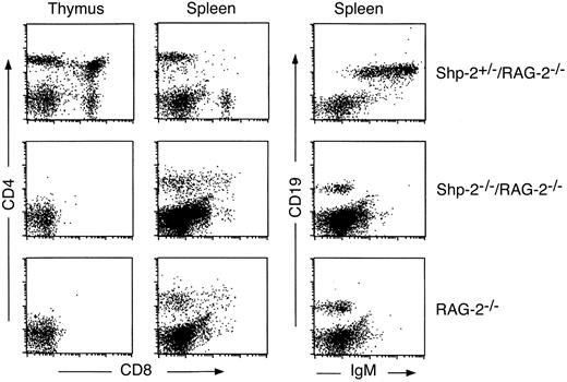
Splenocytes and thymocytes from RAG−/− mice or chimeras generated from Shp-2+/− or Shp-2−/− ES cells were stained for CD4 and CD8 and then analyzed by flow cytometry. In Shp-2+/−/Rag-2−/− chimeras, CD4+or CD8+ mature T cells were readily detected in the spleen, and CD4+, CD8+, and CD4+CD8+ cells were detected in the thymus (Figure 1). Although there were some CD4+ cells in the spleen of Shp-2−/−/Rag-2−/− chimeras, the staining profile was the same as in Rag-2−/− mice. Like Rag-2−/− mice, there were no CD4, CD8 single-positive cells or CD4 and CD8 double-positive cells in the thymus of Shp-2−/−/Rag-2−/− chimeras. Staining of splenocytes with CD19 and IgM revealed the presence of CD19+IgM+ mature B cells in Shp-2+/−/Rag-2−/− chimeras, whereas there were only few CD19+ precursor B cells in Shp-2−/−/Rag-2−/− chimeras or Rag-2−/− mice (Figure 1). Consistently, serum IgM, which is a more sensitive indication of the presence of mature B cells, was not detected by enzyme-linked immunosorbent assay in all 11 chimeric mice generated from Shp-2−/− ES cells (data not shown).
Shp-2 is required for T- and B-lymphocyte development.
Thymocytes or splenocytes were stained with anti-CD4 and anti-CD8a; splenocytes were also stained with anti-IgM and anti-CD19. Live cells (10 000) were collected and analyzed for each sample. Data from one Shp-2+/−/Rag-2−/− and one Shp-2−/−/Rag-2−/− chimera are shown. We examined 4 Shp-2−/−/Rag-2−/− chimeric animals and obtained consistent results.
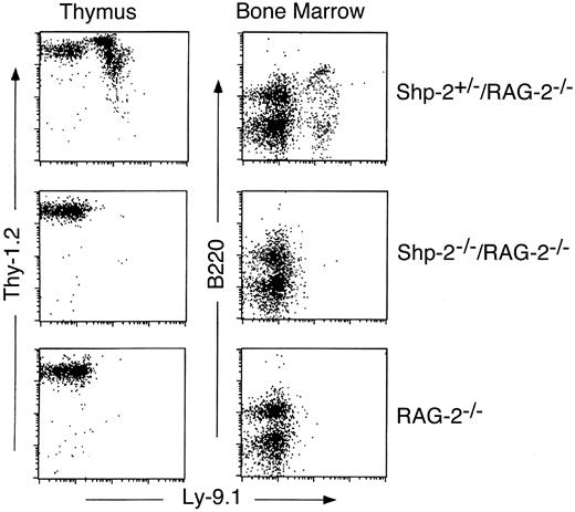
To determine the stage of lymphocyte development that was blocked by the Shp-2 mutation, ES cell–derived precursor T cells in the thymus and precursor B cells in the bone marrow were assayed by staining for Thy-1 plus Ly-9.1 or B220 plus Ly-9.1, respectively. Ly-9.1 staining was able to distinguish the ES and blastocyst-derived precursor lymphocytes because the R1 ES cell line used for Shp-2 targeting was derived from 129/sv strain of mouse and therefore was Ly-9.1+, whereas blastocyst-derived cells were Ly-9.1−. As shown in Figure2, Thy-1+Ly-9.1+T-lineage cells and B220+Ly-9.1+ B-lineage cells were detected in the thymus and bone marrow, respectively, in chimeras generated with Shp-2+/− ES cells. In contrast, no Thy-1+Ly-9.1+ T-lineage cells were detected in the thymus, and no B220+Ly-9.1+ B-lineage cells were detected in bone marrow cells of Shp-2−/−/Rag-2−/− chimeric mice. Together, these data suggest that the Shp-2 mutation causes a block of lymphocyte development at a very early stage, even before precursor T and B cells.
The Shp-2 mutation blocks lymphopoiesis at the precursor stage.
Thymocytes were stained with anti-LY9.1 and anti–THY-1, and bone marrow cells were stained with anti-LY9.1 and anti-B220 and analyzed by flow cytometry. Results from one Shp-2+/−/Rag-2−/− and one Shp-2−/−/Rag-2−/− chimera are shown. Four Shp-2−/−/Rag-2−/− chimeric animals were examined, and consistent results were obtained.
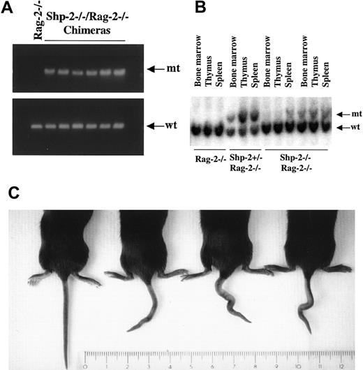
The failure to detect the ES cell–derived lymphocytes in Shp-2−/−/Rag-2−/− chimeras was not due to an inability to generate chimeric mice. First, chimerism was assayed by PCR analysis of tail DNA and was shown by the presence of the Shp-2 mutant allele (Figure 3A). Second, Southern blot analysis clearly detected the Shp-2 mutant allele in nonlymphoid lineage cells in the thymus, spleen, and bone marrow of Shp-2+/−/Rag-2−/− or Shp-2−/−/Rag-2−/− chimeric animals, indicating a reasonable contribution by mutant ES cells (Figure 3B). Third, all Shp-2−/−/Rag-2−/− chimeras, as determined by PCR, had kinky tails, a trait not seen in nonchimeras or chimeras generated from Shp-2+/− ES cells (Figure 3C). This phenotype was observed previously in Shp-2−/−/wild-type chimeric mice. The kinky tail phenotype was also observed in targeted mutant mice lacking fibroblast growth factor receptor-3, due to enhanced and prolonged endochondral bone growth accompanied by expansion of proliferating and hypertrophic chondrocytes within the cartilaginous growth plate.19Shp-2 phosphatase is required for the full and sustained activation of Erk kinase induced by fibroblast growth factor receptor-1.6 Collectively, these data suggest that the Shp-2 mutation has different effects on the development of various cell types: It is required for early lymphopoiesis and is involved in mediating the development of hypertrophic chondrocytes.
Detection of chimerism.
(A) PCR analysis of tail DNA. To detect the contribution of Shp-2−/− ES cells to the body of chimeric animals, tail DNA was extracted from Shp-2−/−/Rag-2−/−chimeras as well as one Rag-2−/− animal as a control. Detection of wild-type (wt) and mutant (mt) Shp-2 alleles was done by PCR analysis. (B) Southern blot analysis of DNA extracted from bone marrow, thymus, and spleen of Shp-2+/−/Rag-2−/− or Shp-2−/−/Rag-2−/− chimeras. Wild-type and mutant Shp-2 alleles were detected using a specific probe as described previously.8 (C) One Rag-2−/− animal (left) and 3 Shp-2−/−/Rag-2−/− chimeric mice with kinky tail are shown.
Shp-1 mutation partially rescues the hematopoietic defects caused by Shp-2 mutation
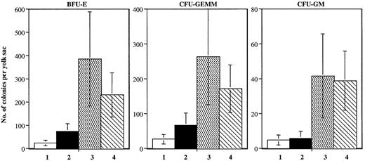
The results described above, together with our previous observations that the Shp-2 mutation suppressed erythroid/myeloid cell differentiation, suggest an important role of Shp-2 in the development of all blood cell lineages. To determine if there is any functional interaction between Shp-1 and Shp-2 in hematopoiesis, we generated double-mutant animals by crossing Shp-2+/− mice with meV/+ mice. Double-mutant mice (Shp-2−/−:meV/meV) also died at midgestation with a phenotype similar to Shp-2−/−embryos, having multiple defects in the mesodermal induction and body organization, particularly the posterior truncation, as described previously.6 Hematopoietic activity was assessed by detecting stem/progenitor cells from the yolk sacs using the CFU assay. As shown in Figure 4, hematopoietic cell development was dramatically suppressed in Shp-2−/− yolk sacs, consistent with the previous result.8 Interestingly, an additional homozygous mutation at the Shp-1 locus partially complemented the hematopoietic defect caused by the Shp-2 mutation. Higher numbers of erythroid lineage (BFU-E) and mixed erythroid-myeloid lineage (CFU-GEMM) progenitors were detected in the double-mutant yolk sacs than in Shp-2−/− littermates. This result suggests an antagonistic effect between Shp-1 and Shp-2 in the development of blood cells, the cell type in which both are normally expressed.
Antagonistic effect of Shp-1 and Shp-2 in mediating embryonic hematopoiesis.
Yolk sacs at 9.0 to 9.5 days postcoitum from intercrosses between mev/+:Shp-2+/− mice were carefully dissected free of maternal tissues in α-MEM supplemented with 15% fetal calf serum. Single-cell suspension was prepared by passing through a 22-gauge needle. Cells were washed once in α-MEM and plated for CFU assay. Genotyping: 1. Shp-2−/−:+/+ or mev/+; 2. Shp-2−/−:mev/mev; 3. Shp-2+/+,+/−:mev/mev; 4. Shp-2+/+,+/−:+/+, mev/+. The results were summarized from 5 independent experiments. There was a significant difference between Shp-2−/−:+/+ or mev/+ and Shp-2−/−:mev/mev mice for BFU-E and CFU-GEMM (P = .03, t test).
As a widely expressed enzyme, the involvement of Shp-2 in cytoplasmic signaling has been demonstrated in a variety of cell types. Shp-2 phosphatase has been implicated as a negative or a positive regulator in different signaling pathways.2 Introduction of a targeted mutation at a tyrosine phosphorylation site (Y759F) in gp130, a coreceptor for interleukin-6 family cytokines, resulted in splenomegaly, lymphadenopathy, and an enhanced acute phase reaction in mutant mice.20 Because Shp-2 binds to phosphorylated Y759, it was reasonable to speculate that Shp-2 acts to negatively regulate the development of T, B, and myeloid cells. However, a more recent report indicates that this tyrosine residue at gp130 is also a docking site for suppressor of cytokine signaling-3 (SOCS-3) and, therefore, the negative regulatory roles previously attritbuted to Shp-2 might be mediated, at least in part, by SOCS-3.21
Using the Rag-2–deficient blastocyst complementation, we have now clearly demonstrated a requirement of Shp-2 for lymphopoiesis in a cell-autonomous manner. Differentiation of lymphoid cell lineages in Shp-2−/−/Rag-2−/− chimeric mice was blocked before pro–T- and pro–B-cell stages. In combination with our previous observations,7,8 our data indicate that Shp-2 is required for the development of erythroid, myeloid, and lymphoid cell lineages. This is consistent with previous data demonstrating a defective expression of PU.1 and interleukin-7 receptor, critical genes for lymphocytes and other cell types, in embryoid bodies differentiated from Shp-2−/− ES cells.7 The block to all blood cell lineages by the Shp-2 mutation strongly suggests that Shp-2 has a role in hematopoietic stem/progenitor cell commitment and differentiation at a very early stage. However, our observation does not rule out the possibility that Shp-2 also has other functions, negative or positive, at later stages of hematopoiesis or in the activities of terminally differentiated myeloid/lymphoid cells. Insight into these aspects of Shp-2 function will be provided by generating conditional Shp-2 knockout alleles in a specific cell lineage at given developmental stages.
Whether Shp-1 and Shp-2 have functional interactions was not clear, although their physiologic roles are at least not completely redundant, because the defective phenotype caused by the Shp-1 mutation was not complemented by the normal expression of Shp-2 inmoth-eaten mice.1 Notably, one recent study identified an activity shared by Shp-1 and Shp-2 in mediating the inhibitory signal relay from paired Ig-like receptor B (PIR-B) in B lymphocytes.22 In contrast, we present evidence here that Shp-1 and Shp-2 have antagonistic effects in mediating embryonic hematopoiesis. The defective yolk sac hematopoiesis caused by theShp-2 mutation was partially rescued by an additionalShp-1 mutation. This finding will help to elucidate the cytoplasmic signaling events mediated by Shp-1 and Shp-2 in hematopoiesis and possibly leukemogenesis.
Acknowledgments
We thank Dr M. Boes for performing the IgM enzyme-linked immunosorbent assay and W. M. Yu for technical assistance.
Supported by grants from National Institutes of Health (CA78606 and GM53660 to G.-S.F. and AI40146 and AI 44478 to J.C.).
The publication costs of this article were defrayed in part by page charge payment. Therefore, and solely to indicate this fact, this article is hereby marked “advertisement” in accordance with 18 U.S.C. section 1734.
References
Author notes
Gen-Sheng Feng, The Burnham Institute, 10901 N Torrey Pines Rd, La Jolla, CA 92037; e-mail:gfeng@burnham-inst.org.


This feature is available to Subscribers Only
Sign In or Create an Account Close Modal