In mammals there are 2 cell types that can phagocytize: macrophages and neutrophils. Malignant cytophagocytosis is a fulminant disease characterized by phagocytosis of bone marrow cellular elements mainly by marrow macrophages (histiocytes) or rarely by other cells (eg, myeloid blasts). Cytophagocytosis by plasma cells in multiple myeloma is an extremely rare condition. Plasma cells are antibody-producing cells and have no phagocytic function. There are only a few reports describing phagocytic plasma cells in patients with multiple myeloma.1-14 None of these reports could explain the mechanism or the clinical importance of phagocytosis by plasma cells. Here we report a myeloma patient with phagocytic myeloma cells expressing CD15 on their surfaces.
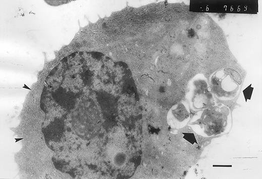
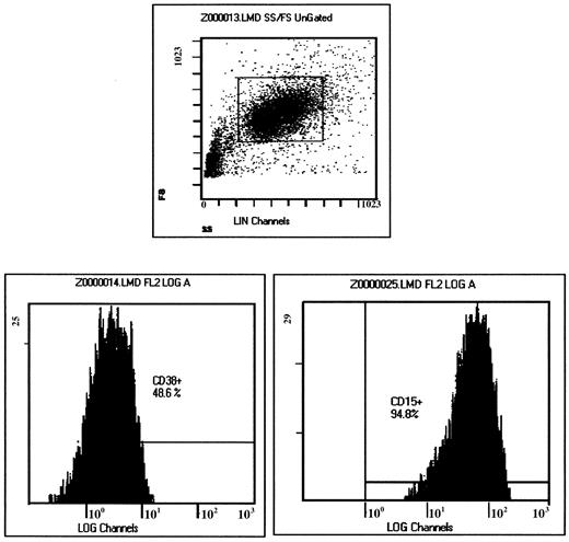
A 52-year-old female patient was admitted to our hospital with complaints of weakness and fatigue. Her medical history was unremarkable. Findings from a physical examination were normal, except that there was pallor. Laboratory findings were as follows: erythrocyte sedimentation rate was 120 mm/h; hemoglobin, 8.9 g/dL; white blood cell count, 3.8 × 10⁹/L; neutrophil count, 1.3 × 10⁹/L; and platelet count, 138 × 109/L. Rouleaux formation was seen on peripheral blood smear. Blood urea nitrogen, uric acid, calcium, aspartate aminotransferase (AST), alanine aminotransferase (ALT), and lactate dehydrogenase levels were normal. Serum protein electrophoresis showed a monoclonal band (4.22 g/dL) in the gamma region. Serum immunoelectrophoresis revealed an abnormal immunoprecipitin with IgG-kappa specificity. Beta-2 microglobulin level was 6478 ng/mL (normal range, 1100-2300 ng/mL). Results of x-ray studies of the extremities, skull, ribs, pelvis, and long bones were normal. Bone marrow aspiration showed normal maturation of the granulocytic, erythrocytic, and megakaryocytic series. The myeloid/erythroid ratio was 2:1. Plasma cells represented 22% of the nucleated cells of bone marrow, and binucleated and atypical plasma cells were seen. In one-third of plasma cells, phagocytosis of mainly erythrocytes and platelets (and rarely, neutrophils, myelocytes, and lymphocytes) was seen (Figure 1). Diffuse plasma-cell infiltration with prominent kappa monoclonality was shown in a bone marrow biopsy. Electron microscopic analysis of bone marrow samples showed atypical plasma cells with phagocytic vacuoles (Figure2). Immunophenotypic characterization of the plasma cells was done by using conjugated monoclonal antibodies against CD45, CD38, CD14, CD15, CD19, CD22, CD23, HLA-DR, and surface kappa and lambda. Antibodies against CD14, CD19, CD22, CD23, HLA-DR, and surface lambda were found to be negative; antibodies against CD38, CD15, and surface and cytoplasmic kappa were positive. The plasma cells were CD15bright and CD45dim+. Surprisingly, CD38+ cells also expressed CD15 on their surfaces (Figure 3). The patient received 6 courses of melphalane and methyl prednisolone therapy. Her clinical condition had improved, the monoclonal band was reduced to 1.2 g/dL, plasma cell concentration was reduced by 60%, and no phagocytic plasma cells were seen in a bone marrow examination.
Phagocytic plasma cells.
May-Grünwald-Giemsa stain (original magnification × 1000).
Phagocytic plasma cells.
May-Grünwald-Giemsa stain (original magnification × 1000).
Atypical plasma cell containing phagocytic vacuoles.
The phagocytic vacuoles are indicated by arrows, and the large amount of rough endoplasmic reticulum, by arrowheads. There is a nuclear chromatin typically resembling the appearance of a “cartwheel” (bar = 1 μm).
Atypical plasma cell containing phagocytic vacuoles.
The phagocytic vacuoles are indicated by arrows, and the large amount of rough endoplasmic reticulum, by arrowheads. There is a nuclear chromatin typically resembling the appearance of a “cartwheel” (bar = 1 μm).
Light scatter properties of analyzed cells (top).
The flow cytometric histograms clearly show that virtually all CD38+ cells (48.6%) in the gated area are positive for CD15 antigen.
Light scatter properties of analyzed cells (top).
The flow cytometric histograms clearly show that virtually all CD38+ cells (48.6%) in the gated area are positive for CD15 antigen.
In the 14 previous multiple myeloma cases with phagocytic plasma cells,1-14 to our knowledge 5 cases had IgG, 2 cases had IgA, 1 case had lambda light chain, and 1 case had nonsecretory-osteosclerotic multiple myeloma. It is noteworthy that, in most of these cases, there was phagocytosis of mainly mature erythrocytes and platelets. Some cases had severe anemia that was thought to be related with erythrophagocytosis.2,8,14Electron microscopic analysis showed phagocytosis in 2 cases,2,6 and in vitro ingestion of latex particles by plasma cells was demonstrated in 1 case.1 Immunophenotypic analysis had not been performed in previous cases. The phagocytosis mechanism could not be explained in these reports. Ludwig et al speculated that the phagocytic plasma cells might result from malignant expansion of one of the rare normal B-lymphocyte clones with phagocytic potential.4 It is not known whether such a normal phagocytic plasma cell clone exists.
We speculated that the aberrant expression of CD15 had a key role in phagocytic function of these plasma cells. CD15 (Leu-M1, Lewis x) is expressed on mature myeloid cells (mainly on neutrophils and monocytes) and is associated with phagocytosis and cell adhesion. Stocks et al showed that antibodies to CD15 suppressed phagocytosis.15Normally, plasma cells do not express CD15 and other myeloid markers. It has been shown that malignant plasma cells might rarely express some non B-cell antigens such as CD13, CD14, CD15, CD33, CD41, glycophorin A, CD2, CD4, and CD56. It has been suggested that aberrant expression of these antigens might be related to poor prognosis.16Although we do not perform routine immunophenotypic analysis in all multiple myeloma patients, we did not find CD15 expression in 12 myeloma patients whose bone marrow aspiration materials were evaluated in our laboratory. This was our first observed phagocytic multiple myeloma patient. Our patient presented with anemia, granulocytopenia, and thrombocytopenia, which were thought to be associated with cytophagocytosis rather than plasma cell infiltration of bone marrow. We believe that aberrant CD15 expression on plasma cells might be related to phagocytic function of plasma cells.


This feature is available to Subscribers Only
Sign In or Create an Account Close Modal