The present study sought to determine the impact of the route of administration of an adeno-associated virus (AAV) vector encoding human factor IX (hFIX) on the induction of an immune response against the vector and its xenogenic transgene product, hFIX. Increasing doses of AAV-hFIX were administered by different routes to C57Bl/6 mice, which typically demonstrate significant immune tolerance to hFIX. The route of delivery had a profound impact on serum hFIX levels as well as the induction of an anti-hFIX humoral immune response. At all dose levels tested, delivery of AAV-hFIX by an intramuscular (IM) route induced an antibody response against the human FIX protein and no hFIX was detected in the serum of animals even at doses of 2 × 10¹¹ DNA viral particles (vp) of AAV-hFIX. This was in stark contrast to the mice that received AAV-hFIX by intraportal vein (IPV) administration. No anti-hFIX inhibitors were observed in any of these mice and therapeutic levels of hFIX were detected in the serum of all mice that received doses of 2 × 10¹⁰ vp AAV-hFIX and higher. When pre-existing neutralizing immunity to AAV was established in mice, AAV-hFIX administration by either the IM or IPV routes did not result in detectable serum hFIX. Although hFIX expression was not observed in mice with pre-existing neutralizing immunity to AAV, an anti-hFIX response was induced in all of the animals that received AAV-hFIX by the IM route. This was not observed in the preimmune mice that received AAV-hFIX by IPV administration. These results suggest that the threshold of inducing an immune response against a secreted transgene product, in this case the xenoprotein hFIX, is lower when the vector is administered by the IM route even in animals with pre-existing immunity to AAV.
Introduction
A concern for those seeking to use gene therapy to correct diseases stemming from genetic deficiencies has been whether the expression of the product of a missing gene will result in an immune response against the “foreign” gene product. This is a complex issue where contributions of the vector, the transgene, and the patients' immunologic background need to be considered. The area that has occupied the greatest amount of time is the immunogenicity of the viral vectors that are used to deliver potentially therapeutic transgenes. Many studies have shown that the immunogenicity of the proteins encoded by endogenous viral genes can stimulate not only the destruction of the transduced cell, but may also lead to the cross-priming of an immune response against the transgene product itself. Recombinant adeno-associated virus (rAAV) vectors are thought to be well suited for gene replacement therapy because they are deleted of all wild-type AAV genes. In addition, AAV serotype 2 vectors are inefficient at transducing potent antigen-presenting dendritic cells (DCs).1 These features greatly reduce the immunogenicity of AAV vectors.
Despite these advantages of AAV vectors, the immune interaction of the host with the transgene and vector is more complex than can be explained by the presence or absence of viral genes. For instance, AAV vectors expressing secreted transgenes, such as ovalbumin (OVA) and factor IX (FIX), have been shown to elicit immune responses against the transgene. Administration of AAV vectors expressing OVA into C57Bl/6 mice by intramuscular (IM) and intravenous (IV) routes induced anti-OVA antibodies.2 IM administration of AAV vector encoding human FIX (hFIX) into C57Bl/6 mice also induced a humoral response directed against the transgene product.3-5 In contrast with these findings, it has been demonstrated that administration of AAV-hFIX by an intravascular route does not induce an anti-hFIX humoral immune response.6-8 Beyond the make-up of the gene therapy vector and the nature of the transgene, the dose of the gene therapy vector and the site of administration of the vector may contribute significantly to the immune response against the transgene product.
To explore these issues we examined the impact of the dose of vector on the induction of an anti-hFIX humoral immune response following IM or intraportal vein (IPV) delivery of AAV-hFIX. We also explored the implications of a pre-existing anti-AAV immune response on AAV-mediated hFIX expression. IM administration of AAV-hFIX vector generated a robust anti-hFIX immune response. This immune response against the transgene blocked hFIX expression in the serum of these mice. In contrast, intravascular administration of AAV-hFIX did not induce anti-hFIX antibodies and significant levels of hFIX were detected in all animals receiving at least 2 × 10¹⁰ DNA viral particles (vp) of vector. In addition, we observed that an anti-AAV immune response, generated with an AAV vector encoding an unrelated transgene, blocked AAV-hFIX transduction by both IM and IPV routes. However, pre-existing humoral immunity to AAV did not prevent the induction of an anti-hFIX immune response in mice receiving AAV-hFIX by an IM route. These results indicate that IPV administration of AAV-hFIX is superior to IM administration.
Materials and methods
Construction and production of AAV vectors
The AAV vectors expressing green fluorescent protein,9 β-galactosidase,10 and hFIX were constructed and generated as described previously.7 The hFIX complementary DNA (cDNA) was expressed from a chimeric cytomegalovirus-Moloney murine leukemia virus (CMV-MMLV) promoter-enhancer coupled to the MLV intervening sequence (IVS). This hFIX expression cassette used the bovine growth hormone polyadenylation signal sequence. AAV titers were determined by dot blot analysis.
Assessment of AAV readministration in mice
The 8-week-old C57BL/6, NIH-3, and Balb/C mice were purchased from Taconic (Germantown, NY). Mice were immunized with various doses of AAV-LacZ IV, and monitored weekly for neutralizing antibodies using serum obtained by retro-orbital bleed every 2 weeks and analyzed for hFIX expression as described below.
Detection of serum hFIX by enzyme-linked immunosorbent assay
Microtiter plates were coated with 100 μL/well of a solution containing 2 mg/mL monoclonal anti-hFIX (Boehringer Mannheim, Indianapolis, IN) diluted in 0.1 M carbonate, pH 9.6. Plates were incubated overnight at 4°C and then washed and blocked for 2 hours at room temperature with 100 μL/well of 1% (wt/vol) nonfat milk in borate-buffered saline (BBM). Test samples (50 μL/well) diluted in BBM (1:5) incubated 2 hours at room temperature and 50 μL/well of a 1:100 dilution of horseradish peroxidase (HRP)–conjugated goat antihuman factor IX antibody (Affinity Biologicals, Hamilton, ON, Canada) was added and incubated for 1.5 hours at room temperature. Color was developed for 25 minutes at room temperature with 50 μL of 1 mg/mL p-nitrophenyl phosphate in 34 mm citric acid, 67 mm dibasic sodium phosphate, 0.1% hydrogen peroxide, (vol/vol), pH 5.0 buffer. Color development was stopped with 50 μL/well 2 M sulfuric acid and the absorbance measured at 490 nm.
Determination of AAV virus-specific serum by enzyme-linked immunosorbent assay
The 96-well MaxiSorp flat surface Nunc-Immuno plates were coated with 5 × 10⁹ vp AAV in 100μL/well 0.1 M carbonate, pH 9.6, incubated overnight at 4°C, washed, and blocked with 100μL 1% bovine serum albumin/phosphate-buffered saline (BSA/PBS; incubated 2 hours at room temperature). One hundred microliters of the mouse test serum at 1:100 was added to each well and washed, and 100 μL 1:5000 rabbit antimouse IgG antibody in 0.01 M Tris-HCl, 0.25 M NaOH, pH 8.0, was added for 2 hours at room temperature. Then 100 μl/well of AKP substrate solution (Bio-Rad, Hercules, CA) was added to each well and the color allowed to develop for half-hour before stopping reactions with 50 μL 0.4 M NaOH. The plates were read at 405 nm. A positive read as more than 0.15 OD405.
Anti-hFIX assay
The 96-well enzyme-linked immunosorbent assay (ELISA) plates were incubated overnight at 4°C with 2 μg/mL Bene-FIX protein in 0.1 M carbonate, pH 9.6, and then washed 5 times in wash buffer. The plate was then blocked with 89 mM boric acid, 90 mM NaCl, 1% (wt/vol) BBM and washed 3 times. Serum samples (50 μL) diluted in BBM were added to each well and the plate incubated 2 hours at room temperature. The plates were washed 3 times before adding 50 μL/well goat antimouse IgG-HRP (1:5000 dilution in BBM) and incubating plate 2 hours at room temperature. The plates were washed with 89 mM boric acid, 90 mM NaCl, pH 8.3, and 50 μL developing solution (o-phenylenediamine dihydrochloride (Sigma, St Louis, MO, P9187) in 10 mL substrate buffer; 10 μL hydrogen peroxide (Sigma, H1009) was added to each well and incubated in the dark for 30 minutes at room temperature. Reactions were stopped with 50 μL 0.4 M NaOH and the plates read at 490 nm.
Results
Immune implications of route of administration of rAAV
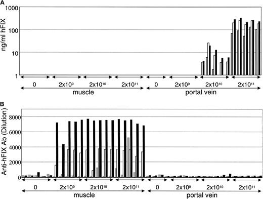
We sought to explore the effect of the route of administration of rAAV and dose of vector on the production of a secreted transgene product, hFIX. Increasing doses of 2 × 10⁹ vp, 2 × 10¹⁰ vp, and 2 × 10¹¹ vp of an AAV vector encoding hFIX were administered to C57Bl/6 mice by either direct IM administration or into the liver by way of a cannula introduced into the portal vein. Serum samples were taken from the animals at various time points following vector administration and the levels of hFIX determined by ELISA (Figure 1, top panel). The levels of hFIX detected in the serum of the mice are shown at various time points following vector administration. Expression of hFIX was not observed in mice that received AAV-hFIX by the IM route. Conversely, hFIX was detected in the serum of mice that received either 2 × 10¹⁰ vp or 2 × 10¹¹ vp AAV-hFIX by the IPV route.
Impact of route of administration of AAV-hFIX on hFIX expression.
Increasing doses, 2 × 10⁹ vp, 2 × 10¹⁰vp, and 2 × 10¹¹ vp of AAV-hFIX were administered to C57Bl/6 mice via cannulization of the portal vein or by direct injection into the quadriceps muscle. Sera were harvested from the animals 11 (gray bars), 25 (white bars), and 41 (black bars) days after vector administration and analyzed for the presence of hFIX (upper panels) and anti-hFIX antibodies (bottom panels).
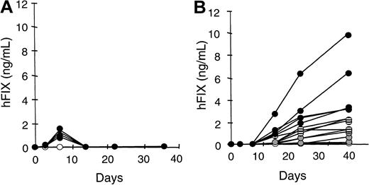
When the mouse sera were tested for the presence of anti-hFIX antibodies, we observed that high titer anti-hFIX antibody could be detected in all mice that received AAV-hFIX by the IM route (Figure 1, lower panel). In addition to being high titer, the anti-hFIX antibody responses were stable and persisted over the course of the experiment (72 days). Anti-mFIX cross-reacting antibodies did not appear to develop because there was no perturbation in serum mFIX levels or in clotting times with retro-orbital bleedings (data not shown). This immune response to hFIX following IM administration of AAV-hFIX was observed in other strains of mice including Balb/C mice (Figure2, left panel). To examine if the anti-hFIX antibodies might be responsible for the absence of serum hFIX expression, the experiment was repeated in immunodeficient NIH-3 mice. hFIX could be readily detected in the serum in NIH-3 mice indicating that the immune response against hFIX was responsible for the absence of hFIX in the sera of mice following AAV-hFIX administered IM. IM administration of AAV-hFIX was more prone to inducing a humoral response than the intravascular IPV route.
Immune response limits hFIX expression following IM administration of AAV-hFIX.
Increasing doses, 1.2 × 10¹⁰ vp (gray circles), 6 × 10¹⁰ vp (stippled circles), and 3 × 10¹¹ vp (solid circles) of AAV-hFIX, were administered to Balb/c (left panel) and NIH-3 (right panel) mice by direct administration into the quadriceps muscle. Sera were harvested from the animals at various time points following vector administration and analyzed for the presence of hFIX.
Impact of pre-existing immunity on rAAV administration
The previous experiment was performed in AAV naive animals. Clearly, most gene therapy patients are likely to have been exposed to AAV in their lifetimes. Indeed, several previous studies have shown that more than 90% of normal blood donors tested displayed anti-AAV antibodies and between 18% and 52% of those individuals possessed neutralizing antibodies to serotype 2–derived AAV vectors.6,11 We have previously shown that this anti-AAV immunity can be reproduced in mice by administering rAAV by an IV route and that preimmune mice are refractory to AAV-mediated gene transfer by a vascular route.6
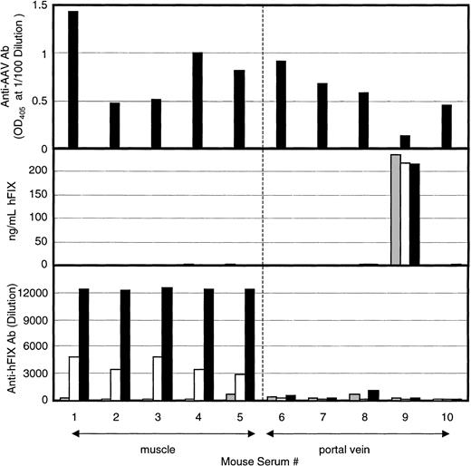
These studies were expanded to examine whether rAAV-hFIX administered by a nonvascular route, specifically, IM, can circumvent pre-existing anti-AAV humoral immunity. C57Bl/6 mice were immunized by tail vein injection with 5 × 10¹⁰ vp AAV encoding lacZ. Thirty-two days after vector administration, sera from the mice were positive for the presence of anti-AAV antibody (Figure3, top panel). As previously observed, the titer of these antibodies varied significantly from mouse to mouse. Also as previously observed, no hFIX expression could be detected after administration of 4 × 10¹¹ vp AAV-hFIX vector by the IPV route into animals that possessed a high anti-AAV antibody titer (Figure 3, middle panels nos. 6, 7, 8, 10). The one mouse of the 5 IPV AAV-hFIX mice that did demonstrate significant levels of hFIX in the sera (Figure 3, middle panels no. 9) manifested a low anti-AAV antibody titer at the time of AAV-FIX administration (Figure 3, upper panels no. 9). A second group of 5 AAV-immune mice was transduced with 1.5 × 10¹¹ vp AAV-hFIX vector by the IM route. Again, hFIX could not be detected in the sera of any of these mice (Figure 3, middle panels nos. 1-5).
Anti-AAV immunity limits hFIX expression.
C57Bl/6 mice were preimmunized to AAV by administering 5 × 10¹⁰ vp of AAV-lacZ by tail vein. Levels of anti-AAV antibodies were tested 1 month following vector administration (top panel). Then, 4 × 10¹¹ vp of AAV-hFIX was administered to AAV-immune mice into the portal vein by way of a cannula or by direct administration into the quadriceps muscle. Sera were harvested from the animals 11 (gray bars), 25 (white bars), and 42 (black bars) days after AAV-hFIX administration and analyzed for the presence of hFIX (middle panels) and anti-hFIX antibodies (bottom panels).
The sera of the IM and IPV AAV-hFIX mice were next tested for the presence of anti-hFIX antibody. None of the IPV AAV-hFIX mice manifested any anti-hFIX antibody. Surprisingly, high anti-hFIX antibody titers were detected in the sera of all of the IM AAV-hFIX mice. This indicates that low levels of hFIX expressed in the IM AAV-hFIX mice were sufficient to prime an anti-hFIX antibody response but insufficient to be detected in the FIX ELISA. The hFIX ELISA can detect levels as low as 0.1 to 0.2 ng hFIX/mL mouse sera (L. Tsui, data not shown). This further suggests that IM administration of AAV-hFIX sensitizes the immune system to respond to hFIX to a far greater extent than vascular administration of AAV-hFIX.
Discussion
We sought to determine the impact of the route of administration and dose of AAV-hFIX on the expression of hFIX in the sera of immunocompetent mice. Two different routes of vector administration were chosen to target 2 tissues for which rAAV has been shown to demonstrate a tropism: muscle and liver. We observed that administration of vector via the portal vein produced higher levels of hFIX than administration of an equal dose of vector to the muscle. All of the mice that received vector by the IM route generated a humoral immune response against the transgene despite expressing barely detectable amounts of FIX in the blood. This is to be contrasted with the observation that none of the mice that received AAV-hFIX vector by the vascular route generated an anti-hFIX antibody response. The target tissue, more so than the levels of protein expression, appeared to direct the nature of the immune response to the xenoprotein. It had been shown previously that administration of AAV encoding OVA, by either a vascular or IM route, induced anti-OVA antibodies.2 These studies, like ours, were performed in C57Bl/6 mice with similar doses of virus. An obvious difference, however, was the transgene used in the 2 studies. We chose to conduct this study in C57Bl/6 mice because they demonstrate significant tolerance to the hFIX protein.12 This does not appear to be the case with OVA, which induced both humoral and T-cell–mediated response by a variety of routes following AAV gene transfer.2
C57Bl/6 mice allow the stable production of hFIX in mice following intraportal administration of AAV-hFIX.7 In long-term studies, hFIX can be observed in these AAV-hFIX mice for well over a year despite the use of a xeno-FIX protein (Brian Donahue, unpublished data, March 1997). This raises the question of whether the IM expression of AAV-FIX administration is more immunogenic than intrahepatic expression of AAV-FIX. Unfortunately, the available data to date in humans and dogs are limited due to the relatively few test subjects and variability in the doses administered. IM administration of 2 × 10¹¹vp/kg AAV-hFIX into 3 hemophiliac patients did not result in the induction of any detectable anti-hFIX antibodies.13 Conversely, IM delivery of a higher dose of AAV vector encoding the canine FIX gene in hemophilic B dogs, induced anti-cFIX antibodies in 2 of 4 dogs that received over 1012 vp/kg.3 In a separate study using hemophilic dogs, anti-cFIX antibodies were not observed in the 2 dogs that received AAV-cFIX vector by the intraportal route.8 It is difficult to draw any firm conclusions from the 2 dog studies because the promoters driving cFIX expression in the 2 vectors differed and the numbers of animals was small. From these data we can conclude that there is a discrepancy between the IM data in normal mice with a xenogenic hFIX, the hemophiliac dog IM data with a homologous cFIX, and the clinical IM data with a homologous hFIX. It is possible that these data reflect the natural differences in the relative immune sensitivity of these different hosts to the IM presentation of FIX.
We have previously shown that a pre-existing anti-AAV neutralizing response can block AAV transduction of the liver following vascular delivery of vector.6 It has been suggested that IM delivery of vector may allow the circumventing of the anti-AAV humoral response. We observed, however, that a pre-existing high-titer anti-AAV antibody response significantly blocked AAV-hFIX transduction of both the liver and muscle and blocked hFIX expression in the blood following either IM or hepatic administration of AAV-hFIX. Although hFIX expression was not detected in the sera of the IM AAV-hFIX mice, all of these mice demonstrated a robust anti-hFIX humoral immune response in these AAV immune mice following administration of the AAV-hFIX vector. This suggests that these mice must have expressed low levels of hFIX at some point to prime this immune response. Because these anti-hFIX immune responses persisted for over 148 days, it is also likely that there was continued low level hFIX expression.
The portal vein has access to approximately two thirds of the liver, which is a normal site of expression of FIX.14-16 IM delivery has the practical benefit of ease of access clinically; however, it does not normally express and secrete FIX. Indeed, the muscle is generally not thought of as a secretory tissue for proteins under normal physiologic conditions. This difference may account for the observed differences in protein expression. How then could the differences in these organs' physiology produce the profound differences in the priming of an anti-hFIX immune response? It has been suggested that muscle expression of FIX protein results in binding of FIX to the muscle.4 This inappropriate presentation of the protein might render the FIX more immunogenic. Cross-priming of immune response can be accomplished by the transfer of both major histocompatibility complex (MHC) class I and class II restricted antigen from nonantigen-presenting cells to professional antigen-presenting cells such as DCs.1 This pathway of loading DCs with hFIX may account for the increased immunogenicity of the IM route of administration of AAV-hFIX. Limiting gene expression to the liver through the route of delivery may produce a more “physiologic” expression of the transgene. This might also be accomplished with the use of a tissue-specific promoter. Consistent with this suggestion is the observation that adenoviral vectors expressing the α1-antitrypsin protein from tissue ubiquitous promoters are more prone to prime an immune response to this secreted transgene than vectors using a liver-specific promoter.17
As gene therapeutics encoding secreted proteins are advanced for a variety of diseases, a better understanding of the implications of the route and dose of vector administered is developing. AAV vectors have many valuable qualities as gene therapy vectors including the absence of any viral genes, the ability to integrate into the host cells, and the absence of viral-mediated pathology. However, most humans have been exposed to wild-type AAV and, in fact, very often the same AAV serotype 2 commonly used in gene therapy vectors. As such, it is critical to examine the interactions of the gene therapeutic and its transgene product in both AAV-naive and AAV-immune animals. These issues become all the more important as gene therapy is applied in the treatment of diseases, such as hemophilia, where the consequences for inducing an immune response to the therapeutic transgene are severe. Our current study indicates that FIX expression following vascular delivery of AAV-hFIX is significantly less immunogenic than IM delivery in both AAV naive and AAV immune mice and that this has a profound impact on hFIX expression.
We would like to thank Drs B. Donahue and T. Harding for their thoughtful input.
All authors are employees of Cell Genesys, which funded the research.
The publication costs of this article were defrayed in part by page charge payment. Therefore, and solely to indicate this fact, this article is hereby marked “advertisement” in accordance with 18 U.S.C. section 1734.
References
Author notes
James G. McArthur, Cell Genesys, Dept of Preclinical Biology and Immunology, 342 Lakeside Dr, Foster City, CA 94404; e-mail: jamesm@cellgenesys.com.


This feature is available to Subscribers Only
Sign In or Create an Account Close Modal