Abstract
Rejection of a graft after human leukocyte antigen (HLA)-identical stem cell transplantation (SCT) can be caused by recipient's immunocompetent T lymphocytes recognizing minor histocompatibility antigens on donor stem cells. During rejection of a male stem cell graft by a female recipient, 2 male (H-Y)–specific cytotoxic T lymphocyte (CTL) clones were isolated from peripheral blood. One CTL clone recognized an HLA-A2–restricted H-Y antigen, encoded by the SMCY gene. Another CTL clone recognized an HLA-B60–restricted H-Y antigen. In this study UTY was identified as the gene coding for the HLA-B60–restricted H-Y antigen. The UTY-derived H-Y antigen was characterized as a 10-amino acid residue peptide, RESEEESVSL. Although the epitope differed by 3 amino acids from its X-homologue, UTX, only 2 polymorphisms were essential for recognition by the CTL clone HLA-B60 HY. These results illustrate that CTLs against several H-Y antigens derived from different proteins can contribute simultaneously to graft rejection after HLA-identical, sex-mismatched SCT. Moreover, RESEEESVSL-specific T cells could be isolated from a female HLA-B60+ patient with myelodysplastic syndrome who has been treated with multiple blood transfusions, but not from control healthy HLA-B60+ female donors. This may indicate that RESEEESVSL-reactive T cells are more common in sensitized patients.
Introduction
Human leukocyte antigen (HLA)-identical hematopoietic stem cell transplantation (SCT) can be complicated by graft-versus-host disease (GVHD) or graft rejection. Graft rejection can be caused by the recognition of minor histocompatibility antigens (mHag) on donor stem cells by immunocompetent HLA-restricted T lymphocytes of recipient origin.1-3 mHag are peptides derived from polymorphic intracellular proteins that differ between donor and recipient.4,5 The peptides are presented on the cell membrane in association with major histocompatibility complex molecules and are capable of eliciting specific T-cell responses.2,4 Specific T-cell responses against male-specific mHag may occur after sex-mismatched blood transfusions and organ or stem cell transplantation.6-8 Clinical studies indicated that female patients who underwent transplantation with HLA-phenotypic identical male stem cells have a higher risk for graft rejection than those who receive stem cells of female origin.6,9 During rejection of male stem cell grafts, H-Y–specific cytotoxic T lymphocyte (CTL) clones could be generated from the peripheral blood of female patients.1,8 10 The H-Y–specific CTL clones are used to identify the H-Y antigens and their genes.
SMCY was the first gene identified that encoded human H-Y antigens.11,12 HLA-A2– and HLA-B7–restricted H-Y epitopes derived from the SMCY protein were characterized through peptide elution of male cells, and microcapillary liquid chromatography–electrospray ionization mass spectrometry combined with T-cell epitope reconstitution. A molecular method can be explored as well. The DNA sequences of 8 Y-specific genes are known.13 Each gene has a ubiquitous tissue expression and a homologue on the X chromosome encoding a similar, but nonidentical, protein isoform. The amino acid sequence identity of the X-Y isoforms varies from 85% to 97%. Because these ubiquitously expressed Y-specific genes have sufficient polymorphisms to generate male-specific peptides, they are potential candidates to encode H-Y antigens.
DFFRY was identified as a second gene encoding a human H-Y antigen.14,15 The DFFRY-derived H-Y peptide was recognized by an HLA-A1–restricted CTL clone, which was generated from a female patient with acute myeloid leukemia who rejected her HLA-identical male stem cells.1 We identified the HLA-A1–restricted H-Y epitope by DFFRY deletion mutants andDFFRY minigenes encoding small peptides. Synthetic peptides were used to identify this H-Y antigen recognized by the HLA-A1 HY–specific CTL clone. Recently, the UTY gene was identified as a gene coding for an H-Y epitope, which was recognized by an HLA-B8–restricted CTL clone generated during GVHD.16
The first indication that multiple H-Y antigens may be involved in male stem cell graft rejection in a female patient was the isolation of HLA-A2– and HLA-B60–restricted cytotoxic and proliferative H-Y–specific CTL clones from the patient, a woman with aplastic anemia who rejected an HLA-genotypic identical male stem cell graft.17,18 The H-Y epitope recognized by the HLA-A2–restricted CTL clone was previously characterized as a peptide derived from the SMCY gene.11 The H-Y–specific HLA-B60–restricted CD8+ CTL clone showed no reactivity to the SMCY protein, indicating that another Y-specific gene was also involved in the male stem cell graft rejection.
In this study, we identified UTY as the gene encoding the H-Y epitope recognized by the HLA-B60–restricted CTL clone. The H-Y epitope was localized on the UTY gene using UTYdeletion mutants and UTY minigenes. Synthetic peptides confirmed that the amino acid sequence of the UTY–derived H-Y epitope was RESEEESVSL. Although the epitope contained 3 different amino acids compared to its UTX-homologue protein, only 2 amino acids were shown to be essential residues for recognition by the HLA-B60 H-Y CTL clone. Furthermore, we demonstrated that RESEEESVSL-specific T cells could be isolated from a female HLA-B60+ patient with myelodysplastic syndrome (MDS) treated with multiple blood transfusions, and not from 2 healthy control HLA-B60+ female donors. This may reflect increased frequencies of RESEEESVSL-specific T cells in female HLA-B60+ patients sensitized by frequent stimulation with male cells.
Materials and methods
CTL and cell lines
The CD8+ CTL clone HLA-B60 HY was derived by limiting dilution from peripheral blood mononuclear cells (PBMC) of a female patient with aplastic anemia, who had rejected HLA-identical male stem cells.17 18 The CTL clone was cultured by stimulation with irradiated allogeneic PBMC and donor-derived Epstein-Barr virus (EBV)-transformed B cells (EBV-LCL) in RPMI-1640 medium (Bio-Whittaker, Verviers, Belgium) containing 15% human serum, 3 mmol/L L-glutamine, 1% leukoagglutinin (Difco Laboratories, Detroit, MI), and 120 U/mL recombinant IL-2 (Roussel Uclaf, Paris, France). HeLa cells and WEHI-164 clone 13 cells were obtained from American Type Culture Collection (Rockville, MD) and maintained in complete culture medium consisting of RPMI-1640 medium containing 10% fetal calf serum (FCS) (Bio-Whittaker).
Cloning of Y-specific genes
Total RNA was isolated from male EBV-LCL with Trizol (Gibco BRL, Gaithersburg, MD) according to the manufacturer's instructions. cDNA was prepared from RNA using M-MLV BRL reverse transcriptase (Gibco, BRL) for 60 minutes at 37°C as described.19,20 One fiftieth of the cDNA reaction was amplified using specific primers for each Y gene identified by a systematic search of the nonrecombinant region of the human Y chromosome.14 BecauseSMCY and DFFRY had large open reading frames, each gene was cloned in 3 overlapping cDNA constructs. With the exception of SMCY, all genes were amplified using the expanded long template PCR system (Boehringer Mannheim GmbH, Mannheim, Germany). The amplification was started with a denaturation step of 2 minutes at 92°C, followed by 30 cycles, with each cycle consisting of 20 seconds at 92°C, 1 minute at 60°C, and 1 minute at 68°C.SMCY was amplified using the Marathon cDNA amplification kit (Clontech Laboratories, Palo Alto, CA) according to the manufacturer's procedure. After amplification, each Y-specific cDNA was visualized on an ethidium bromide–stained low-melt agarose gel (Sigma Chemical, St Louis, MO). cDNA was isolated from the low-melt agarose gel using agarose (Boehringer Mannheim GmbH) according to the manufacturer's procedure. Each individual cDNA was cloned in the expression vector PCR3.1 using the eukaryotic TA cloning kit (Invitrogen, Carlsbad, CA).
Cloning of subgenic fragments or minigenes of theUTY gene
Restriction analysis on the UTY gene revealed anHpaI site on position 3306, an NheI site on position 2458, and a PstI site on position 1569 of theUTY cDNA. NheI and PstI also have a recognition site in the multiple cloning site of the PCR3.1 plasmid, directly after the 5′ and 3′ ends of the UTY cDNA, respectively. Deletion mutants UTY/NheI andUTY/PstI were generated by incubating the plasmid PCR3.1 containing the UTY gene with the restriction enzymesNheI and PstI, deleting 2472-bp and 2493-bp long fragments at the 5′ and 3′ sites of the UTY gene, respectively. Deletion mutant UTY/HpaI was generated by digesting UTY with HpaI and the PCR3.1 plasmid with EcoRV directly after the 3′ end of theUTY cDNA, deleting a 756-bp long fragment of theUTY gene. After digestion, the linear truncated constructs were visualized on an ethidium bromide–stained low-melt agarose gel. After isolating the constructs with agarose, the linear constructs were ligated with the rapid DNA ligation kit (Boehringer Mannheim GmbH) to form circular plasmid DNA consisting of vector PCR3.1 with the truncated UTY/HpaI,UTY/NheI, andUTY/PstI cDNAs.
The oligonucleotides (5′ → 3′) used for generation of theUTY minigenes are listed in Table1. After oligonucleotide synthesis, each pair of oligonucleotides formed double-stranded DNA minigenes with aBamHI site and a HindIII site after hybridization. These minigenes were cloned into theBamHI/HindIII-digested PCR3.1 expression vector.
Oligonucleotides (5′ → 3′) used for generation of UTY minigenes
| position UTY gene (bp) . | Sequence (5′ → 3′) . |
|---|---|
| 79-120 | For: AGCTTCACCATGGCGAGCCGCGAGAGTGAAGAGGAGTCTGTTAGCCTGACAGTCTAG Rev: GATCCTAGACTGTCAGGCTAACAGACTCCTCTTCACTCTCGCGGCTCGCCATGGTGA |
| 172-216 | For: AGCTTCACCATGAGGCTTCATGAAGATGGCGCCAGAACGAAGACCCTACTAGGCAAGTAG Rev: GATCCTACTTGCCTAGTAGGGTCTTCGTTCTGGCGCCATCTTCATGAAGCCTCATGGTGA |
| 484-522 | For: AGCTTCACCATGCGAGCCAAGGAAATTCATTTACGACTTGGGCTCATGTTCTAG Rev: GATCCTAGAACATGAGCCCAAGTCGTAAATGAATTTCCTTGGCTCGCATGGTGA |
Transfection of HeLa cells and screening of transfectants
Y-specific cDNA was transfected into HeLa cells by the DEAE-dextran-chloroquine method as described.21 One day before transfection, HeLa cells were seeded in 96-well flat-bottom microtiter plates at 15 000 cells/well in 100 μL complete culture medium. Before transfection, medium was discarded and replaced by 45 μL DEAE-dextran/DNA mixtures. These mixtures were prepared for duplicate transfections in 96-well V-bottom microtiter plates by sequentially adding: 98 μL RPMI-1640 supplemented with 10% decomplemented NuSerum IV (Collaborative Biomedical Products, Bedford, MA) containing 0.3 mg/mL DEAE-dextran (Sigma Chemical) and 100 μmol/L chloroquine, 1 μL of TE (10 mmol/L Tris, 1 mmol/L EDTA, pH 7.4) containing 200 ng Y-specific cDNA in plasmid PCR3.1 and 1 μL TE containing 200 ng plasmid PCR3.1-B60 (plasmid PCR3.1 containing the HLA-B60 gene) or without HLA-B60 construct as a control. The HeLa cells were incubated for 4 hours at 37°C, after which the DEAE-dextran/DNA was discarded and replaced by 80 μL phosphate-buffered saline (PBS) containing 10% dimethyl sulfoxide (DMSO). After 2 minutes at room temperature, PBS-DMSO was replaced by 200 μL complete culture medium. Transfected HeLa cells were incubated for 48 hours at 37°C. After removing the medium, 4000 HLA-B60 HY CTL were added in 200 μL RPMI-1640 containing 10% pooled human serum and 300 IU IL-2 per milliliter. After 24 hours, the tumor necrosis factor (TNF) content in 50 μL supernatant was determined by adding it to 50 μL complete culture medium containing 50 000 WEHI-164 clone 13 cells, sensitive to a cytolytic effect by TNF.22 After 18 hours, 10 μL cell proliferation reagent WST I (Boehringer Mannheim GmbH) was added, and the TNF concentration was calculated in comparison with recombinant TNF standards measured in the same assay.
Peptide synthesis and peptide recognition assays
Peptides were synthesized by solid-phase strategies on an automated multiple peptide synthesizer (SyroII; MultiSynTech, Witten, Germany) and characterized by mass spectrometry. The purity of the peptides was determined by analytical reversed-phase high-pressure liquid chromatography and it proved to be at least 80%.51Cr-release assays were used to determine lysis of target cells. EBV-LCL target cells were labeled with 100 μCi of Na51CrO4. After 1 hour of incubation at 37°C, the cells were washed 3 times with RPMI supplemented with 2% FCS. Then 2000 Na51CrO4-labeled EBV-LCL cells/well were plated in a 96-well V-bottom microtiter plate in 100 μL RPMI-1640 + 10% pooled human serum containing various concentrations of peptide. After 1 hour of incubation at 37°C, 20 000 CTLs were added in 100 μL RPMI-1640 + 10% pooled human serum.51Cr-release was measured after incubation at 37°C for 4 hours.
Screening of UTY protein for HLA-B60–binding peptides
The UTY protein was screened for HLA-B60–binding peptides by using the program HLA peptide–binding predictions (http://bimas.dcrt.nih.gov/molbio/hla_bind/). The scores are predictions of half-time of dissociation to HLA-B60 molecules for the indicated peptides.
HLA-B60 binding assay
Homozygous HLA-B60+ EBV cells were incubated at 4°C for 90 seconds in citric acid, pH 3.1, to elute peptides from the HLA class I complexes. After washing the cells twice with RPMI-1640 medium containing 2% FCS, the EBV cells were resuspended at a concentration of 400 000 cells/mL in Iscove minimum Dulbecco medium (IMDM) containing 2% FCS and 1.5 μg/mL β2-microglobulin. A total of 100 μL EBV cells were added to 25 μL 150 nmol/L of fluorescence-labeled HLA-B60–binding peptide KESTC(FL)HLVL in a 96-well, V-bottom microtiter plate. For evaluation of maximal binding of the fluorescent peptide to the EBV cells, 25 μL PBS was added to the wells. For determination of peptide binding to HLA-B60 molecules, 25 μL PBS containing peptide concentrations of 0.4 μg/mL to 50 μg/mL was added to the wells. KESTLHLVL without fluorescence label was used as a control. After incubation for 24 hours at 4°C, the cells were washed twice with PBS, and the binding of the fluorescence-labeled peptide to the EBV cells was determined by FACS analysis. The concentration of a given peptide that induced a decrease of 50% of the mean channel of fluorescence of maximal binding (IC50) was calculated.
Isolation of IFNγ-secreting cells
After informed consent, bone marrow and peripheral blood were collected from an HLA-B60+ patient with MDS and 2 healthy HLA-B60+ female donors. Mononuclear cells were isolated by washing and Ficoll-Isopaque (1.077 g/mL) density-gradient centrifugation. A total of 40 × 106 PBMC were stimulated with irradiated 20 × 106 autologous PBMC loaded with 1 μg/mL RESEEESVSL peptide in IMDM medium (Bio-Whittaker) containing 10% human serum and 3 mmol/L L-glutamine. After 16 hours of incubation at 37°C, IFNγ-producing cells were isolated using the MACS IFNγ secretion assay (Miltenyi Biotec, Auburn, CA) according to the manufacturer's procedure. After isolation, IFNγ− and IFNγ+ T cells were stimulated with RESEEESVSL peptide-loaded autologous mononuclear bone marrow cells in IMDM medium containing 10% human serum, 3 mmol/L L-glutamine, and 120 U/mL recombinant IL-2. The T cells were restimulated on days 7 and 14 with RESEEESVSL-loaded irradiated autologous mononuclear bone marrow cells. On day 21, T-cell reactivity against the RESEEESVSL epitope was determined in a 51Cr release assay.
Results
Cloning of Y-specific genes
After isolation of RNA from male EBV-LCL cells, Y-gene–specific primers were used in a PCR reaction to amplify only the Y-specific genes and not their X-homologues. After cloning the PCR products in an expression vector, restriction and sequence analysis of the amplified Y-specific cDNAs confirmed their Y specificity (data not shown). As described previously, all Y-specific cDNAs were appropriately transcribed and translated as determined in an in vitro transcription/translation assay.14
Cloning and identification of the gene encoding the HLA-B60–restricted H-Y T-cell epitope
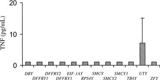
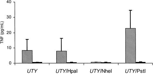
To determine whether any of the Y-specific genes encode the HLA-B60–restricted H-Y T-cell epitope, the cloned Y-cDNAs were cotransfected with HLA-B60 cDNA into HeLa cells. As shown in Figure1, transfection of UTY cDNA resulted in a significant amount of TNF production by HLA-B60 HY CTL, whereas transfection of all other Y genes induced a TNF release of 1 pg/mL. Deletion mutants of the UTY sequence were obtained by digestion with restriction enzymes HpaI and PstI, resulting in a deletion at the 3′ site of the UTY gene of 756 bp (UTY/HpaI) and 2493 bp (UTY/PstI), respectively (Figure2). An additional deletion of 2472 bp at the 5′ site of the UTY gene (UTY/NheI) was generated using the restriction enzyme NheI. As is demonstrated in Figure 3, no TNF production by the HLA-B60 HY CTL clone was measured when stimulated with HeLa transfected with UTY/NheI and HLA-B60. Transfection of UTY/PstI in HeLa resulted in TNF release by the HLA-B60 HY CTL, illustrating that the HLA-B60–restricted H-Y peptide was encoded by an amino acid sequence between positions 1 and 523 of the UTY protein. To confirm that the TNF release by the HLA-B60 HY CTL was HLA-B60 restricted, theUTY cDNAs were also transfected without HLA-B60 into HeLa cells. No significant TNF release by the HLA-B60 HY CTL was found when the UTY cDNAs were transfected in the absence of the HLA-B60 molecule (Figure 3).
TNF production by HLA-B60 HY CTL after stimulation with HeLa transfected with UTY and HLA-B60 cDNA.
HeLa cells were cotransfected with indicated Y-specific cDNA and HLA-B60 cDNA. Forty-eight hours after transfection, HLA-B60 HY CTL was added. Culture supernatants were harvested 1 day later and tested for TNF production.
Location of UTY deletion mutants and HLA-B60 HY CTL epitope-encoding minigenes.
TNF production by HLA-B60 HY CTL after stimulation with HeLa transfected with UTY cDNA.
HeLa cells were transfected with UTY or UTYdeletion mutants cDNA and HLA-B60 cDNA (▧), or without HLA-B60 cDNA (▪). Forty-eight hours after transfection, HLA-B60 HY CTL was added. Culture supernatants were harvested 1 day later and tested for TNF production.
Localization of the HLA-B60–restricted H-Y epitope in the UTY gene
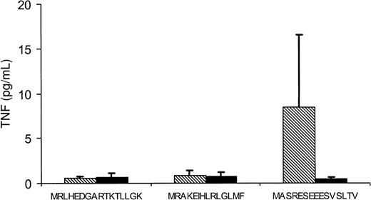
To identify the H-Y peptide coding sequence, the 1-523 amino acid (AA) region of the UTY protein was screened for HLA-B60–binding peptides that differ by at least one amino acid from its X homologue. Twelve peptides (9 or 10 residues) with scores varying from 17 600 to 704 000 were observed (Table2). Three minigenes coding for 8 HLA-B60–binding peptides were constructed and cotransfected with HLA-B60 into HeLa cells (Figure 2). As is demonstrated in Figure4, only minigene 79-120, coding for peptide MASRESEEESVSLTV, induced TNF production by HLA-B60 HY CTL, indicating that this minigene encoded the H-Y epitope recognized by the HLA-B60 HY CTL clone. Transfection of the minigene without cotransfection of the HLA-B60 molecule into HeLa cells did not result in TNF production.
HLA-B60–binding peptides derived from the 1-523 AA region of the UTY protein
| AA position UTY protein . | Peptide . | Score . |
|---|---|---|
| 164-172 | KEIHLRLGL | 704 000 |
| 29-38 | RESEEESVSL | 640 000 |
| 60-69 | HEDGARTKTL | 160 000 |
| 164-173 | KEIHLRLGLM | 40 000 |
| 41-49 | EEREALGGM | 20 000 |
| 61-69 | EDGARTKTL | 20 000 |
| 398-407 | SDNWNGGQSL | 20 000 |
| 40-49 | VEEREALGGM | 20 000 |
| 121-130 | ADYWKNAAFL | 20 000 |
| 61-70 | EDGARTKTLL | 20 000 |
| 32-40 | EEESVSLTV | 17 600 |
| 31-40 | SEEESVSLTV | 17 600 |
The region between amino acids 1 and 523 of the UTY protein was screened for HLA-B60–binding peptides. Scores are predictions of half-time of dissociation to HLA-B60 molecules for the indicated peptides. Underlined amino acids are mutations compared to the UTX sequence.
Minigene encoding H-Y epitope is recognized by the HLA-B60 HY CTL.
HeLa cells were transfected with UTY minigene cDNA 79-120, 172-216, or 484-522 together with HLA-B60 cDNA (▧), or with UTY minigene cDNA only (▪). Forty-eight hours after transfection, HLA-B60 HY CTL was added. Culture supernatants were harvested 1 day later and tested for TNF production.
Identification of the H-Y epitope recognized by CTL clone HLA-B60 HY
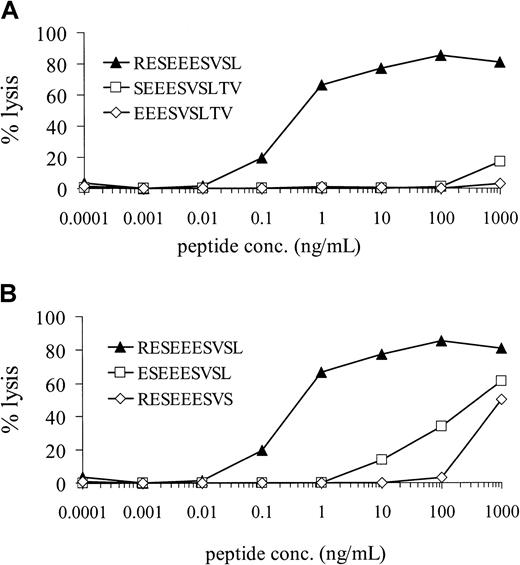
Minigene 79-120, coding for peptide MASRESEEESVSLTV, coded for 3 HLA-B60–binding peptides (Table 2). The relevant peptides were synthesized and loaded at various concentrations on female HLA-B60+ EBV-LCL cells. Recognition of the peptide loaded EBV-LCL cells by the CTL clone HLA-B60 HY was determined in a 51Cr-release assay. As is shown in Figure 5A, the RESEEESVSL-loaded EBV-LCL cells were efficiently lysed by HLA-B60 HY CTL with half-maximal target cell lysis at a peptide concentration of 300 pg/mL, whereas neither EEESVSLTV nor the SEEESVSLTV peptide was recognized by the HLA-B60 HY CTL. To determine whether the 10-residue peptide RESEEESVSL was the minimal epitope recognized by HLA-B60 HY CTL, 2 peptides lacking the N- or C-terminal amino acid of RESEEESVSL were synthesized and tested for recognition by HLA-B60 HY CTL in a51Cr release assay. As demonstrated in Figure 5B, EBV-LCL cells loaded with the 10-residue peptide RESEEESVSL were most efficiently lysed by the HLA-B60 HY CTL.
Specific lysis of RESEEESVSL-loaded female EBV-LCL target cells by the HLA-B60 HY CTL.
Female HLA-B60+ EBV-LCL cells were 51Cr-labeled for 1 hour. After washing, the cells were incubated for 1 hour with UTY-derived peptides at various concentrations. HLA-B60 HY CTL was added at an effector-to-target ratio of 10:1, and 51Cr release was measured after 4 hours. (A) Lysis of female EBV-LCL target cells by the HLA-B60 HY CTL after loading with different peptides encoded by minigene 79-120. (B) Lysis of female EBV-LCL target cells by the HLA-B60 HY CTL after loading with the 10-residue H-Y peptide RESEEESVSL or the 9-residue peptides lacking its N- or C-terminus.
Two polymorphisms in H-Y epitope are essential for T-cell recognition
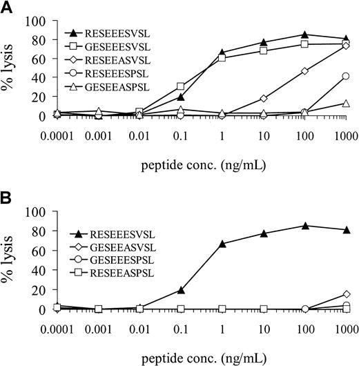
The H-Y epitope RESEEESVSL differed by 3 amino acids from its X homologue GESEEASPSL, which was not recognized by the HLA-B60 HY CTL clone (Figure 6A). To determine whether all 3 polymorphisms were equally important for recognition by the HLA-B60 HY CTL, all 3 peptides—each containing one UTX-homologue amino acid on position 1, 6, or 8—were synthesized. As shown in Figure 6A, EBV-LCL cells loaded with peptide GESEEESVSL were well recognized by HLA-B60 HY CTL. In contrast, an X-homologue amino acid on position 6 or 8 of the H-Y epitope resulted in a reduced lysis of EBV-LCL cells by the HLA-B60 HY CTL, suggesting that these polymorphisms were essential for T-cell recognition. The peptides GESEEASVSL and GESEEESPSL, each containing 2 X-homologue amino acids, were not recognized by HLA-B60 HY CTL (Figure 6B). To determine whether the mutated peptides bind equally well to HLA-B60 molecules as the original RESEEESVSL peptide, an assay based on competition for binding to HLA-B60 between a peptide of interest and a fluorescence-labeled standard peptide was used. Similar to the standard peptide without fluorescence label, 0.8 μg/mL RESEEESVSL competitor peptide induced 50% inhibition of fluorescence-labeled peptide binding (IC50), suggesting a strong binding of RESEEESVSL to HLA-B60 molecules. A control irrelevant peptide did not compete for binding of the fluorescence-labeled standard peptide to the HLA-B60 molecule. All mutated variants of RESEEESVSL induced IC50 at concentrations varying from 0.6 to 0.8 μg/mL, indicating that the altered recognition of the mutated peptides by HLA-B60 HY CTL was not a consequence of altered binding of the peptides to HLA-B60 molecules.
Polymorphisms in H-Y epitope differentially contributed to HLA-B60 recognition.
Female HLA-B60+ EBV-LCL cells were 51Cr-labeled for 1 hour. After washing, the cells were incubated for 1 hour with indicated peptides at various concentrations. HLA-B60 HY CTL was added at an effector-to-target ratio of 10:1, and 51Cr release was measured after 4 hours. (A) Lysis of female EBV-LCL target cells by HLA-B60 HY CTL after loading with the 10-residue UTY-derived peptide RESEEESVSL or 3 peptides each containing 1 UTX-homologue amino acid on position 1, 6, or 8. (B) Lysis of female EBV-LCL target cells by HLA-B60 HY CTL after loading with the 10-residue UTY-derived peptide RESEEESVSL or 3 peptides each containing 2 UTX-homologue amino acids.
Isolation of HLA-B60 HY CTL from a female patient with MDS
To determine whether HLA-B60 HY CTL were present in female patients, PBMC of 2 healthy donors and a patient with MDS who was sensitized by multiple blood transfusions were stimulated with the RESEEESVSL epitope. After stimulation, activated T cells (IFNγ+) were separated from nonreactive T cells (IFNγ−) based on their IFNγ production. The frequency of IFNγ-producing T cells in PBMC of donors and patient was below the detection level, as measured by flow cytometry (less than 0.05%; data not shown). After 20 days of culture, FACS analysis showed that 20% to 30% of the T cells in all T-cell lines were CD3+ and CD8+ (data not shown). Reactivity of the T-cell lines against the RESEEESVSL epitope was tested in a 51Cr-release assay. The IFNγ T-cell line generated from PBMC of the patient with MDS lysed RESEEESVSL-loaded autologous or allogeneic HLA-B60 female EBV cells, whereas no reactivity could be observed against the control peptide (Figure7). Moreover, HLA-B60+ EBV cells from a male were also efficiently lysed by the T-cell line. The IFNγ− T-cell line derived from the same patient showed no reactivity against the RESEEESVSL epitope (Figure 7). The IFNγ+ and IFNγ− T-cell lines derived from the 2 donors exhibited no reactivity against the RESEEESVSL epitope (data not shown).
Specific lysis of RESEEESVSL-loaded female EBV-LCL target cells by HLA-B60 HY CTL isolated from a patient with MDS.
IFNγ-secreting (IFNγ+) T cells and IFNγ-nonsecreting (IFNγ−) T cells were isolated from the PBMC of the patient with MDS after stimulation with peptide RESEEESVSL and were tested for specific reactivity. EBV-LCL cells were 51Cr-labeled for 1 hour. After washing, the female EBV cells were incubated for 1 hour with either the HYB60 peptide RESEEESVSL or the HYA2 peptide FIDSYICQV. IFNγ+ and IFNγ− T cells were added at the indicated effector-to-target ratios, and 51Cr release was measured after 4 hours. Auto EBV cells were autologous HLA-B60 and HLA-A2+ EBV cells derived from the female patient with MDS. Allo EBV cells were allogeneic HLA-B60 and HLA-A2+ female EBV cells.
Discussion
The involvement of male-specific mHag antigens in graft rejection was indicated by the observation that female patients who underwent transplantation with HLA-phenotypic identical male stem cells had a higher risk for graft rejection than those who underwent transplantation using stem cell grafts of female origin.6,9 T-cell clones recognizing male cells could be generated during stem cell graft rejection after sex-mismatched transplantation. H-Y–specific T cells can be used to identify the H-Y antigens and their genes.1 17
Two human H-Y epitopes have been identified. The H-Y epitopes, recognized by HLA-A2– and HLA-B7–restricted CTL clones, were shown to be encoded by the ubiquitously expressed SMCYgene.11,12 Recently, we identified DFFRY as a second male-specific protein capable of inducing an HLA-A1–restricted CTL response during stem cell graft rejection in humans.14 In this study we characterized UTY as the gene coding for the H-Y epitope recognized by an HLA-B60–restricted CTL clone. The CTL clone was generated from the same female patient, during rejection of the HLA-identical male stem cell graft, as the HLA-A2–restricted H-Y–specific CTL clone recognizing the SMCY-derived epitope. This illustrates that the immunogenic peptides from both the SMCYand the UTY gene were involved in the male stem cell graft rejection in one female patient.
UTY maps to proximal Yq11 within the H-Y antigen-controlling locus HY.13 The complete coding region of UTY shows 86% identity at the amino acid level to the X-homologue UTX. TheUTY gene is widely expressed and encodes a tetratricopeptide repeat protein.23 Tetratricopeptide motifs are found in a variety of functionally distinct proteins and are believed to mediate protein–protein interaction. Differential splicing of theUTY gene may generate 3 UTY isoforms differing at their C-termini. The N-terminus of the UTY isoforms is conserved. Because the HLA-B60–restricted H-Y epitope is located at the N-terminus, the 3 UTY isoforms encode the same H-Y epitope. Although UTY is ubiquitously expressed, the expression level in different tissues may be variable. An HLA-B8–restricted H-Y–specific CTL clone recognizing a UTY-encoding epitope lysed male hematopoietic cells efficiently, whereas no or limited reactivity could be detected against HLA-B8+ male fibroblasts.16 24 The reactivity of the HLA-B60–restricted H-Y–specific CTL clone against different cells and tissues has to be further evaluated.
Although the UTY epitope contained 3 different amino acids compared with its UTX homologue, only 2 polymorphisms were essential residues for recognition by the HLA-B60 HY CTL. Substitution of the N-terminal amino acid of the H-Y epitope by its X-homologue amino acid did not affect recognition by the H-Y–specific CTL. Substitution of the other 2 polymorphisms resulted in a decreased CTL recognition of the mutated H-Y peptides. This was not due to a decreased binding of the mutated peptides to HLA-B60, as was demonstrated in HLA-B60 binding studies. The single mutated H-Y epitopes were only recognized when high peptide concentrations were used. Additional substitution of the N-terminal amino acid in the latter peptides by its X-homologue completely abolished T-cell recognition. This indicates that the N-terminal amino acid does contribute to the 3-dimensional structure of the HLA-peptide complex recognized by the specific CTL clone. This contribution was only detectable using suboptimal single mutated epitopes.
The identification of UTY as a third gene encoding an H-Y epitope demonstrates that multiple male antigens derived from different Y-specific genes are involved in male stem cell graft rejection. The role of these different H-Y antigens in stem cell graft rejection must be further evaluated. H-Y–specific T cells present in female patients during male graft rejection can be monitored by using HLA/HY peptide tetramerics.25 Tetrameric complexes of HLA-A2 or HLA-B7 with SMCY-derived H-Y epitopes were used to monitor H-Y–specific T cells during GVHD of male patients treated with female stem cells. A significant increase of H-Y–specific CTL during acute and chronic GVHD could be visualized.25
Identification of H-Y–specific T cells in female patients before SCT may be useful in the selection of donor–recipient combinations. This would especially be relevant in female patients who may be sensitized against male antigens by multiple blood transfusions before SCT.1 26 We demonstrated that RESEEESVSL-specific T cells could be isolated from an HLA-B60+ female patient with MDS who received multiple blood transfusions, and not from 2 healthy control HLA-B60+ female donors. Because the frequency of RESEEESVSL-specific T cells in the patient was very low (less than 0.05%), direct analysis of the frequencies of H-Y–specific T cells by flow cytometry was not possible. However, we were able to isolate and culture RESEEESVSL-specific T cells within 3 weeks from the patients PBMC and not from the control donors, indicating an increased frequency of RESEEESVSL-specific T cells in the patient. Transplantation of HLA-identical male stem cells in such patients may lead to activation of these RESEEESVSL-specific T cells, which may induce rejection of a male stem cell graft.
Acknowledgments
We thank Bregje Mommaas and Jan Wouter Drijfhout (Immunohematology and Bloodbank, Leiden University Medical Center, Leiden, The Netherlands) for providing the fluorescence-labeled HLA-B60–binding peptide.
Supported by the Dutch Cancer Society (grant RUL 99-2028) and the J.A. Cohen Institute for Radiopathology and Radiation Protection.
The publication costs of this article were defrayed in part by page charge payment. Therefore, and solely to indicate this fact, this article is hereby marked “advertisement” in accordance with 18 U.S.C. section 1734.
References
Author notes
Mario H. J. Vogt, Department of Hematology, Leiden University Medical Center, C2-R, PO Box 9600, 2300 RC Leiden, The Netherlands; e-mail:m.h.j.vogt@lumc.nl.



This feature is available to Subscribers Only
Sign In or Create an Account Close Modal