Abstract
Augmentation of the fetal hemoglobin (HbF) levels is of therapeutic benefit in patients with sickle cell anemia. Hydroxyurea (HU), by increasing HbF, lowers rates of pain crisis, episodes of acute chest syndrome, and requirements for blood transfusions. For patients with no HbF elevation after HU treatment, augmentation of HbF levels by 5-aza-2′-deoxycytidine (5-aza-CdR, decitabine) could serve as an alternate mode of treatment. Eight adult patients participated in a dose-escalating phase I/II study with 5-aza-CdR at doses ranging from 0.15 to 0.30 mg/kg given 5 days a week for 2 weeks. HbF, F cell, F/F cell, γ-globin synthesis ratio, complete blood count, and chemistry were measured. The average γ-globin synthesis relative to non-α-globin synthesis prior to therapy was 3.19% ± 1.43% and increased to 13.66% ± 4.35% after treatment. HbF increased from 3.55% ± 2.47% to 13.45% ± 3.69%. F cells increased from 21% ± 14.8% to 55% ± 13.5% and HbF/F cell increased from 17% to 24%. In the HU nonresponders HbF levels increased from 2.28% ± 1.61% to 2.6% ± 2.15% on HU, whereas on 5-aza-CdR HbF increased to 12.70% ± 1.81%. Total hemoglobin increased by 1 g/dL in 6 of 8 patients with only minor reversible toxicities, and all patients tolerated the drug. Maximum HbF was attained within 4 weeks of treatment and persisted for 2 weeks before falling below 90% of the maximum. Therefore 5-aza-CdR could be effective in increasing HbF in patients with sickle cell anemia who failed to increase HbF with HU. Demonstration of sustained F levels with additional treatment cycles without toxicity is currently being performed.
Introduction
Sickle cell anemia occurs as a result of a mutation in the β-globin gene, affecting 1 in 500 African-Americans. Despite advances in supportive care and treatment for persons with the disease, unacceptable morbidity and mortality are still seen. Survival of patients with sickle cell anemia remains 25 to 30 years less than that of the general African-American population.1
The abnormal hemoglobin (α2βS2) tetramer, in its deoxygenated state, forms intracellular polymers, with subsequent “sickling” of the erythrocyte. Patients with sickle cell anemia suffer from frequent episodes of severe, debilitating pain, requiring repeated acute intervention in medical care facilities.2 The recurring vaso-occlusive episodes ultimately result in bacterial infections, cerebrovascular accidents, infarctions of several tissues, life-threatening events, and premature death.3-5
The clinical severity is altered by β-haplotype, presence of α or β-thalassemia, and the presence of fetal hemoglobin (HbF). Children with sickle cell anemia developed symptoms after 3 to 6 months of age, when HbF decreased to levels seen in adults.6Heterozygotes for sickle cell and hereditary persistence of fetal hemoglobin with 70% hemoglobin S were nonanemic and remained relatively asymtomatic because of the pancellular distribution of HbF.7 8
Laboratory and clinical studies using hydroxyurea (HU) in primates and in patients with sickle cell anemia demonstrated a modest increase in HbF.9,10 Patients randomized to HU in a clinical had lower rates of painful crises, fewer episodes of the acute chest syndrome, and fewer requirements for blood transfusions.11-14 Some patients had an insignificant change in HbF and reported clinical well-being. However, the reports of secondary malignancies in patients with polycythemia rubra vera treated with HU have raised concerns about its long-term safety.15-18
Other agents that enhance HbF production include erythropoietin, as a single agent or in combination with HU. Butyrate and other short-chain fatty acids also stimulate fetal globin expression with increases in γ-globin messenger RNA, γ-globin chain synthesis, HbF, and F reticulocytes.19-21 Butyrate therapy resulted in sustained HbF production and elevated hemoglobin in 11 of 16 patients with sickle cell anemia.22 Three of the nonresponders had increased HbF with HU, but there was no reference to treatment of the HU nonresponders.
DeSimone and coworkers demonstrated, in primates, the augmentation of HbF to levels of 70% of total hemoglobin using 5-azacytidine (5-aza-C), and a similar response in HbF with the analog 5-aza-2′-deoxycytidine (5-aza-CdR).23,24 These primate studies were followed by phase I clinical trials with 5-aza-C in patients with sickle cell disease and β-thalassemia major.25-27 Lowrey and Nienhuis treated 2 patients with 5-aza-C for 30 months with improvement in clinical status.28 Further studies were abandoned because of an apparent increase in the incidence of tumors in rats treated with 5-aza-C, despite a subsequent report that the analog, 5-aza-CdR, was nontumorigenic and that it may even inhibit cancer development in this animal model.29,30 Ten of the 49 control Fischer rats developed cancer, whereas no cancers were found in 10 animals treated with 5-aza-CdR (P = .107). Tumor suppressor activity was also demonstrated in a mouse intestinal neoplasia model, using weekly injections of 5-aza-CdR. In these genetically susceptible mice, the average number of intestinal adenomas was reduced from 113 in control mice to only 2 in treated mice.31
The insufficient documentation of adverse events led us to conduct a clinical trial (phase I/II) with 5-aza-CdR in patients who were nonresponders to HU, because HU serves as the standard of comparison for drugs capable of inducing HbF.13 32 The purpose of the study was to determine the dose of 5-aza-CdR required to increase the HbF level using the end points of γ-globin chain synthesis ratio γ/(β + γ) more than 0.2, or a HbF level more than 20% or the maximum tolerated dose.
Patients, materials, and methods
Patient selection, inclusion and exclusion criteria
This was a single-arm, dose escalation trial of 5-aza-CdR primarily in patients who showed no or minimal increase in HbF when treated with HU. Eight patients over the age of 18 years with sickle cell anemia participated in the study, between March 1998 and January 1999, after providing written informed consent. Five patients had been on HU for more than 1 year in the double-blind, randomized trial and failed to demonstrate a response. Two of the patients had a moderate but unsustained increase in HbF, and one patient was never treated with HU. Compliance was documented by blood levels of drug and pill counts. All continued to experience painful episodes. All patients underwent detailed medical history, physical examination, and baseline laboratory tests for biochemical and hematologic parameters, and were required to be off HU for 4 weeks before entry into the study.
Patients were excluded from participation if they were pregnant or of child-bearing age and were unwilling to use contraception. Other reasons for exclusion were liver or renal disease (alanine transaminase > 300 IU or albumin < 2.0 g/dL; creatinine > 2.5 mg/dL); history of malignancy; positive for human immunodeficiency virus or acquired immunodeficiency syndrome; cerebrovascular accident within the last year; proven response to HU with increase in HbF or clinical improvement. Patients on chronic blood transfusion therapy were also excluded. Patients were allowed to withdraw from the study at any time for reasons of their own.
Drug administration
The study was conducted in the ambulatory care center in the outpatient clinic. The 5-aza-CdR was supplied as a lyophilized powder for injection in 50-mg vials (Pharmachemie BV, Haarlem, The Netherlands). (Since completion of the study, the ownership for the decitabine has been transferred from Pharmachemie BV to SuperGen of California as of September 20, 1999.) Each vial was reconstituted with 10 mL sterile water for injection, USP. The calculated dose was diluted with sodium chloride for injection, USP to a final volume of 25 mL and administered by direct intravenous injection over 4 to 7 minutes. Patients were allowed to leave the clinic as soon as assessment was completed after the infusion; assessment included a physical examination, vital signs, and inspection of the local infusion site.
Treatment
Eligible patients were enrolled in cohorts of 3. Patients were allowed to re-enter successive cohorts after laboratory tests showed a return to baseline γ-chain synthesis. The starting dose of 5-aza-CdR was 0.15 mg/kg per day, based on preclinical studies in our laboratory.24 Patients in cohorts 1 and 2 received this dose intravenously for 5 days a week for 2 consecutive weeks, followed by 2 weeks off therapy. Treatment was repeated for 2 consecutive weeks, followed by 6 weeks of observation. The dose was escalated in subsequent cohorts by 0.05 mg/kg per day. Patients in cohorts 3 and 4 were treated for 2 weeks followed by 6 weeks of observation. Baseline HbF, F cells, F reticulocytes, and γ/(βS+γ) synthesis ratios were determined on all patients. HbF and F cells were repeated weekly, and the γ-chain synthesis ratios were repeated on days 15 and 22 of each cycle.
Laboratory testing and monitoring
Clinical evaluation and laboratory tests for toxicity were repeated weekly and as necessary. Toxicity was evaluated using the National Cancer Institute Common Toxicity Criteria. Complete blood cell and reticulocyte counts were obtained by standard procedures.
The F cell number was determined by the acid elution method.33 HbF was measured by alkali denaturation in 2 independent laboratories.34 In some samples containing more than 5% HbF, it was also quantified by high-performance liquid chromatography (HPLC). Globin chain synthesis was measured by incorporation of [3H]-leucine into globin chains of reticulocytes followed by separation of globin chains by HPLC using a C4 column.35
Results
Three men and 5 women ranging in age from 18 to 46 years completed the trial. The starting dose was 0.15 mg/kg per day; the dose was increased in subsequent cohorts by 0.05 mg/kg per day, and the highest dose tested was 0.30 mg/kg per day. Six patients received both single- and 2-cycle treatment regimens and 2 patients were treated only once at the 0.15-mg dose level. Patients receiving 0.15 and 0.20 mg/kg per day were treated for 2 cycles. Patients receiving higher doses were treated for only a single cycle. The 2-cycle protocol was replaced by single-cycle therapy after completion of the 0.2-mg/kg dose to achieve a more rapid dose escalation.
Toxicity
None of the patients experienced any untoward effects during administration of the drug or of nausea or vomiting at any time during the treatment period. There was no report of use of antiemetics. All patients showed a decrease in white blood count and 2 developed absolute neutropenia (absolute neutrophil count < 500), with resolution within 3 days and none developed fever or infection. Hemoglobin levels increased by at least 1 g/dL in 6 patients (Table1). Platelet counts were stable during treatment and subsequently increased an average increment of 220% by the end of the cycle.
Summary of hematologic responses to 5-aza-CdR
| Patient . | Dose . | HbF % . | F cells % . | γ/β + γ . | Hb g/dL . | Neutrophils . | |||||
|---|---|---|---|---|---|---|---|---|---|---|---|
| Pre . | Post . | Pre . | Post . | Pre . | Post . | Pre . | Post . | Pre . | Nadir . | ||
| 1 | 0.15 | 2.3 | 9.6 | 25 | 49 | 3.3 | 9.4 | 7.3 | 8.1 | 2.7 | 1.7 |
| 0.25 | 10.7 | 48 | 13.1 | 7.4 | 8.5 | 4.0 | 0.7 | ||||
| 2 | 0.15 | 4.3 | 12.4 | 2.4 | 13.7 | 8.5 | 9.6 | 7.0 | 0.7 | ||
| 0.25 | 15 | 22.0 | 8.7 | 10.0 | 6.3 | 0.3 | |||||
| 3 | 0.20 | 0.9 | 11.3 | 8 | 51 | 1.6 | 11.6 | 7.7 | 9.3 | 9.6 | 3.8 |
| 0.30 | 13.9 | 54 | 12.5 | 8.6 | 9.0 | 7.8 | 2.1 | ||||
| 4 | 0.20 | 0.5 | 8.6 | 3 | 36 | <1.0 | 10.1 | 7.5 | 8.6 | 12.1 | 2.9 |
| 0.30 | 11.2 | 47 | 12.0 | 8.5 | 9.0 | 12.5 | 2.3 | ||||
| 5 | 0.15 | 3.4 | 9.5 | 18 | 51 | 3.4 | 6.4 | NA | NA | NA | NA |
| 0.25 | 13.1 | 56 | 14.4 | 8.8 | 9.4 | 4.5 | 1.8 | ||||
| 6 | 0.15 | 4.3 | 15.9 | 5.0 | 12.4 | 8.8 | 10.6 | 5.4 | 2.8 | ||
| 7 | 0.20 | 3.4 | 18.8 | 28 | 77 | 4.1 | 14.7 | 9.2 | 9.5 | 4.3 | 1.6 |
| 0.30 | 20.2 | 74 | 17.0 | 8.5 | 10.1 | 5.0 | 0.4 | ||||
| 8 | 0.15 | 8.3 | 18.1 | 44 | 76 | 4.7 | 22.0 | 8.6 | 9.1 | 3.7 | 1.8 |
| ± SD | 3.6 ± 2.4 | 13.5 ± 3.7 | 21 ± 14.8 | 55.3 ± 13.5 | 3.2 ± 1.4 | 13.7 ± 4.4 | 8.3 ± 0.6 | 9.3 ± 0.7* | 6.2 ± 2.9 | 1.4 ± 1.0 | |
Effect of 5-aza-CdR on HbF
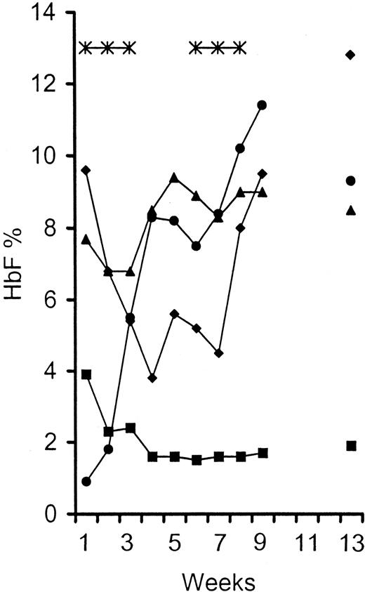
Table 1 shows the maximum HbF and other hematologic parameters achieved at each dose level. A typical temporal HbF response to 5-aza-CdR (patient 3, treated at 0.20 mg/kg per day for 2 cycles) is presented in Figure 1. Also included in the figure are absolute neutrophil count, hemoglobin, and absolute reticulocyte count. The HbF response begins after only 1 week of treatment and is biphasic, with the first peak occurring 3 to 4 weeks after beginning the cycle. The maximum HbF is attained after the second cycle and is approximately 35% greater than the initial peak.
Temporal changes in fetal hemoglobin, hemoglobin, and absolute neutrophil and absolute reticulocyte counts of patient 3 in response to 5-aza-CdR treatment.
An HU nonresponder was treated with 5-aza-CdR in a dose of 0.2 mg/kg per day for 2 treatment cycles. HbF peaked at 8.3% during the first cycle and increased to 11.3% during the second cycle. Maximum HbF was attained 3 to 4 weeks after the beginning of each cycle. Note that the neutrophil count following the second cycle of treatment does not reach a lower level than that resulting from the first cycle. ♦, neutrophils/1000; ▴, Hb g/dL; *, treatment with 0.2 mg/mL; ●, HbF %; ▪, absolute reticulocytes/10-5.
The average γ-globin synthesis relative to total non-α-globin synthesis before 5-aza-CdR treatment was 3.19% ± 1.43% and increased to 13.66% ± 4.35% after treatment (Table 1). This increase in γ-globin synthesis was associated with an average increase in HbF from 3.55% ± 2.47% to 13.45% ± 3.69%. The elevation in HbF could be attributed to increases in both the percentage of F cells and the amount of HbF per F cell. F cells increased from 21% ± 14.8% to 55% ± 13.5% and the HbF/F cell increased from 17% to 24.4%.
Table 2 shows the maximum HbF for each of the patients who were nonresponsive to HU. These 5 patients had little or no elevations in HbF despite compliance with HU. The HbF (percentage of total hemoglobin ± SD) measured before and during compliant HU therapy remained unchanged. However, after 5-aza-CdR administration, the average maximum HbF was 12.7% ± 1.8%, a 5.6-fold increase over baseline and a 35-fold greater increase over that seen during HU therapy. The peak HbF was observed 5 weeks after the start of 5-aza-CdR therapy as compared to the lack of response after more than 24 weeks with HU.
Differences in maximum HbF levels attained following HU and 5-aza-CdR treatment in patients who did not increase their HbF levels after compliant HU therapy
| Patient . | Pre Rx HbF % . | Post Rx HU HbF % . | Post Rx 5-aza-CdR HbF % . |
|---|---|---|---|
| 1 | 2.3 | 1.3 | 10.7 (0.25) |
| 2 | 4.3 | 5.5 | 15.0 (0.25) |
| 3 | 0.9 | 1.1 | 13.9 (0.30) |
| 4 | 0.5 | 0.3 | 11.2 (0.30) |
| 5 | 3.4 | 4.3 | 13.1 (0.25) |
| ± SD | 2.28 ± 1.61 | 2.60 ± 2.15 | 12.70 ± 1.81* |
Values within parentheses are doses of 5-aza-CdR (mg/kg per day).
P < .001, paired t-test.
The 2 HU responders who were unable to sustain the levels (patients 6 and 7, Table 1) were treated with 5-aza-CdR and attained HbF of 15.9% (0.15 mg/kg) and 20.2% (0.30 mg/kg), respectively. This represents an average 52% increase over that seen with HU. Only patient 8 in the study had not been previously treated with HU. This patient achieved a maximum HbF of 18.1% with the lowest dose of 5-aza-CdR (0.15 mg/kg).
Six patients were treated at 2 dose levels of 5-aza-CdR (Figure2). Because there were large individual variations in baseline HbF and response to therapy, the effect of the higher dose level was always compared with each patient's response at the lower dose. Although a dose-response relationship was not clearly demonstrated, increasing the dose of 5-aza-CdR consistently resulted in an increased maximum HbF within a given patient.
The 5-aza-CdR dose and HbF response.
Six patients were treated with 2 doses of decitabine. In each case, the greatest elevation of HbF occurred at the highest dose. ■, before treatment; □, hydroxurea; , 0.15 mg/kg; ▧, 0.20 mg/kg; ▤, 0.25 mg/kg; ▨, 0.30 mg/kg.
The 5-aza-CdR dose and HbF response.
Six patients were treated with 2 doses of decitabine. In each case, the greatest elevation of HbF occurred at the highest dose. ■, before treatment; □, hydroxurea; , 0.15 mg/kg; ▧, 0.20 mg/kg; ▤, 0.25 mg/kg; ▨, 0.30 mg/kg.
Effect of 5-aza-CdR on platelets, neutrophils, and reticulocytes
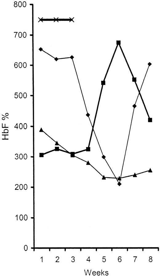
Generally, 5-aza-CdR is considered to be a cytotoxic drug and has been reported to have a marked effect on myelopoiesis at the chemotherapeutic concentrations used for the treatment of hematologic malignancy (> 10−4 mol/L).36 To define the toxicity of low-dose 5-aza-CdR on blood cell production we analyzed the changes in platelet, neutrophil, and absolute reticulocyte counts after 5-aza-CdR treatment. Figure 3 presents the temporal changes in these blood cell elements during the course of 5-aza-CdR treatment. Each point on each curve is the average value for that element obtained at weekly intervals for all patients and all treatment doses. Unlike other cytotoxic agents, low-dose 5-aza-CdR treatment resulted in a transient increase in platelet count that peaked at 220% above baseline 5 weeks (week 6) after initiation of treatment (P < .0001), and returned toward baseline by the seventh week. This increase in platelets was mirrored by a transient decline in neutrophils with a nadir of 32% of baseline at 6 weeks (P < .0001). Table 1shows the individual neutrophil counts at the nadir. In patients whose neutrophil counts decreased below 1000, the nadir lasted only 1 to 3 days, and baseline values were re-established by the seventh week even when they were treated with a 2-cycle regimen (Figure 2). The changes in platelet and neutrophil counts first appeared at about the third week of a cycle. There was a reduction in absolute reticulocyte count, with a nadir of 59% of baseline at 6 weeks. The decrease in absolute reticulocyte count, although not as steep as that seen in neutrophil count, was observed after only 1 week of treatment. Total hemoglobin levels increased (a mean of 1 g/dL approximately 12%) by the 6th week (Table 1, P < .0001 compared to baseline). None of the patients developed any sickle cell disease-related events during the period of study.
Temporal changes in platelet, absolute neutrophil, and absolute reticulocyte counts during 5-aza-CdR treatment.
Each point on a curve is the average count obtained at weekly intervals for all patients at all doses. ♦, neutrophils/10; ▪, platelets/1000; ▴, absolute reticulocyte/1000; ×, treatment.
Temporal changes in platelet, absolute neutrophil, and absolute reticulocyte counts during 5-aza-CdR treatment.
Each point on a curve is the average count obtained at weekly intervals for all patients at all doses. ♦, neutrophils/10; ▪, platelets/1000; ▴, absolute reticulocyte/1000; ×, treatment.
Discussion
We have demonstrated that low-dose 5-aza-CdR is effective in augmenting HbF in the 8 patients studied. The only observed drug-related adverse change was a transient neutropenia, which was not accompanied by any clinical sequelae. The surprising finding is that 5-aza-CdR can augment HbF in patients who do not respond to HU. The maximum HbF attained in these patients was 6-fold higher than baseline.
The mechanism of action of 5-aza-CdR is likely to be different from mere cytotoxicity. 5-aza-CdR has been shown to cause DNA hypomethylation through inhibition of DNA methyltransferase (DMT).37 Hypomethylation is correlated with changes in gene expression, and increased γ-gene expression could be the direct result of hypomethylation of the γ-promoter.38 The difference in mechanism can also be seen by the greater increase in HbF production compared to that observed as a result of cytotoxicity. The 2.2-fold increase in platelet count is not consistent with cytotoxicity but rather with preferential differentiation of pluripotential stem cells into the megakaryocytic lineage at the expense of neutrophil and red cell production. This is a plausible explanation because these cells arise through a common progenitor cell.39,40 This explanation is underscored by the observation that primitive myeloid cells in culture treated with 5-aza-CdR underwent megakaryocytic differentiation.41 Serial bone marrow sampling for progenitor cell analysis during 5-aza-CdR treatment may help clarify the mechanism of the transient neutropenia, that is, preferential differentiation versus cytotoxicity. Some of the reduction in absolute number of reticulocytes might also reflect decreased hypoxic stress as a result of increased hemoglobin level. Although the differences in HbF among patients appear to be correlated with differences in γ-globin synthesis, the ultimate HbF attained is probably also the result of prolonged F cell survival.42
Administration of low doses of 5-aza-CdR, as used in our patients, may be essential for optimal stimulation of HbF production and optimal therapeutic effect. Higher doses may result in greater neutropenia and may also blunt the HbF response. Jutterman and colleagues have recently demonstrated that after incorporation into DNA, 5-aza-CdR binds covalently to DMT blocking DNA polymerase progression, and at high level, adduct formation results in growth arrest and cell death.43 The limited covalent adduct formation occurring during low-dose 5-aza-CdR therapy can be efficiently repaired allowing DNA polymerase progression, and survival of hypomethylated cells.
Although 5-aza-CdR was effective in augmenting HbF in our patients, we do not advocate its use in patients who are responding well to HU. We do see a potential role for 5-aza-CdR in patients who are not responding to HU and in those with pretreatment HbF below 2.0%, for the latter patients appear to be the majority of the nonresponders to HU.12 14 Patients who are candidates for elective surgery and have multiple red blood cell antibodies may benefit from preoperative treatment with 5-aza-CdR, because of the rapid escalation in both hemoglobin and HbF. Although only a small number of patients were studied, our results are encouraging. We have started a second phase of the study to determine whether HbF levels can be maintained optimally for 36 weeks by increasing or decreasing doses and cycle length according to duration of effect and toxicity in each patient.
This phase I/II dose escalation trial demonstrated the ability of 5-aza-CdR to stimulate HbF production with minimal toxicity and in patients who had not responded to HU. Studies to demonstrate a sustained effect of 5-aza-CdR by repeated intermittent doses are in progress. An oral formulation of 5-aza-C in combination with tetrahydrouridine to ensure bioavailability would be a welcome alternate drug for patients with sickle cell anemia.24Obviously, long-term safety of the drug should continue to be a consideration before any drug can be offered for routine treatment of patients.
The publication costs of this article were defrayed in part by page charge payment. Therefore, and solely to indicate this fact, this article is hereby marked “advertisement” in accordance with 18 U.S.C. section 1734.
References
Author notes
Mabel Koshy, University of Illinois at Chicago, 840 South Wood St (M/C 787), Chicago, IL 60612; e-mail:mkoshy@uic.edu.


This feature is available to Subscribers Only
Sign In or Create an Account Close Modal