Abstract
Cloning, expression, and genotype studies of the defective gene for δ-aminolevulinate dehydratase (ALAD) in a patient with an unusual late onset of ALAD deficiency porphyria (ADP) were carried out. This patient was unique in that he developed the inherited disease, together with polycythemia, at the age of 63. ALAD activity in erythrocytes of the patient was less than 1% of the normal control level. ALAD complementary DNA (cDNA) isolated from the patient's Epstein-Barr virus (EBV)–transformed lymphoblastoid cells had 2 base transitions in the same allele, G177 to C and G397 to A, resulting in amino acid substitutions K59N and G133R, respectively. It has been verified that the patient had no other ALAD mutations in this and in the other allele. By restriction fragment length polymorphism (RFLP) analysis, all family members of the proband who had one-half ALAD activity compared with the ALAD activity of the healthy control were shown to have the same set of base transitions. Expression of ALAD cDNA in CHO cells revealed that K59N cDNA produced a protein with normal ALAD activity, while G133R and K59N/G133R cDNA produced proteins with 8% and 16% ALAD activity, respectively, compared with that expressed by the wild type cDNA. These findings indicate that while the proband was heterozygous for ALAD deficiency, the G397 to A transition resulting in the G133R substitution is responsible for ADP, and the clinical porphyria developed presumably due to an expansion of the polycythemic clone in erythrocytes that carried the mutant aladallele.
Introduction
δ-Aminolevulinate dehydratase (E.C.4.2.1.24, ALAD) is a cytosolic enzyme in the heme biosynthetic pathway that catalyzes the condensation of 2 molecules of δ-aminolevulinic acid (ALA) to form a monopyrrole porphobilinogen (PBG).1 The enzyme activity is present in great excess in normal cells; thus a partial inhibition of the enzyme activity is not usually accompanied by clinical consequences.2-4 Four unrelated cases of ALAD deficiency porphyria (ADP) have been reported to date; 2 German male patients developed the disease at adolescence,5 1 Swedish boy has suffered from severe ADP since birth,6 and 1 Belgian male patient7developed the inherited disease at the age of 63 years. The latter patient is the topic of this study. Molecular analysis of the gene defects has been previously reported for the 2 German patients8,9 and the Swedish boy,10 but not for the Belgian male. The molecular analysis of ALAD cDNA in the Belgian patient and the defect found in the patient are described herein. Sequence analysis of ALAD cDNA isolated from the proband's Epstein-Barr virus (EBV)–transformed lymphoblastoid cells had 2 base transitions in one allele, G177 to C and G397to A, resulting in amino acid substitutions K59N and G133R, respectively. The other allele was entirely normal, indicating that the proband is heterozygous for ALAD deficiency. The expression of ALAD cDNA in CHO cells revealed that both G133R and K59N/G133R cDNA produced proteins with abnormally reduced ALAD activity. These findings indicate that the Belgian patient with ADP is heterozygous for ALAD deficiency, and clinical ADP may have been precipitated by other pathophysiological factors, such as polycythemia, in this patient.
Materials and methods
Proband and family members studied
The proband is a male with ADP whose clinical and biochemical features were previously reported.7 At the age of 63 years, he developed polycythemia (erythrocytes, 7.27 × 10¹²/L; hematocrit, 0.55; hemoglobin, 171 g/L; and erythroid hyperplasia in the marrow) of an unknown origin. The patient also developed a loss of strength in both arms, which gradually became worse.11 He showed a markedly elevated plasma ALA concentration (110 μg/dL) compared to the normal value of less than 10 μg/dL, while the plasma monopyrrole porphobilinogen (PBG) was undetectable. The urinary ALA excretion of 85 mg/dL was abnormally elevated compared with the normal value of less than 5 mg/dL, without an increase in PBG concentration. The erythrocyte7 and lymphocyte ALAD activity12 of the proband was less than 1% and approximately 20% of the activity of healthy controls, respectively. Other causes of ALAD deficiency, such as lead poisoning, tyrosinemia, alcoholism, smoking, cirrhosis, renal insufficiency, and diabetes mellitus, have been excluded by specific testing for each disorder or from the proband's past history.7 Both oral and intravenous glucose treatments were effective in ameliorating biochemical and clinical abnormalities, which are typical responses of acute hepatic porphyrias to such a treatment.13,14 Family studies showed that except for a brother, who had normal levels of enzyme activity, a sister, a daughter, and a granddaughter of the patient had approximately 50% ALAD activity compared with the activity of healthy controls,7 indicating that these subjects are heterozygous carriers of ALAD deficiency.
Cell cultures
Isolation of mononucleated cells from blood, transformation of cells to lymphoblastoid cells with EBV, and cultivation of transformed cells were carried out as described previously.12
Cloning and sequence analysis of ALAD cDNA
Total RNA was prepared from EBV-transformed lymphoblastoid cells according to the method of Cathala et al.15 The first-strand DNA was synthesized using SuperScript RnaseH−reverse transcriptase (Gibco BRL, Gaithersburg, MD) primed with oligo (dT) (oligodeoxythymidylic acid)12-18. The first PCR amplification of ALAD cDNA was performed by using oligomers ALAD21 and ALAD31 (Table 1). ALAD21 corresponded to the beginning portion of exon 1A, and ALAD31 corresponded to the 3′-untranslated region of ALAD cDNA. The PCR product was purified using the Geneclean kit (BIO 101, La Jolla, CA). It was then used as a template for the second PCR amplification, which was carried out using ALAD23 and ALAD2 as the primer (Table 1), corresponding to the 5′-untranslated region (downstream of ALAD21) and the 3′-untranslated region (upstream of ALAD31) of ALAD cDNA, respectively. Amplification and further steps were performed independently 3 times in separate experiments. PCR was carried out using a DNA Thermal Cycler (Perkin Elmer-Cetus, Norwalk, CT) employing a thermal cycle program. PCR products were purified using a Geneclean kit followed by cloning into pGEM-T Easy Vector (Promega Company, Madison, WI). DNA sequencing was carried out by the dideoxy chain-termination method16 using a genetically engineered T7 DNA polymerase (SequiTherm Long-Read Cycle sequencing kits; LC Epicentre-Technologies, Madison, WI).
Synthetic oligonucleotides for PCR
| Primer . | Sequence . | Location . |
|---|---|---|
| ALAD 2 | 5′-GTTCTAGAGGGCCTGGCACTGTCCTCCA-3′ | 3′-untranslated |
| ALAD 3 | 5′-GCAATCCATCTGTTGAGGA-3′ | exon 5 |
| ALAD 4 | 5′-CACAGGCAGACATCACAGGCC-3′ | exon 5 |
| ALAD 17 | 5′-TACAGCCTATCACCAGCCTC-3′ | exon 3 |
| ALAD 21 | 5′-GGGAGACCGGAGCGGGAGACA-3′ | exon 1A |
| ALAD 23 | 5′-CAATGCCCCAGGAGCCCTCG-3′ | exon 2 |
| ALAD 29 | 5′-CTGTAGCTCATCACCGATAC-3′ | exon 8 |
| ALAD 31 | 5′-GGTTGTCACTTGTCTGAGGC-3′ | 3′-untranslated |
ALAD cDNA or genomic DNA was amplified by PCR using a pair of synthetic oligonucleotides as primers, as described in “Materials and methods.”
Nucleotide sequence analysis of genomic DNA
Genomic DNA was prepared from lymphoblastoid cells of the proband. Regions containing the base transitions identified by cDNA analysis were amplified by PCR. PCR was carried out using a set of primers, ALAD17 and ALAD4 (Table 1), to detect the G177 to C transition, and another set of primers, ALAD3 and ALAD29 (Table 1), to detect the G397 to A transition. Purification of PCR products, cloning, and sequence analysis were carried out as described above.
ALAD genotype analysis in family members of the proband
ALAD genotype analysis in the proband's family were carried out by restriction fragment length polymorphism (RFLP) usingCfr10I and PstI for the G177 to C and G397 to A transition of ALAD cDNA, respectively. ALAD cDNA from all the family members having less than 50% of normal ALAD activity was PCR-amplified with primers ALAD23 and ALAD29 (Table 1) for 40 cycles using an annealing temperature of 50°C. The PCR products were purified using the Geneclean kit, diluted in an appropriate buffer, and then digested with Cfr10I and PstI. RFLP products were analyzed by electrophoresis in a 2% agarose gel.
Expression of mutant ALAD cDNA
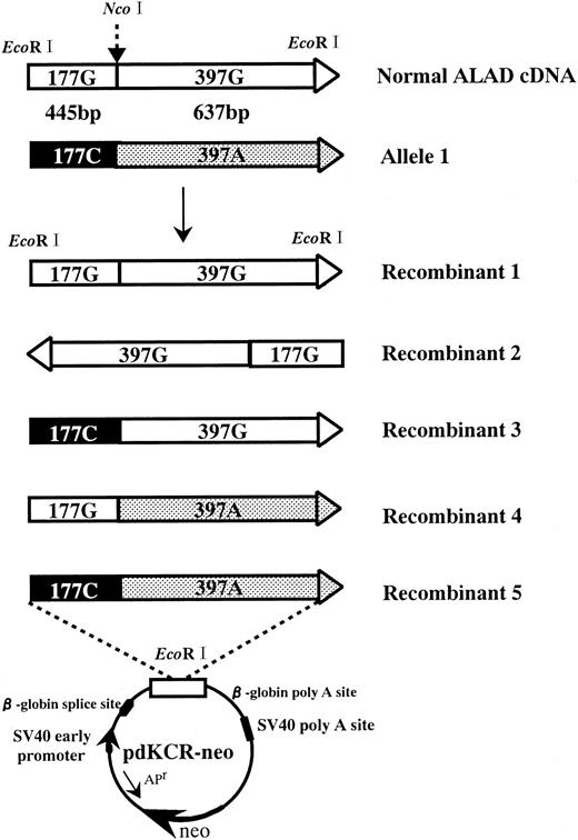
For expression of ALAD cDNA, the pdKCR-Neo vector, which contains the SV40 early gene promoter, donor and acceptor sites, and polyadenylation sites derived from the rabbit β-globin gene and SV40 early gene, was used. Cloned ALAD cDNA from the proband was digested with NcoI and ligated with the normal cDNA to prepare ALAD cDNA containing the G177 to C transition or the G397 to A transition (see Figure 3). Recombinant ALAD cDNA was restricted with EcoRI followed by purification with the Geneclean kit and subcloned into theEcoRI site of the pdKCR-Neo vector (see Figure 3). Recombinant plasmids were introduced into CHO cells by coprecipitation with calcium phosphate (CellPhect Transfection kit; Pharmacia Fine Chemicals, Uppsala, Sweden) followed by selection with 800 μg/mL G418 (Geneticin, Gibco BRL, Gaithersburg, MD) starting 24 hours after transfection.
Western blot analysis
Human ALAD expressed in CHO cells was specifically detected by Western blot analysis using 0.5 × 10⁶ CHO cells. The antibody used in this study was rabbit immunoglobulin G (IgG) purified from a polyclonal antibody against human ALAD, which cross-reacted with the human ALAD but not with the CHO enzyme.17 Detection of the immune complex was carried out using the ECL Western blotting detection system (Amersham Life Sciences, Buckinghamshire, England).
Assay of ALAD activity
Allele-specific oligonucleotide hybridization analysis
Genomic DNA was extracted from peripheral blood mononuclear cells of a healthy subject and EBV-transformed lymphoblastoid cells of the patient, and PCR was performed to amplify exon 5 of thealad gene using the specific primers. After application of denatured amplicons to a sheet of nylon filter, membranes were prehybridized in a buffer18 for 4 hours followed by hybridization overnight with an end-labeled mutant oligonucleotide, 5′-GGTCAGTGCAGTGAGTTC-3′, or the normal oligonucleotide, 5′-GGTCACTGCGGTGAGTTC-3′. The membranes were then washed as described previously18 and exposed to an x-ray film.
Fluorescence in-situ hybridization
Bone marrow slides were deparaffinized using the Paraffin Pretreatment Reagent kit (Vysis, Downers Grove, IL). Fluorescence in-situ hybridization (FISH) analysis was performed using the Locus Specific Identifiers (LSI) kit (Vysis) according to the manufacturer's protocol. For detection of the chromosomal alad gene, a LSI bcr/abl (breakpoint cluster region/) dual color translocation probe, which is labeled with green fluorescence for bcr and red fluorescence for abl, was used. Because the abl gene is also located to 9q34, 2 signals are detected at chromosome 9q34 of normal specimens.
Results
Cloning of the mutant ALAD cDNA
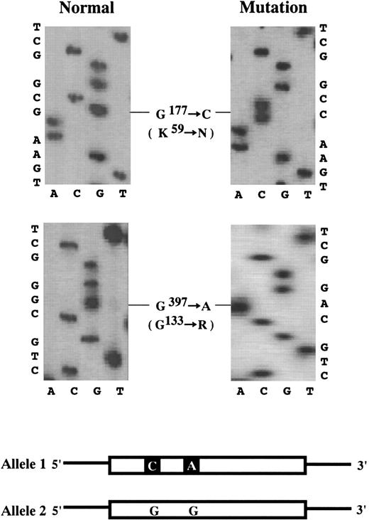
By nucleotide sequence analysis, a set of 2 base transitions was identified in one allele (Allele 1, Figure1). One transition was a G177to C transition, resulting in amino acid substitution K59N, while the other transition was a G397 to A transition, resulting in amino acid substitution G133R. The coding region of the otheralad allele (Allele 2, Figure 1) was shown to be entirely normal. The 2 base transitions were also confirmed in the genomic DNA of the proband (data not shown).
Nucleotide sequence analysis of ALAD cDNA from the proband.
G177 to C transition, producing K59N substitution, and G397 to A transition, producing G133R substitution. Scheme: Schematic representation of the proband's ALAD cDNA. Allele 1 has 2 base transitions, G177 to C and G397 to A, while Allele 2 is entirely normal. Thick boxes and thin horizontal lines denote translated and untranslated regions, respectively.
ALAD activity of the proband was less than 1% of the normal ALAD activity in erythrocytes, and the ALAD activity was approximately 20% in the patient's EBV-lymphoblastoid cells, which suggests that the defect is only partial in lymphoid cells but almost complete in erythroid cells. Sequence analysis of a region of the aladgene from −444 base pair (bp) of intron 1 to 25 bp of exon 1B, including the erythroid-specific promoter,19 revealed that there was no abnormality, which indicates that the marked ALAD deficiency in erythrocytes is not due to a lack of expression of the erythroid-specific ALAD messenger RNA (mRNA).
ALAD genotype studies
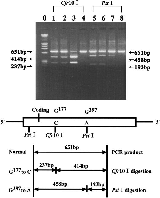
Because both G177 to C and G397 to A base transitions created new restriction sites, ie, Cfr10I andPstI sites, respectively, RFLP analysis withCfr10I and PstI was used to identify the sites in family members of the proband. As shown in Figure2, cDNA from the proband, the sister, and the daughter were positive for the Cfr10I andPstI cleavage sites, which indicates that they carry both of these base transitions, whereas a healthy subject was negative for the sites. RFLP analysis with Cfr10I showed that one-half of the PCR product from the proband and his sister remained uncut, indicating that their other alad allele was normal at the position of 177. On the other hand, the PCR product from the proband's daughter was digested completely, indicating that she was homozygous with the G177 to C transition.
ALAD genotype analysis.
ALAD genotype analysis in the family was carried out byCfr10I and PstI RFLP for the G177 to C transition and the G397 to A transition, respectively. Lanes 1 and 5, the proband; lanes 2 and 6, the sister; lanes 3 and 5, the daughter; lanes 4 and 8, healthy control.
ALAD genotype analysis.
ALAD genotype analysis in the family was carried out byCfr10I and PstI RFLP for the G177 to C transition and the G397 to A transition, respectively. Lanes 1 and 5, the proband; lanes 2 and 6, the sister; lanes 3 and 5, the daughter; lanes 4 and 8, healthy control.
ASO analysis
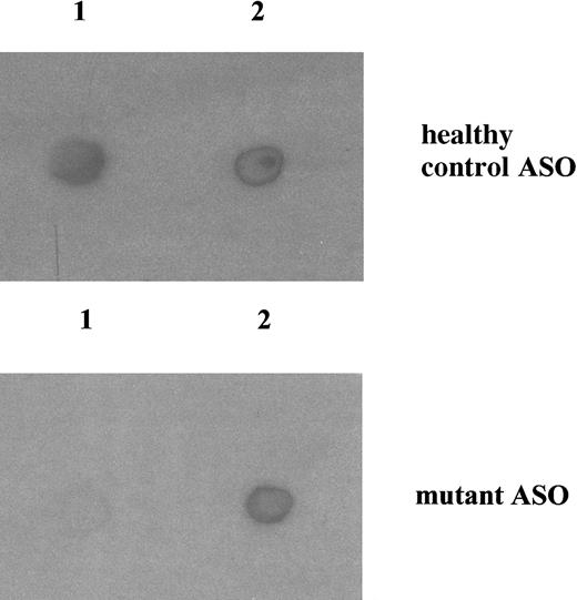
ASO analysis was performed using genomic DNA isolated from EBV-lymphoblastoid cells. As shown in Figure3, an amplicon from exon 5 of a healthy subject hybridized only with the normal oligonucleotide, while that of the patient equally hybridized with both the normal and the mutant oligonucleotide. This finding indicates that the lymphoblastoid cells of the patient were heterozygous for the ALAD mutation.
ASO analysis of genomic DNA.
Amplicons from exon 5 of genomic DNA prepared from mononuclear cells of a healthy subject and EBV-lymphoblastoid cells from the proband were spotted onto a sheet of nylon filter and hybridized with the normal or the mutant nucleotide as described in “Materials and methods.” Top panel, with the normal oligonucleotide; bottom panel, with the mutant oligonucleotide. Lane 1, healthy subject; lane 2, the patient.
ALAD activity expressed by the mutant ALAD cDNAs
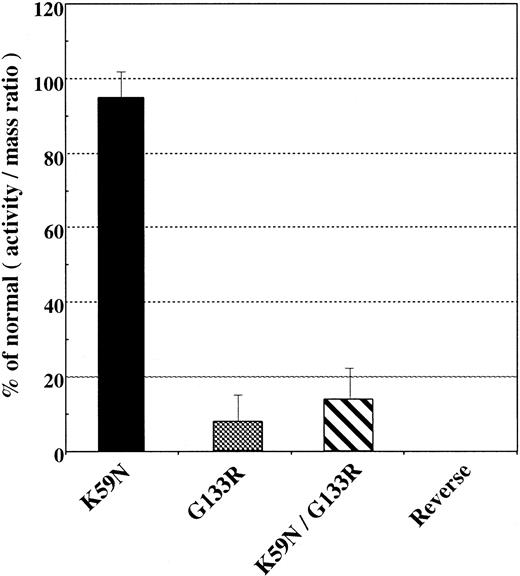
To determine the effect of these 2 base transitions on enzyme activity, mutant ALAD cDNA was expressed by transfection into CHO cells. Construction of the expression vector with ALAD cDNA is shown in Figure 4. The expressed ALAD activities were compared with the activities obtained by transfection of the normal ALAD cDNA. Human ALAD protein expressed in CHO cells was specifically detected and quantified by Western blot analysis using a polyclonal rabbit IgG versus a human ALAD, which reacts with the human ALAD but not the hamster ALAD. Human-specific ALAD activity was calculated as the difference between the total enzyme activity of CHO cells transfected with ALAD cDNA and the activity of CHO cells transfected with a plasmid in which ALAD cDNA was inserted in the reverse orientation (mock transfection). Because transformed CHO cells with recombinant ALAD cDNA, except for mock transfection, expressed immunoreactive human ALAD proteins, human-specific ALAD activity was calculated as an A/M ratio, as described previously.8 The K59N/G133R mutant showed 16.0% ± 8.1% of the A/M ratio compared with that expressed by the normal ALAD cDNA (Figure5). Mutant ALAD carrying a single G133R substitution showed a similar decrease in the A/M ratio (8.1% ± 7.0%), while the K59N protein expressed a normal A/M ratio (94.6% ± 6.6%) (Figure 5). These findings indicate that the G397 to A transition is responsible for the decrease of ALAD activity in this family and for ADP in the proband, and the K59N represents a polymorphic ALAD.
Construction of the expression vector with ALAD cDNA.
Recombinant 1: Normal ALAD cDNA cloned from EBV-transformed lymphoblastoid cells of the patient (allele 2). Recombinant 2: Normal ALAD cDNA introduced in the opposite direction into the pdKCR-Neo vector. Recombinant 3: G177 to C transition ALAD cDNA, reconstituted by replacing EcoRI-NcoI fragment (637 bp) from normal ALAD cDNA with theEcoRI-NcoI fragment (444 bp) of G177to C/G397 to A transition of ALAD cDNA. Recombinant 4: G397 to A transition of ALAD cDNA, reconstituted by replacing EcoRI-NcoI fragment (444 bp) from normal ALAD cDNA with the EcoRI-NcoI fragment (637 bp) of G177 to C/G397 to A transition of ALAD cDNA. Recombinant 5: G177 to C and G397 to A transition of ALAD cDNA, cloned from lymphoblastoid cells of the proband (allele 1). These recombinant cDNA were introduced into theEcoRI site of the third exon of rabbit β-globin in the pdKCR-Neo expression vector.
Expression of ALAD cDNA in CHO cells.
Transfection of CHO cells with ALAD cDNA, quantitation of human ALAD protein expressed in cells, and assays on ALAD activity were carried out as described previously.8 Specific ALAD activity (A/M ratio) expressed as a percent of the activity expressed by normal ALAD cDNA. Data are the mean of quadruple assays.
FISH analysis
To examine the possibility that the normal allele may have been lost by chromosomal deletion during the progression of polycythemia, FISH analysis was performed on bone marrow slides of the patient. A FISH probe for the abl gene was additionally used in this analysis by taking advantage of the fact that the abl gene is also located on chromosome 9q34, where the alad gene is present. Thus 2 signals detecting the 9q34 locus should be detected in normal samples. While this was clearly shown in a freshly prepared sample from mononuclear cells of a healthy individual, there was no signal detected in the patient's bone marrow samples prepared from paraffin blocks (data not shown). FISH analysis was also performed using bone marrow slides of the patient, obtained at 3 different occasions, (1) before and (2) after the onset of PV, and (3) at autopsy, but failed to show any signal in all samples. The failure of detection of these signals, however, does not suggest selective deletion of both alleles at 9p34 because FISH analysis using a probe for the bcr gene also failed to show any signal (data not shown). Thus the lack of signals in the proband's samples is more likely due to a degeneration of DNA than to a specific loss of these alleles, which is presumably due to the fact that the proband's specimens were stored for more than 10 years at room temperature.
Discussion
This study describes the first complete molecular analysis of a defective alad gene in a patient with an unusual late-onset acute hepatic porphyria. The patient was first reported to have clinically homozygous ALAD deficiency because the activity of erythrocyte ALAD was nearly completely lost, and it was not restored to normal by incubation with dithiothreitol or zinc ion.7 It was also reported that decreased activity of ALAD was observed in his pedigree spanning over 3 generations, which clearly indicates the inherited nature of the gene defect.7 However, ALAD deficiency in the patient's EBV-transformed lymphoblastoid cells was only partial (approximately 20% of normal),12 but it was almost complete in erythrocytes (less than 1% of normal). Molecular analysis of ALAD cDNA demonstrated that the proband carried a single defective alad allele (allele 1); the other allele (allele 2) was been shown to be completely normal. Allele 1 contained a set of 2 base transitions, G177 to C and G397 to A (Figure 1). Genotype analysis of family members of the proband indicated that all the family members having less than 50% of normal ALAD activity carried the same set of base transitions (Figure 2). These findings established the fact that there were no other base transitions of the alad gene in the proband, and the molecular defect in family members of the proband could be unequivocally demonstrated by the existence of a set of 2 base transitions of the alad gene in allele 1.
The G397 to A transition, which resulted in a G133R substitution, was previously reported occurring in a Swedish boy with ADP,10 although that study did not examine a phenotypic property of the mutant protein. We have included a phenotypic characterization in this current study. The G397to A transition was located in the highly conserved zinc-binding site in the enzyme,20-22 and thus it may likely influence enzyme activity, but this question was not examined. Our expression studies in CHO cells clearly showed that the mutant ALAD with G133R substitution had significantly decreased enzyme activity (Figure 5). Furthermore, a mutant protein having both K59N and G133R substitutions also showed a similar decrease in ALAD activity as G133R (Figure 5), indicating that the G397 to A transition resulting in G133R substitution was responsible for the deficiency of ALAD activity.
The G177 to C transition, which resulted in K59N substitution, was previously reported to exist in a polymorphic ALAD termed ALAD2.23 It has been reported that the gene frequencies of ALAD2 in white and Japanese populations were approximately 10% and 6%, respectively.24 The fact that K59N substitution occurs at a significant frequency suggests that this amino acid substitution probably does not result in a significant deficiency of enzyme activity, but it has never been evaluated. Our findings in this study, using transfection of K59N cDNA into CHO cells and studying expressed enzyme activity, demonstrated that K59N substitution does not significantly influence enzyme activity (Figure5). Our study also demonstrated that the daughter of the proband, who was clinically unaffected, had one-half normal ALAD activity in her erythrocytes and was compound heterozygous for K59N and K59N/G133R substitutions (Figure 2). Decreased ALAD activity in her erythrocytes therefore must be due to the G133R substitution, which had little enzyme activity.
The proband in this study was unique in several aspects. First, there is a marked difference in ALAD activity in erythrocytes (less than 1% of normal) and lymphoblastoid cells (approximately 20% of normal). The human alad gene has 2 different exon 1s, each having its specific promoter sequence and its transcription regulated in a tissue-specific manner.19,25 Namely, the housekeeping transcript includes exon 1A, but not 1B, while the erythroid-specific transcript contains exon 1B, but not 1A. The promoter region upstream of the erythroid-specific exon 1B contains several CACCC boxes and 2 potential GATA-1 binding sites.19 25 Sequence analysis of the promoter region of exon 1B of the alad gene of the proband confirmed the normal sequence for these ciselements, excluding the possibility that erythroid-specific ALAD transcription is impaired. Secondly, the proband was unusual in that he developed this inherited disease very late in his life. Thirdly, ADP symptoms occurred following the development of polycythemia. Thus the late onset of the inherited disease in the proband may be due to the fact that, while the heterozygosity of ALAD deficiency itself was not sufficient for the development of ADP, it was precipitated with ADP when it was combined with another pathophysiological factor, such as polycythemia, that facilitates its expression.
The association of ADP with polycythemia in the proband is of particular interest. The close temporal relationship between the onset of ADP and thrombocytopenia and between the relapse and the polycythemia following treatment with phosphorous 32 (32P) strongly favors a causal relationship between the polycythemic myeloproliferative disease and the ADP in this patient.11The fact that polycythemia or myelodysplastic syndrome are clonal disorders is well documented.26-28 Thus progeny of a single cell gains ascendancy in the bone marrow. This fact is consistent with our finding that the proband's erythrocytes showed an extremely low ALAD activity (less than 1%) despite the fact that he was heterozygous for ALAD deficiency. In a study of 2 patients with polycythemia vera, Adamson et al27 demonstrated the clonal origin of erythrocytes, granulocytes, and platelets but not of peripheral blood lymphocytes. Our findings that the proband's erythrocytes had less than 1% ALAD activity while lymphocytes had approximately 20% activity are entirely consistent with the above view and suggest that the proband's erythrocytes are represented by the polycythemia clone which carried the mutant alad allele. Thus our study demonstrates that heterogeneous ALAD deficiency, which usually has no clinical consequences, can manifest itself as ADP if the defect is clonally expanded, as in the case of polycythemia.
Acknowledgments
We are grateful to Mr Yasuhiko Obara for performing FISH analysis.
The publication costs of this article were defrayed in part by page charge payment. Therefore, and solely to indicate this fact, this article is hereby marked “advertisement” in accordance with 18 U.S.C. section 1734.
References
Author notes
Shigeru Sassa, Laboratory for Biochemical Hematology, The Rockefeller University, New York, NY; e-mail:sassa@rockvax.rockefeller.edu.

This feature is available to Subscribers Only
Sign In or Create an Account Close Modal