Fifty-eight acute promyelocytic leukemia (APL) patients (11 newly diagnosed and 47 relapsed) were studied for arsenic trioxide (As2O3) treatment. Clinical complete remission (CR) was obtained in 8 of 11 (72.7%) newly diagnosed cases. However, As2O3 treatment resulted in hepatic toxicity in 7 cases including 2 deaths, in contrast to the mild liver dysfunction in one third of the relapsed patients. Forty of forty-seven (85.1%) relapsed patients achieved CR. Two of three nonresponders showed clonal evolution at relapse, with disappearance of t(15;17) and PML-RAR fusion gene in 1 and shift to a dominant AML-1-ETO population in another, suggesting a correlation between PML-RAR expression and therapeutic response. In a follow-up of 33 relapsed cases over 7 to 48 months, the estimated disease-free survival (DFS) rates for 1 and 2 years were 63.6% and 41.6%, respectively, and the actual median DFS was 17 months. Patients with white blood cell (WBC) count below 10 × 109/L at relapse had better survival than those with WBC count over 10 × 109/L (P = .038). The duration of As2O3-induced CR was related to postremission therapy, because there was only 2 of 11 relapses in patients treated with As2O3 combined with chemotherapy, compared with 12 of 18 relapses with As2O3 alone (P = .01). Reverse transcription polymerase chain reaction (RT-PCR) analysis in both newly diagnosed and relapsed groups showed long-term use of As2O3 could lead to a molecular remission in some patients. We thus recommend that ATRA be used as first choice for remission induction in newly diagnosed APL cases, whereas As2O3 can be either used as a rescue for relapsed cases or included into multidrug consolidation/maintenance clinical trials.
MOST ACUTE PROMYELOCYTIC leukemia (APL) patients have the characteristic chromosome translocation t(15;17) that juxtaposes PML gene on chromosome 15 and RARα gene on chromosome 17, forming a PML-RARα fusion gene.1-3 The PML-RARα protein encoded by the fusion gene plays an important role in the pathogenesis of APL.4 Although 70% to 85% of APL patients achieved complete remission (CR) by cytotoxic chemotherapy,4 a significant part of patients experienced severe complications, necessitating intensive supportive care, which is hardly available in most developing countries. The use of differentiation therapy with all-trans retinoic acid (ATRA) has not only opened a new approach in cancer treatment, but also rendered remission induction relatively easy in most of the APL patients.5,6 Moreover, the overall disease-free survival in the patients with CR achieved by ATRA and consolidated as well as maintained by chemotherapy has been significantly higher than those treated with chemotherapy alone for remission induction, consolidation, and maintenance treatment. In spite of these advances, 30% to 40% of patients would relapse within 5 years after CR. The majority of these patients lost sensitivity to ATRA and chemotherapy and died shortly thereafter.
In early 1970s, Zhang et al7-9, from Harbin Medical University in Northeast China, found that intravenous administration of arsenic trioxide (As2O3 ) with relatively small doses (10 mg/d) was effective in treating patients with APL, lymphoma, and liver cancer. However, it was only recently that the therapeutic effect of As2O3 was proven in APL patients by several groups in China.10-13 Recently, Soignet et al14 confirmed in Western population that As2O3 treatment achieved CR in 11 out of 12 relapsed APL patients.
In vitro studies indicate that As2O3 may exert biphasic action on APL cells, induction of apoptosis at higher concentrations (0.5 to 2 μmol/L), and partial differentiation at lower concentrations (0.1 to 0.5μmol/L). As2O3, at both high and low concentrations, is able to trigger the degradation of PML-RARα fusion protein.15 Interestingly, the drug was equally effective in inducing apoptosis in ATRA–sensitive and –resistant APL cells.16,17
Several important issues, nevertheless, remain to be addressed for As2O3 treatment in APL. Is As2O3 treatment equally effective for newly diagnosed APL patients? Could the drug induce a molecular remission in these patients as well as in relapsed cases? What could be the possible prognostic parameters and suitable postremission treatment in relapsed APL patients rescued with As2O3? In this report, we describe the results of a comprehensive study on 11 previously untreated and 47 relapsed APL cases.
PATIENTS AND METHODS
Patients.
The diagnosis of APL was established on the basis of clinical presentation, morphological criteria of the French-American-British (FAB) classification, cytogenetic evaluation for t(15;17), and reverse transcription polymerase chain reaction (RT-PCR) analysis for PML-RARα transcripts. Two groups of patients, newly diagnosed and relapsed APL, entered into this As2O3 treatment multicenter study including five hospitals in Shanghai (Rui-Jin Hospital, Ren-Ji Hospital, Xin-Hua Hospital, Zhong-Shan Hospital, and Gan-Quan Hospital). Between July 1996 and April 1998, 11 cases of newly diagnosed APL without exposure to any antileukemia treatment were included into this study. Between December 1994 and February 1998, 47 relapsed patients (2 at the second relapse, 2 at the third relapse, and 43 cases at the first relapse) who had received ATRA for the remission induction and chemotherapy/ATRA in the following consolidation and maintenance therapy were enrolled. However, none of them were still taking ATRA at the time of relapse. Informed consent was obtained for every patient entering into this study. The main clinical and hematological characteristics of the evaluated patients are shown in Table 1. Of note, part of the data concerning As2O3 induction therapy in 15 cases in the relapsed group was presented in our previous report.12
Characteristics of Patients With APL in the Present Study
| . | Newly Diagnosed APL . | Relapsed APL . |
|---|---|---|
| Evaluated cases | 11 | 47 |
| Sex (male/female) | 3/8 | 29/18 |
| Median age (range, years) | 41 (24 to 60) | 38 (7 to 55) |
| Median WBC (range, ×109/L) | 2.2 (1.1 to 65.0) | 3.4 (0.6 to 56.0) |
| <2 | 4 | 14 |
| 2 to 10 | 4 | 24 |
| 10 to 20 | 1 | 3 |
| >20 | 2 | 6 |
| Median RBC (range, ×1012/L) | 2.33 (1.03 to 2.96) | 3.65 (1.78 to 5.31) |
| Median hemoglobin (range, g/L) | 70 (51 to 97) | 109 (56 to 180) |
| Median platelet (range, ×109/L) | 20 (10 to 50) | 38 (4 to 236) |
| Median percentage of blasts and promyelocytes in BM (range) | 87.5 (38.0 to 95.5) | 66.0 (12.5 to 95.0) |
Induction therapy.
As2O3 solution was prepared by the Pharmacy of Traditional Chinese Medicine in the First Hospital affiliated with Harbin Medical University of China. The following protocol was used: 10 mg As2O3 (10 mL, 0.1% aqua solution) was diluted in 500 mL of 5% glucose-normal saline solution for intravenous drip over 2 to 3 hours per day, for 6-weeks duration. If necessary, a second course was performed after an interval of 7 days. Those patients who failed to reach CR after 2 courses were considered as nonresponders (NR) and were treated with chemotherapy.
Supportive care.
Sequential measurements of complete blood cell count (every other day), bone marrow (BM) cytology (every 10 days), renal functions, and hepatic functions (every 1 to 2 weeks) were performed during As2O3 remission induction treatment. Measurement of coagulation and fibrinolysis parameters, including fibrinogen, DD dimers, fibrin degradation product (FDP), prothrombin time, and activated partial thromboplastin time was performed by standard methods for each patient before and during the As2O3 treatment. Coagulopathy was treated at the physician’s discretion using low-dose heparin, platelet transfusion, and fresh plasma. Patients were administered hydroxyurea or moderate chemotherapy (Daunarubicin: 40 mg/m2/d × 3 d; Ara-C: 100 mg/m2/d × 3 to 5 d) when their white blood cell (WBC) counts were over 30 to 40 × 109/L, based on the observation that there seems to be less clinical syndrome associated with As2O3-induced hyperleukocytosis as compared with ATRA–induced ones. Symptomatic therapy was performed without discontinuation of As2O3 when moderate side effects occurred while As2O3 was withdrawn in the case of serious toxic effects.
Definition of outcomes.
Achievement of CR required patients to have no clinical evidence of APL, untransfused hemoglobin greater than 10g/dL, neutrophils greater than 1.5×10⁹/L, platelets greater than 100×109/L, BM to be normocellular or moderately hypocellular with less than 5% promyelocytes, and absence of leukemic cells with cytoplasmic Auer rods. Disease-free survival (DFS) was defined as the time from CR to relapse, death from any cause, or censoring of the data on the patients.
Follow-up.
After CR was achieved with As2O3, the patients were treated by three different therapeutic protocols for consolidation: (1) chemotherapy group: continuous treatment with chemotherapy DA/MA monthly [Daunorubicin(D): 45mg/m2/d on day 1 to 3 or Mitoxantrone(M) 8 mg/d on day 1 to 3, and Ara-C(A) 100 to 200 mg/d on day 1 to 7]. One course every 2 months in the first year, every 3 months in the second year, and every 4 months in the third year. (2) As2O3 group: 10 mg As2O3 daily continuing 28 to 30 days as a course with approximately 30 to 60 days interval between two cycles within the first year, approximately 7 to 14 days as a course every 2 months over the second and third year. (3) Chemotherapy and As2O3 combination group: chemotherapy was administered as group (1) while As2O3 was used as group (2), but during the interval of chemotherapy. No randomization was performed due to the short supply of As2O3for some of the patients and the refusal of some patients to further use of chemotherapy or As2O3. Follow-up was terminated on March 31, 1999.
Statistical analysis.
Association between pairs of patients’ covariates, including individual characteristics and the treatment indicator, was evaluated using Fisher’s exact test and generalized exact test. Analysis of DFS and overall survival were performed with Kaplan-Meier product-limit estimation.
Cytogenetic studies.
Metaphase chromosomes were prepared from BM cells after short-term culture (24 hours). RHG-banding technique was used and karyotype analysis was performed according to International System for Human Cytogenic Nomenclature (ISCN).18
RT-PCR.
RT-PCR analysis for PML-RARα transcripts was performed according to our previously described methods.19
Fluorescence in situ hybridization (FISH).
Dual-color FISH was performed with the probe YAC 185B2, P1 164 (biotinylated), and YAC 417D9, Co664 (digoxigenin-labeled) prepared by nick translation for PML-RARα and AML1-ETO detection, respectively. Chromosome painting was performed employing whole chromosome painting (WCP) probes for chromosome 5,15,17, and 22 (Cambio, Cambridge, UK, & Oncor, Gaithersburg, MD). The procedure was as recommended by the manufacturer. In brief, slides were denatured in 70% formamide/2 × SSC (1 × SSC: 0.15 mol/L NaCl and 0.015 mol/L natriumcitrate, pH 7.0) at 70°C. Then, the probes were applied to denatured slides and hybridized at 37°C overnight. After posthybridization washing, probes were detected using avidin conjugated Texas Red and fluorescein isothiocyanate (FITC)-labeled antibodies. DAPI was used as a counterstain.
Combination of Wright’s staining and dual-color FISH.
BM samples collected before and 20 days after As2O3 treatment from five patients (case 21,22,29,35, and 37) were subjected to simultaneous morphological and FISH analysis. The procedures described by Haferlach et al20 were used with modification. Briefly, mononuclear cell fractions were isolated from BM aspirate by Ficoll-Hypaque density gradient centrifugation, and were spun onto slides by cytospin (Shandon, Runcorn, UK; 800 rpm, 4 minutes). For morphological observation, Wright’s staining was performed. Leukemic promyelocytes or myelocyte-like cells were photographed by conventional microphotography (Olympus, Tokyo, Japan), and the location of the cells was documented. Slides were incubated in xylene for 5 minutes to remove the cedar wood oil, fixed in Carnoy’s fixation (methanol: acetic acid, 3:1) for 15 minutes, washed in phosphate-buffer solution for 1 minute, and fixed again in paraformaldehyde for 1 minute. The PML-RARα fusion gene was detected by using t(15;17) translocation DNA probe (Oncor, Gaithersburg, MD). Dual-color FISH was performed following the manufacturer’s instructions. The results were observed through a triple-bandpass filter (Olympus) equipped on an Olympus microscope and pictured with Kodak 400 film (Eastman Kodak, Rochester, NY).
RESULTS
Newly Diagnosed Patient Group
CR.
There were 11 newly diagnosed APL patients entered into this study, 7 cases were treated with As2O3 and 4 with combined As2O3 and chemotherapy. Eight (72.7%) entered into CR, and the median time to obtain CR was 35 days (range, 30 to 36 days) with a median dosage of 295 mg (Table 2). One patient died of cerebral hemorrhage on day 1 of As2O3 treatment. The other 2 patients (cases 2 and 8) died on day 15 after As2O3 treatment.
Efficacy of As2O3 Treatment in APL
| Group . | Treatment . | Case Numbers . | CR (%) . | Days to CR (Medium) . |
|---|---|---|---|---|
| Newly diagnosed patients | As2O3 As2O3 + Chemo | 7 4 | 6 (85.7%) 2 (50.0%) | 30 to 36 (35) 36 to 36 (36) |
| Relapsed patients | As2O3 As2O3 + Chemo | 31 11 | 26 (83.9%) 9 (81.8%) | 17 to 76 (30) 25 to 63 (35) |
| As2O3 + ATRA | 5 | 5 (100.0%) | 19 to 46 (39) |
Hyperleukocytosis.
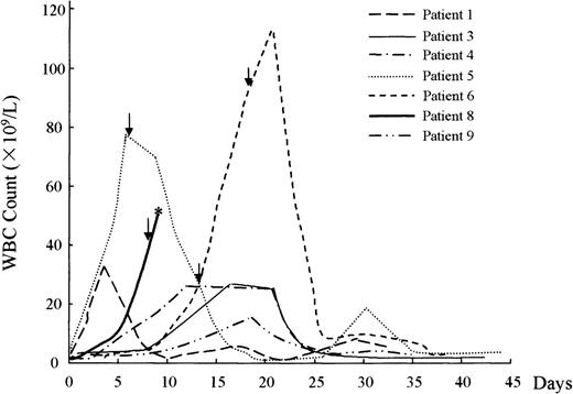
Hyperleukocytosis, as defined by WBC count superior to 10 × 109/L, developed in 8 of the 11 (72.7%) newly diagnosed patients with WBC counts from 26 to 183 × 109/L (median, 41.5 × 10⁹/L), before, or 5 to 20 days after receiving As2O3(Fig 1). WBC counts declined with moderate chemotherapy in 2 cases and spontaneously in 4 cases, without occurrence of the related adult respiratory distress syndrome (ARDS) clinical syndromes. Among the others, 2 cases died, hyperleukocytosis may be one of the causes leading to treatment failure in 1 patient.
Hyperleukocytosis developed in newly diagnosed patients during As2O3 treatment. * represents that patient died on day 15 after As2O3 treatment. Arrow indicates the time when chemotherapy was administered. Case 11 is not shown here because the patient died on day 1 of As2O3 treatment.
Hyperleukocytosis developed in newly diagnosed patients during As2O3 treatment. * represents that patient died on day 15 after As2O3 treatment. Arrow indicates the time when chemotherapy was administered. Case 11 is not shown here because the patient died on day 1 of As2O3 treatment.
Toxic effects.
The major As2O3-related toxicities, as listed in Table 3, were skin reactions (rash, itching, erythema) (3 of 11), gastrointestinal reactions (vomiting, nausea, and diarrhea) (4 of 11), cardiac dysfunction (1 of 11), similar to those in the relapsed group. However, hepatic damage occurred in 7 of 11 patients with elevation of the serum glutamic pyruvic transaminase (SGPT) ranging from 82 to 918 IU/L (median, 266 IU/L; normal range, 10 to 64 IU/L) and serum glutamic oxaloacetic transaminase (SGOT) 58 to 934 IU /L (median, 114 IU/L; normal range, 10 to 42 IU/L). Among these 7 patients, symptomatic medication was administered and withdrawal of As2O3 was indicated when severe liver dysfunction occurred. Five patients recovered and the other 2 failed. These 2 patients are worth particular attention because of the development of lethal hepatic damage as described below.
Side Effects of As2O3 in 58 APL Patients
| Side Effects . | Group . | Newly Diagnosed Patients . | Relapsed Patients . | P Value . | ||||||
|---|---|---|---|---|---|---|---|---|---|---|
| Grade . | 1 . | 2 . | 3 . | 4 . | 1 . | 2 . | 3 . | 4 . | ||
| Skin reaction | 1 | 2 | 0 | 0 | 8 | 43-150 | 0 | 0 | NS | |
| Gastrointestinal reaction | 4 | 0 | 0 | 0 | 103-151 | 0 | 0 | 0 | NS | |
| Cardiac dysfunction | 1 | 0 | 0 | 0 | 8 | 0 | 0 | 0 | NS | |
| Facial edema and neuropathy | 1 | 0 | 0 | 0 | 5 | 0 | 0 | 0 | NS | |
| Liver dysfunction | 2 | 3 | 2 | 0 | 14 | 1 | 0 | 0 | .001 | |
Abbreviation: NS, not significant.
Including 1 patient received As2O3 + ATRA concurrently.
Including 2 patients received As2O3 + chemotherapy.
Case 2: a 33-year-old woman with previously untreated APL was treated with 10 mg As2O3 daily and continued for 10 days. No previous history of hepatitis was noted and the tests for hepatitis B virus (HBV) and hepatitis C virus (HCV) were negative. The percentage of promyelocytes in BM decreased from 78.5% to 40%, WBC count in peripheral blood increased from 1.1 × 10⁹/L to 9.5 × 10⁹/L, whereas sequential measurement showed that SGPT and SGOT increased from 52 IU/L and 53 IU/L to 633 IU/L and 576 IU/L, respectively. This patient died on day 15 with liver failure: SGPT 918 IU/L, SGOT 934 IU/L, bilirubin 20.6μmol/L, and alkaline phosphatase 65 IU/L in spite of intensive supportive care. Liver biopsy was not performed because of patient and relatives’ refusal. Renal function test was normal during the treatment. RT-PCR showed positive results of S-type PML-RARα transcript.
Case 8: a 34-year-old woman, with no history of liver dysfunction (SGPT 54 IU/L, SGOT 49 IU/L, bilirubin 4.7 μmol/L, alkaline phosphatase 76 IU/L), received As2O3 as induction therapy. One week later, liver toxicity occurred with SGPT (255 IU/L) and SGOT (305 IU/L) increased significantly, the WBC count and percentage of APL cells in PB reached the highest level (50×109/L and 93%, respectively). She died of cerebral hemorrhage accompanied with severe liver impairment (SGPT 900 IU/L; SGOT 905 IU/L bilirubin 14.1 μmol/L, alkaline phosphatase 73 IU/L) and hyperleukocytosis (50×109/L), despite timely withdrawal of As2O3 and hepatic supportive treatments. Renal function index was normal during the whole As2O3 treatment. She had L-type fusion gene by RT-PCR.
Disease-free survival.
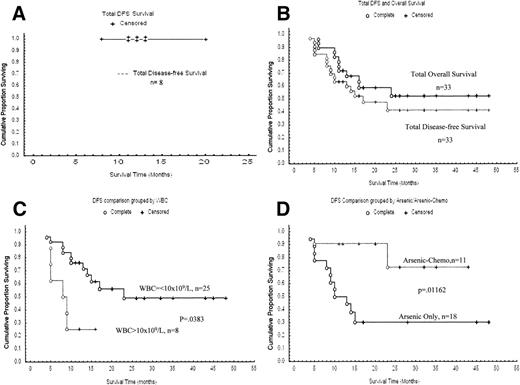
After CR, five patients received chemotherapy for maintenance whereas three patients received As2O3, one (case 9) of the three latter cases being shifted to chemotherapy after two courses of As2O3. With a median follow-up of 12 months, all these eight patients are still in CR (range: 8 to 20 months) (Fig 2A).
(A) Kaplan-Meier product-limit estimate of DFS from the time of CR for newly diagnosed patients. (B) Kaplan-Meier product-limit estimate of DFS and overall survival from the time of CR for relapsed patients. (C) DFS in relapsed APL patients with regard to WBC count at relapse. (D) DFS in relapsed APL patients between arsenic group and combination group.
(A) Kaplan-Meier product-limit estimate of DFS from the time of CR for newly diagnosed patients. (B) Kaplan-Meier product-limit estimate of DFS and overall survival from the time of CR for relapsed patients. (C) DFS in relapsed APL patients with regard to WBC count at relapse. (D) DFS in relapsed APL patients between arsenic group and combination group.
Cytogenetics and molecular genetics data.
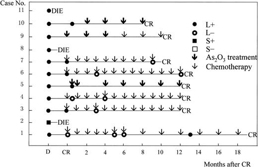
t(15;17) was found in cases 2 to 9 and 11 when diagnosed, whereas cytogenetic analysis failed in cases 1 and 10 due to lack of metaphase cells. All of the 11 newly diagnosed APL patients were PML-RARα positive by RT-PCR at diagnosis. RT-PCR remained positive in four of five patients when CR was obtained, but became negative after 1 to 3 months of consolidation with chemotherapy in three cases (Fig 3).
RT-PCR and follow-up data of 11 newly diagnosed patients. D, at diagnosis; CR, complete remission; L/S, long/short-type isoform of PML-RAR transcripts. Arrows indicate treatment protocol, As2O3 or chemotherapy, as postremission therapy.
RT-PCR and follow-up data of 11 newly diagnosed patients. D, at diagnosis; CR, complete remission; L/S, long/short-type isoform of PML-RAR transcripts. Arrows indicate treatment protocol, As2O3 or chemotherapy, as postremission therapy.
RELAPSED PATIENT GROUP
Remission induction.
Among the 47 patients treated with As2O3, 4 died of cerebral hemorrhage at early days of treatment (one on day 7 and three on day 8) due to low platelet and low fibrinogen (3 cases) or tumor cell infiltration into the central nervous system (1 case). A total of 31 were treated with As2O3 alone, 11 with combination of As2O3 and moderate chemotherapy, and 5 with As2O3 and ATRA. A total of 40 of 47 (85.1%) patients went into CR, whereas the CR rate was 83.9% (26 of 31) when 31 patients receiving As2O3 alone were analyzed (Table 2). The overall median time for getting CR was 31 days, with a median dosage, correspondingly, of 310 mg. In the 3 resistant patients (cases 10, 27, and 33), case 10 had 85% APL cells at the onset of relapse and the leukemic cells in BM rose to 71.5% after a transient drop to 38% with initial As2O3 treatment, losing response to As2O3 or chemotherapy. There was no decrease in blast percentage in case 27 after two courses of As2O3 treatment, then the patient was treated with chemotherapy but failed to respond to either. In case 33, the percentage of promyelocytes in the BM increased from 19.5% to 94% after nearly 2 months of treatment with As2O3and the patient finally died.
Hyperleukocytosis.
Hyperleukocytosis developed during As2O3treatment in 26 of the 47 relapsed patients (55%) with the WBC counts ranging from 11.9 × 10⁹/L to 167 × 109/L (median, 38 × 109/L) after 1 to 43 days (median, 17 days) of treatment. The WBC counts in 11 of 26 patients returned to normal after chemotherapy, including 1 patient who developed ARDS on day 22 of As2O3 treatment when the WBC count was 67.0 × 10⁹/L, whereas those in the other 14 cases fell to normal spontaneously. One patient presenting hyperleukocytosis died of cerebral hemorrhage with low platelet count and low fibrinogen on day 7.
Side effects.
As2O3-related toxicities occurred in 12 of 47 with skin reactions, 10 of 47 with gastrointestinal reactions (vomiting, nausea, diarrhea), 15 of 47 with liver dysfunction, 8 of 47 with cardiac dysfunction, and 5 of 47 with facial edema and neuropathy (Table 3). Most of the side effects were modest and responded to symptomatic treatment, further confirming our previous report.12 There was no difference in frequency or in extent of side effects between patients treated with As2O3 alone and those with combination therapy (As2O3 + chemotherapy or As2O3 + ATRA).
Disease-free survival.
The follow-up data were available in 33 patients, the other 7 were out of follow-up. As shown in Fig 2B, the estimated DFS rates at 1 and 2 years of the 33 patients followed were 63.6% and 41.6%, respectively, and the median DFS was 17 months. The estimated 1 and 2 year overall survival rates were 72.1% and 50.2%, respectively, whereas the actual median overall survival was 25 months (Fig 2B).
A number of factors with possible influence on the DFS were analyzed. The disease status before As2O3-induced CR was at first relapse in 29, at second relapse in 2, and at third relapse in 2 cases. Of note, 3 of 4 patients treated with As2O3 at advanced stage (the second or third relapse) relapsed again, compared with 14 of 29 treated at the first relapse. More importantly, patients presenting for As2O3 treatment with WBC lower than 10 × 109/L had DFS significantly better than those with WBC higher than 10 × 109/L (P = .038)(Fig 2C). For postremission therapy after As2O3-induced CR, 4 were treated with chemotherapy alone (from 8 to 17 months), 18 with As2O3 alone (from 7 to 48 months), and 11 with combination therapy (from 11 to 44 months). Disease recurrence developed in 3 of 4 cases treated with chemotherapy alone, 12 of 18 with As2O3 alone, and 2 of 11 with the combination, respectively. Therefore, the duration of CR also tended to be related to postremission treatment protocols, with combination of As2O3 and chemotherapy giving better DFS compared with As2O3 alone (P = .01) (Fig 2D).
Table 4 shows the outcomes of the 17 cases that relapsed again after As2O3 treatment. Only one (case 17) patient has regained durable CR for 20 months with chemotherapy and ATRA as the maintenance treatment, whereas others unfortunately died, although a short CR was obtained among 4 cases.
Response of 17 Patients Relapsed after As2O3 to Different Reinduction Protocols
| Reinduction Protocol . | CT . | ATRA . | As2O3 . | Combination . | No Antileukemic Treatment . | |||
|---|---|---|---|---|---|---|---|---|
| As2O3 + ATRA . | As2O3 Followed by CT . | ATRA Followed by CT . | As2O3 + ATRA Followed by CT . | |||||
| Cases | Case 24 | Case 14 | Case 1 | Case 12 | Case 5 | Case 9 | Case 21 | Case 11 |
| Case 35 | Case 17 | Case 4 | Case 16 | Case 8 | Case 39 | Case 23 | Case 32 | |
| Case 43 | ||||||||
| Response | 2 NR | 1 NR | 2 NR | 2 NR | 1 NR | 2 NR | 2 CR | 2 NR |
| 1 CR | 2 CR | |||||||
| Follow-up | 2 died | 1 died 1 CR | 2 died | 2 died | 2 died 1 out of follow-up | 2 died | 2 relapsed and died | 2 died |
Abbreviation: CT, chemotherapy.
Cytogenetics and molecular genetics.
Karyotyping was performed successfully in 22 cases at diagnosis of relapse. A total of 19 of 22 had t(15;17) and 3 cases had not. In addition, a complex karyotype, 46,XY, t(5;15)(q14;q22), t(15;17)(q22;q11-21), ins(16;17)(p11p12;q?), was observed in case 11 when relapse occurred after As2O3-induced CR, which was confirmed by dual-color painting with WCP probes for chromosome 5, 15, 16, and 17.
RT-PCR results were obtained in 29 patients at the time of relapse before As2O3 treatment. Among them, case 10 was PCR negative for PML-RARα in spite of the fact that fusion gene transcript was positive at first disease presentation. In case 27, both PML-RARα and AML1-ETO were amplified by RT-PCR though only AML1-ETO was detected by FISH. These two patients showed no response to As2O3 induction as described above. In another nonresponder (case 33), RT-PCR analysis was not obtained due to lack of material.
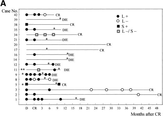
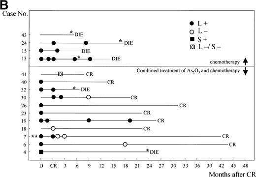
In the other patients, four cases (cases 4, 5, 25, and 38) had S-type fusion genes, one of whom remained in CR until now, one died, and the other two were lost to follow-up. The remaining 23 cases all showed RT-PCR positivity for L-type transcript, among whom 9 remained in CR, 9 died. Follow-up data were not available in the remaining 5 cases. RT-PCR data immediately after As2O3-induced hematological CR were available only in 15 cases. Positive PML-RARα fusion transcripts were detected in 14 of 15 cases, indicating that As2O3 induction of clinical CR was not associated with molecular CR in the majority of patients (Fig 4). It is worth noting, however, that RT-PCR negative results were obtained in two patients (cases 2 and 3, Fig 4) after long-time maintenance therapy (41 and 37 months, respectively) with As2O3 alone.
RT-PCR and follow-up data after postremission treatment with As2O3 alone (A), or chemotherapy alone, or chemotherapy/As2O3 combination (B) in 43 relapsed APL patients. D, at diagnosis; CR, complete remission; L/S, long/short-type isoform of PML-RAR transcripts. * Indicates each time of relapse.
RT-PCR and follow-up data after postremission treatment with As2O3 alone (A), or chemotherapy alone, or chemotherapy/As2O3 combination (B) in 43 relapsed APL patients. D, at diagnosis; CR, complete remission; L/S, long/short-type isoform of PML-RAR transcripts. * Indicates each time of relapse.
Presence of t (15;17) in partially differentiated APL cells occurs during As2O3 treatment.
To evaluate the possible in vivo partial differentiation of APL cells induced by As2O3, as suggested by our previous report,12 fresh APL cells in BM were obtained from five cases during remission induction and analyzed by morphological examination and combination of Wright’s staining and dual-color FISH. These patients all had t(15;17) on chromosome karyotyping and PML-RARα transcripts by RT-PCR at diagnosis of relapse. The percentages of leukemic promyelocytes in BM was over 64.5% in four cases, whereas the remaining one had only 11% promyelocytes. However, the percentage of promyelocytes in BM decreased gradually in all the five cases during As2O3 treatment. In contrast, after 15 to 20 days of treatment, increased number of myelocyte-like cells and many degenerating cells with condensed or coarse nuclei with scanty cytoplasm (“nude” nucleus) was observed in both BM and peripheral blood. The percentage of myelocyte-like cells in BM was highest 20 to 25 days after the initiation of As2O3 treatment (Table 5), however, terminally differentiated elements such as polynucleated granulocytes did not increase with As2O3 treatment.
Clinical and Laboratory Results in Five Patients After As2O3 Treatment
| Before As2O3Treatment . | Days of Receiving As2O3 Treatment . | After As2O3 Treatment . | |||||
|---|---|---|---|---|---|---|---|
| Case No. . | BM . | BM . | |||||
| Promyelocyte (%) . | Myelocyte (%) . | Metamyelocyte (%) . | Promyelocyte (%) . | Myelocyte (%) . | Metamyelocyte (%) . | ||
| 21 | 93 | 1.5 | 1 | 28 | 0.5 | 15.5 | 18.5 |
| 22 | 73 | 1 | 0 | 12 | 5.5 | 44.5 | 27 |
| 29 | 75 | 8.5 | 2 | 18 | 79 | 5 | 0 |
| 35 | 11 | 22.5 | 0 | 25 | 0.5 | 54.5 | 0 |
| 37 | 64.5 | 10 | 3 | 22 | 6 | 45 | 13 |
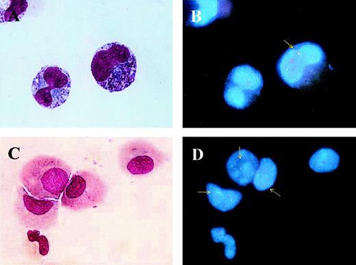
Dual-color FISH generated two red and two green spots in normal interphase cells, corresponding to PML and RARα genes, respectively. It can be expected that in APL cells with t(15;17) have a yellow signal due to the fusion of one PML and one RARα allele in addition to one red and one green signal (Fig 5A). This three-color signal complex was observed not only in typical leukemic promyelocytes before As2O3 (Fig 5B), but also in more differentiated elements, such as myelocyte-like cells (Fig 5C), during the treatment (Fig 5D), confirming that the partially differentiated granulocytes were indeed derived from the leukemia clone and not from the residual normal hematopoietic precursors.
BM samples before (A and B) and during (C and D) As2O3 treatment were collected for analyzing the origin of differentiated myeloid cells. Morphological examination (A and C) showed promyelocytes (A) and myelocyte-like cells (C), in which one red, one green, and one yellow fusion signal could be observed in the same cells (B and D). Arrows pointed yellow signals that represented PML- RAR fusion gene.
BM samples before (A and B) and during (C and D) As2O3 treatment were collected for analyzing the origin of differentiated myeloid cells. Morphological examination (A and C) showed promyelocytes (A) and myelocyte-like cells (C), in which one red, one green, and one yellow fusion signal could be observed in the same cells (B and D). Arrows pointed yellow signals that represented PML- RAR fusion gene.
DISCUSSION
In the present study, we were able to conduct a multicenter clinical research on 11 previously untreated and 47 relapsed patients. Eight of eleven (72.7%) newly diagnosed patients achieved CR with the median time of 35 days, similar to a previous report in China with a CR rate of 73% in newly diagnosed APL patients.10 These results are comparable to that obtained by ATRA in newly diagnosed patients. On the other hand, 40 of 47 (85.1%) relapsed APL patients achieved CR. Hence, As2O3 is able to induce high CR rate in both newly diagnosed and relapsed APL patients. Among the 51 patients (8 primary and 43 relapsed) who received enough long As2O3 remission induction, 3 cases should be considered as nonresponders because they failed to achieve CR even after two courses of the drug. Among these 3 patients, 1 had no available cytogenetic and molecular data, the other 2 (cases 10 and 27), although presenting with t(15;17) and PML-RARα at first diagnosis, had altered genotype of leukemic cells at relapse. Case 10 was negative for PML-RARα by RT-PCR, whereas case 27 developed a new malignant clone with AML1-ETO fusion gene, which is characteristic of AML-M2, in addition to PML-RARα. However, the clone with AML1-ETO transcript was dominant because cytogenetically only t(8;21), but not t(15;17), was detectable using the FISH method. The in vivo sensitivity to As2O3 appears to require the expression of PML-RARα, although a recent in vitro study showed that As2O3- induced apoptosis of APL cells was not dependent on PML or PML-RARα expression.21 In APL cells, PML and/or nuclear body (NB) functions are lost because PML-RARα displaces PML and other NB components to nuclear microspeckles, whereas As2O3 appears to target PML and PML-RARα onto NB and induce their degradation.22Though As2O3 at relatively high concentrations (1 to 2 μmol/L) exerts apoptotic effect on a wide range of cell lines including lymphoid lineage, in vitro differentiation-inducing effects at low dose (0.1 to 0.5 μmol/L) were observed selectively in APL cells (unpublished data). In accordance with our findings, partially differentiated myeloid cells occurred in 8 of 11 newly diagnosed and 26 of 47 relapsed patients with hyperleukocytosis, and a large number of myelocyte-like cells containing PML-RARα fusion gene emerged after 15 to 20 days of in vivo treatment with As2O3 (Fig5). Notably, our previous pharmacokinetic data showed that except for a short period (2 to 3 hours) due to intravenous drip, the in vivo plasma concentration of As2O3 was low (<0.5 to 1 μmol/L) most of the time over the treatment course.12
One sensible issue on As2O3 treatment is the adverse effects. In this study, when the newly diagnosed and relapsed APL groups were compared, they showed almost similar incidence for hyperleukocytosis, skin reaction, gastrointestinal reaction, and cardiovascular system dysfunction. However, an unexpected finding was that the hepatotoxicity was much higher in newly diagnosed patients than in relapsed ones, because 7 of 11 (63.6%) (2 cases in grade 1, 3 cases in grade 2, and 2 cases in grade 3) newly diagnosed APL patients developed hepatic dysfunction in contrast to 15 of 47 (31.9%) (14 cases in grade 1 and 1 case in grade 2) in relapsed cases (P = .001). None of the 7 newly diagnosed patients with liver toxicity presented abnormal liver function tests or HBV or HBC antigens and antibodies before As2O3 treatment. More importantly, among these patients, 2 died with highest SGPT and SGOT levels, 918 IU/L and 934 IU/L (case 2) and 900 IU/L and 905 IU/L (case 8), respectively. Because both patients died on day 15 of As2O3 induction, a period not long enough to achieve CR, it is difficult to evaluate the response with regard to the reduction of leukemia cell population, though hyperleukocytosis appeared in one case, suggesting a possible differentiation-inducing effect of As2O3. Meanwhile, we did not find severe hepatic damage happening in a group of relapsed APL patients treated during the same period with the same batch of As2O3, eliminating the possibility of variation in drug quality.
The significant difference between the two groups could be ascribed to their distinct sensitivity towards the toxic effects of the drug. Recent data suggested that intracellular antioxidant levels may be involved in the defense of cells against arsenite genotoxicity.23 It was found that the activities of two antioxidant enzymes, catalase and glutathione peroxidase, were 5.4-fold and 5.8-fold lower in xrs-5 cells than those in Chinese hamster ovary (CHO)-k1 cells. The xrs-5 cells are x-ray hypersensitive CHO subclone with higher sensitivity to sodium arsenite inhibition of cell growth and micronuclei induction compared with CHO-k1 cells.24Therefore, we postulate that in relapsed patients, long-term treatment with ATRA and/or chemotherapeutic drugs could induce or modify some antioxidant enzymatic system and enhance the antioxidant ability, so they had better tolerance to arsenic than newly diagnosed patients. Another possibility is that patients with higher susceptibility to As2O3-induced damage may belong to a special group with reduced capacity of drug detoxication and could be already selected out through previous ATRA/chemotherapy. The shortage of materials from our newly diagnosed patient group, unfortunately, did not allow us to perform further study on the enzymes related to the toxicity of As2O3. Because remission induction with ATRA in newly diagnosed APL patients never gives rise to such a severe liver toxicity and the retinoic acid syndrome now can be easily handled, we believe that ATRA should be used as the first-line drug for remission induction, whereas As2O3, until further evaluation of its toxicity, should be incorporated into a multidrug consolidation/maintenance therapy during remission or as a rescue in relapsed patients. Additionally, drugs can be used to relieve the side effects of As2O3 in cases of severe intoxication. As Moore et al25 reported, 2,3-dimercaptopropanesulphonate (DMPS) was able to reduce toxicity of As2O3. Dimercaptosuccinic acid (DMSA) analogues were also put forward to decreasing the tissue content of arsenic in acute As2O3 poisoning in NMRI male mice,26 suggesting that potentially protective measures are available while using As2O3 to treat the malignancies.
ATRA has been proven to have a highly specific effect on the newly diagnosed APL patients with t(15;17) and PML-RARα expression. Previous studies showed, however, when ATRA was used as the maintenance treatment alone after CR achieved with the same drug, most patients relapsed within 6 months, mainly because of the development of drug resistance. On the contrary, long-time remission was reported in a series of 32 APL patients treated with As2O3 as single therapeutic agent, among whom one fourth had a survival time of more than 10 years,9 suggesting long-term treatment with As2O3 may induce molecular remission. In the present work, we analyzed this issue by using RT-PCR before and after As2O3-induced CR. It was found that immediately after CR, the leukemic clone persisted in 4 of 5 newly diagnosed patients and 14 of 15 relapsed patients investigated. Therefore, As2O3 induction is not sufficient to induce a molecular remission. Nevertheless, a relatively long DFS (48 and 44 months in cases 2 and 3, respectively) with negative RT-PCR was observed in 2 cases in the relapsed group, indicating that long-term use of As2O3 alone could indeed lead to a molecular remission in some patients. This result suggests that As2O3 may be more potent than ATRA in terms of maintaining molecular/clinical remission and justifies inclusion of As2O3 into multidrug postremission treatment in future clinical trials.
The reason that the effect of ATRA is less durable than As2O3 in treating APL patients could be multiple. As a vitamin-like hormone, ATRA may induce more easily a metabolic resistance than As2O3, an inorganic small compound. Secondly, ATRA mainly induces differentiation of APL cells, whereas As2O3 could induce both apoptosis and partial differentiation of these leukemia cells. Thirdly, although the two drugs share a common target, PML-RARα, As2O3 may exert an effect on a wider spectrum of proteins, whereas the action of ATRA is limited to RA receptors only. For example, As2O3 could modify the phosphorylation of transcription factors such as AP1 or DNA hypomethylation27 or cause downregulation of bcl-2 protein.15
One of the major purposes of this study was to evaluate the outcome of relapsed APL patients after CR achieved with As2O3, to find out possible prognostic factors and what could be the best postremission treatment. Among the 33 relapsed APL patients available for follow-up, the median DFS time was 17 months, whereas relapse occurred in 17 patients. As expected, patients at the second or third relapse before As2O3-induced CR seemed to relapse more frequently (3 of 4) than those at the first relapse before As2O3-induced CR (14 of 29), although more cases should be studied. Next, patients with lower tumor burden as reflected by low WBC counts (below 10 × 109/L) showed statistically better DFS than those with higher tumor burden (WBC > 10 × 109/L) (P = .038). When different treatment protocols were compared, clinical outcome seems to be associated with postremission therapy, since there was only 2 relapses of 11 cases in the combination therapy group, compared with the 12 of 18 with As2O3 treatment (P = .01). These results indicate that combination therapy may be the choice of treatment to achieve a longer survival. Furthermore, ATRA could still have a role to play even in patients relapsed after As2O3, because among 17 cases who relapsed again after As2O3 induced-CR, only one achieved a DFS for 20 months after reinduction with ATRA and with combined chemotherapy/ATRA as maintenance treatment. It is possible that APL cells in this case restored sensitivity to ATRA, as suggested by a recent study that As2O3 could affect the RA response through changes in proteins involved in the RA pathway.28 If this is true, then a new strategy to prolong the survival could be designed in which ATRA is incorporated into an As2O3 + chemotherapy + ATRA triple combination after As2O3-induction in relapsed APL patients. An important issue is how to use the three medications. Adverse reactions were similar among relapsed patients who received the combination of As2O3 with ATRA or chemotherapy or As2O3 alone. However, in vitro studies showed that the use of ATRA and As2O3 might interfere with the full effect of each drug. Shao et al29found that the ATRA-induced cell differentiation was interfered by As2O3 addition, and the addition of ATRA reduced As2O3-induced cell apoptosis. Hence, sequential use of the two drugs may be better than simultaneous use, as also suggested in SCID mouse-APL model (Chen et al, in preparation).
In conclusion, As2O3 treatment can lead to a high CR rate in both newly diagnosed and relapsed APL patients. However, the severe hepatic adverse effect in some patients does not support As2O3 to be used as a first-line remission-inducing drug. Finally, the As2O3/chemotherapy combination after As2O3–induced CR in relapsed patients yielded better DFS than As2O3 or chemotherapy alone. Long-term use of As2O3 alone is able, at least in part of the patients, to induce a molecular remission, justifying further investigation of its use in a multidrug maintenance therapy after ATRA induced CR and chemotherapy consolidation, in a hope that the DFS in APL could be further improved.
ACKNOWLEDGMENT
The authors thank all members of Shanghai Institute of Hematology for their support and encouragement and thank Prof Anne Hagemeijer for kindly giving us the P1 164 and Co664 clones that were used as probes for FISH in the detection of AML1-ETO. We also thank Prof Dao-Pei Lu from Beijing Medical University, Beijing, China, Prof. Laurent Degos from Saint-Louis Hospital, Paris, France, and Prof Ryuzo Ohno from Hamamatsu University School of Medicine, Japan, for constructive discussion.
C.N. and H.Y. contributed equally to this work.
Supported in part by the Chinese Climbing Project, National Natural Sciences Foundation of China, Shanghai Municipal Commission for Sciences and Technologies, Samuel Waxman Cancer Research Foundation, and the Clyde Wu Foundation of Shanghai Institute of Hematology.
The publication costs of this article were defrayed in part by page charge payment. This article must therefore be hereby marked “advertisement” in accordance with 18 U.S.C. section 1734 solely to indicate this fact.
REFERENCES
NOTE ADDED IN PROOF
The data listed as unpublished in Discussion were published in J Natl Cancer Inst 91:772, 1999.
Author notes
Address reprint requests to Sai-Juan Chen, MD, PhD, or Jiong Hu, MD, Shanghai Institute of Hematology, Department of Hematology/Oncology, Rui Jin Hospital, Shanghai Second Medical University, 197 Rui Jin Road II, 200025, Shanghai, China; email:zchen@ms.stn.sh.cn.

This feature is available to Subscribers Only
Sign In or Create an Account Close Modal