To the Editor:
Band 3 (anion exchanger 1, AE1) is the most abundant integral protein of the red blood cell membrane. It is composed of 911 amino acids encoded by the EPB3 gene. Several mutations in the EPB3 gene have been found associated to different diseases, eg, hereditary spherocytosis, Southeast Asian ovalocytosis, chorea-acanthocytosis, and familial distal renal tubular acidosis, thus demonstrating that the effects of EPB3 mutation are dependent on type and location.1,2 The function of the band 3 carboxyl terminus tail that protrudes on the cytoplasmic side of the erythrocyte membrane is unknown.3We describe the first example of a disease-causing mutation located in the extreme 3′ end of the AE1 coding sequence.
Hereditary spherocytosis (HS) is a common hemolytic anemia in which the spheroidal, osmotically fragile erythrocytes are destroyed in the spleen. HS due to band 3 deficiency has an autosomal dominant mode of transmission and is characterized by mild to moderate anemia.1
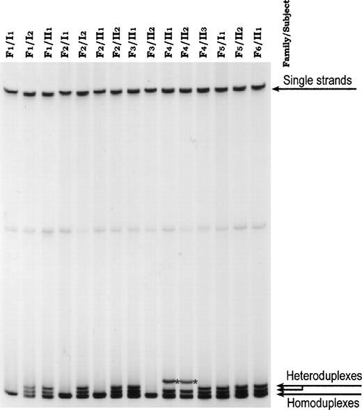
Six children, belonging to unrelated Italian families from the Vesuvian area (Southern Italy), presented a homogeneous clinical picture showing moderate hemolytic anemia and splenomegaly. HS was diagnosed on the basis of clinical findings, familial history, and routine hematological investigations. The protein phenotype, performed by sodium dodecyl sulfate-polyacrylamide gel electrophoresis (SDS-PAGE), was typically that of band 3-deficient HS (band 3/spectrin: −21% ± 2% compared with that of the controls).4 In all patients, single/strand conformation polymorphism (SSCP) screening of band 3 gene showed an alteration in exon 20 (not shown).4 Direct sequencing of the abnormal fragments constantly showed a deletional frameshift mutation in codon 894 (ACC → AC; not shown). We designated this mutation with the area of origin as Vesuvio. Heteroduplex analysis confirmed this mutation in 5 additional patients in the 6 families. This analysis failed to find the mutation in 4 healthy members (Fig 1). In 2 brothers of family 3 we also detected a previously described silent polymorphism (codon 904, TAC → TAT)5 occurring in trans (Fig 1 and not shown). Analysis of the PstI polymorphic site of the EPB3 gene indicated that HS was invariably associated with aPstI (+) allele (not shown). Semiquantitative reverse transcription-polymerase chain reaction (RT-PCR) amplification with primers flanking the deletion (primer sense in exon 19 and primer antisense in exon 20) and with lower numbers of cycles produced heteroduplexes, suggesting that mRNA corresponding to allele Vesuvio was expressed (Fig 2).
Heteroduplex analysis of exon 20 of band 3 genomic DNA. In all 11 HS patients, heteroduplexes were visible (they were generated on PCR between normal fragments and fragments carrying the C deletion). *Band shows heteroduplex produced by silent polymorphism occurring in trans.
Heteroduplex analysis of exon 20 of band 3 genomic DNA. In all 11 HS patients, heteroduplexes were visible (they were generated on PCR between normal fragments and fragments carrying the C deletion). *Band shows heteroduplex produced by silent polymorphism occurring in trans.
Mutant band 3 mRNA is detected in patient II1 of family 1. The presence of the cDNA Vesuvio was verified by PCR amplification of reverse-transcribed total reticulocyte RNA. Aliquots of the PCR reaction after 14, 16, 18, and 20 cycles were screened using heteroduplex analysis.
Mutant band 3 mRNA is detected in patient II1 of family 1. The presence of the cDNA Vesuvio was verified by PCR amplification of reverse-transcribed total reticulocyte RNA. Aliquots of the PCR reaction after 14, 16, 18, and 20 cycles were screened using heteroduplex analysis.
Urinary pH and acid excretion were in the normal range in all patients.
Band 3 Vesuvio is the first mutation located in the cytoplasmic COOH terminal tail. The singularity of this mutation is the opening of the reading frame for an extra 133 codons (instead of 18), before the new stop codon at position 1027. The presence of the corresponding mRNA suggests that neither the transcription of the mutant gene nor the stability of the mutant mRNA is affected. Nevertheless, because no elongated additional band 3 was seen by Western blotting (not shown), we offer one of the following possibilities to explain the band 3 deficiency: (1) abnormal band 3 insertion in the endoplasmic reticulum; (2) defective translocation of mutant band 3 from the endoplasmic reticulum to the plasma membrane; and (3) premature band 3 degradation in the bone marrow erythroblasts. The COOH-terminal 18 amino acids of the normal band 3 are predicted to be replaced by an abnormal sequence in band 3 Vesuvio. Alignment of the 13 known sequences pertaining to different members of the three anion exchanger families (AE1, AE2, and AE3) shows that the amino acids in positions 897, 901, 905, and 906 are highly conserved through evolution. As reported by Anderson and von Heijne,6 these affected residues could act as stop transfer signals during cotranslational insertion of band 3 protein into the endoplasmic reticulum membrane of erythroid precursor cells. Furthermore, alternatively and/or additionally, the presence of a much longer COOH terminus could be the hindrance to the band 3 insertion. Band 3 Vesuvio mutation shows that the cytoplasmic COOH terminal tail is also very critical for the assembly and/or maintaining of band 3 in the mature erythrocyte membrane. Finally, we did not evaluate the unlikely possibility of a mutant band 3 insertion into the plasma with proteolytically truncation to a size similar to normal band 3.
We found band 3 Vesuvio in 6 unrelated families from the same geographic area. Because this mutation is located on the samePst1 allele and does not occur in a hot spot area, the presence of a founder effect is strongly suspected.


This feature is available to Subscribers Only
Sign In or Create an Account Close Modal