Abstract
Because CD28-mediated T-cell costimulation has a pivotal role in the initiation and maintenance of T-cell responses, we tested the hypothesis that CD28 is critical for the development of graft-versus-host disease (GVHD). We compared the in vivo effects of CD28−/− T cells transplanted from B6 donor with the CD28 gene deleted by homologous recombination with those of CD28+/+ T cells transplanted from wild-type C57BL/6 (B6) donor. Fifty million CD28−/− or CD28+/+ splenocytes from B6 mice were transplanted into unirradiated (B6 × DBA/2)F1 (BDF1) recipients. Unlike CD28+/+, CD28−/− T cells from B6 mice had lower levels of proliferation and interleukin-2 production, had a limited ability to generate cytotoxic T lymphocytes against the recipient, and did not induce immune deficiency, despite survival in the recipient for at least 28 days. The ability to prevent rejection was reduced by the absence of CD28, because as many as 1.0 × 10⁷ CD28−/− CD8+ cells were needed to prevent rejection of major histocompatibility complex (MHC) class-I incompatible marrow in sublethally irradiated (550 cGy) bm1 recipients, whereas 8.0 × 10⁵ CD28+/+CD8+ T cells were sufficient to produce a similar effect, indicating that CD28 on donor CD8+ cells helps to eliminate host immunity. Two million CD4+CD28−/− or CD28+/+ T cells were transplanted into sublethally irradiated (750 cGy), MHC class-II incompatible (B6 × bm12)F1 recipients. With CD28−/−cells, 44% of the recipients died at a median of 20 days compared with 94% at a median of 15 days with CD28+/+ cells (P < .001). Two million CD8+CD28−/− or CD28+/+ T cells were transplanted into sublethally irradiated (750 cGy), MHC class-I incompatible (B6 × bm1) F1 recipients. With CD28−/−cells, 25% of the recipients died at a median of 41 days compared with 100% at a median of 15 days with CD28+/+ cells (P < .001). (B6 × bm12)F1 and (B6 × bm1)F1 mice surviving after transplantation of CD28−/− cells recovered thymocytes, T cells, and B cells in numbers and function comparable with that of irradiation-control F1 mice. We conclude that CD28 contributes to the pathogenesis and the severity of GVHD. Our results suggest that the severity of GVHD could be decreased by the administration of agents that block CD28 function in T lymphocytes.
© 1998 by The American Society of Hematology.
ACUTE GRAFT-VERSUS-host disease (GVHD) continues to be the major complication of allogenic bone marrow transplantation, producing immune deficiency, infection, organ damage, and death. GVHD is initiated by mature donor T cells that recognize minor or major histocompatibility antigens of the recipient.1 Efficient T-cell activation requires antigen recognition and costimulation.2,3 Antigen recognition is mediated by the interaction between T-cell receptor (TCR) and antigen peptides presented by major histocompatiblity complex (MHC) molecules on antigen-presenting cells. The best characterized costimulatory system involves the CD28 molecule on T cells and its ligands, B7-1 and B7-2 on antigen-presenting cells. CTLA-4, a structural homologue of CD28, shares the ability to bind B7 molecules.4,5 The B7/CD28 interaction delivers a positive signal, whereas the B7/CTLA-4 interaction delivers a negative signal for T-cell activation.6-8 Blocking CD28 costimulation can inhibit T-cell responses in a variety of in vitro and in vivo systems.9-14 CTLA-4Ig or anti-B7 antibodies, which bind to B7 and block the interaction of B7 with CD28 and CTLA-4, have been shown to ameliorate GVHD in a variety of murine models, suggesting that CD28 activation is involved in the pathogenesis of GVHD.15-19
To avoid the limitation that treatment with CTLA-4Ig or anti-B7 antibodies might not completely block the interaction of B7 with CD28, or might sustain GVHD by blocking the interaction of B7 with CTLA-4, we and others have used T cells from donors with a deletion of the CD28 gene produced by homologous recombination.20 Recently, Speiser et al21 reported that splenocytes from CD28−/− donors were comparable with those from CD28+/+ donors in their capacity to cause acute lethal GVHD in MHC-mismatched recipients, suggesting that CD28 costimulation is not necessary for GVHD lethality. Thus, the role of CD28 in GVHD remains controversial. We have compared the effects of CD28−/− and CD28+/+ T cells from C57BL/6 (B6) donors in unirradiated H2-incompatible (B6 × DBA2)F1 (BDF1) recipients and in irradiated H2 class-I–incompatible (B6 × bm1)F1 and H2 class-II–incompatible (B6 × bm12)F1 recipients. We found that CD28−/− donor T cells had markedly reduced ability to cause GVHD.
MATERIALS AND METHODS
Mice.
B6, BDF1, B6.C-H2bm12 (bm12), and B6.C-H2bm1 (bm1) mice were purchased from the Jackson Laboratory (Bar Harbor, ME). (B6 × bm12)F1 and (B6 × bm1)F1 mice were bred at Fred Hutchinson Cancer Research Center (FHCRC, Seattle, WA). Founders for the Ly5-congenic B6.SJL-Ly5a Ptprca Pep3b(B6-Ly5.1) strain were provided by Dr David Myers (Sloan Kettering Institute, New York, NY). B6 CD28-deficient mice were generated through homologous recombination in embryonic stem cells by Shahinian et al.20 CD28-deficient mice were bred onto a B6 background for five generations. Homozygous CD28−/− mice were kindly donated to us by Dr Craig Thompson (University of Chicago, Chicago, IL). All the mice used in this report were housed in microisolator cages.
T-cell purification.
For purification of CD4+ or CD8+ T cells by positive selection, a magnetic cell separation system was used according to the manufacturer’s instructions (Miltenyi Biotech Inc, Auburn, CA). Lymph node cells were incubated with biotin-conjugated anti-CD4 monoclonal antibodies (MoAb) (hybridoma GK 1.5; ATCC, Rockville, MD) or anti-CD8 MoAb (hybridoma 2.45; ATCC) for 15 minutes at 4°C. After two washes, cells were incubated with streptavidin-MicroBeads for 15 minutes at 4°C. After washing, the cell suspension was passed through a VS+ separation column placed in the magnetic field. The VS+ column was washed two to three times with 0.5% bovine serum albumin (BSA)/phosphate-buffered saline (PBS) and then removed from the magnetic field. Retained cells were flushed from the VS+ column with 0.5% BSA/PBS. The isolated cells were passed over a second VS+ column to increase the purity of enriched cells. After separation, cells were stained with phycoerythrin-conjugated streptavidin and analyzed by flow cytometry. The purity of CD4+ or CD8+ cells ranged from 94% to 99%.
Transplantation.
Fifty million spleen cells were resuspended in PBS and injected via the tail vein into 8- to 11-week-old unirradiated BDF1 recipients. (B6 × bm12)F1 or (B6 × bm1)F1 recipients mice were exposed to 700 cGy of irradiation (60Co source) at 20 cGy per minute. Two million CD4+ or CD8+ cells from CD28−/− or CD28+/+ B6 donors were suspended in PBS and injected via the tail vein into 8- to 9-week-old irradiated (B6 x bm12)F1 or (B6 x bm1)F1 recipients within 24 hours after irradiation. Mice were monitored twice weekly for weight loss and manifestations of GVHD. For experiments in which engraftment was an endpoint, marrow was depleted of T cells by complement-mediated lysis with antibodies specific for Thy-1, CD4, and CD8 as described previously.22 Five million T-cell–depleted marrow cells from B6-Ly5.1 donors were injected into bm1 recipients exposed to 550 cGy of irradiation with no T cells added to the graft or with graded numbers of CD8+ cells from CD28−/−or CD28+/+ donors added to the graft.
Flow cytometry.
Spleen cell suspensions from BDF1 recipients were stained with FITC-conjugated anti-CD4, anti-CD8, or anti-B220 MoAbs (Pharmingen, San Diego, CA) and biotin-conjugated anti-H2Dd (hybridoma 34-5-8S; ATCC), followed by phycoerythrin-conjugated streptavidin (Southern Biotechnology, Birmingham, AL). Two-color flow cytometric analysis was performed on a FACScan using LYSIS II software (Becton Dickinson, San Jose, CA). Recipient-derived cells were distinguished from donor cells by H2Dd expression. For analysis of engraftment by flow cytometry, peripheral-blood lymphocytes (PBL) were stained with FITC-conjugated anti-CD3 MoAb and biotinylated anti-Ly5.1 MoAb followed by phycoerythrin-conjugated streptavidin as described previously.22
Detection of the neomycin-resistance gene by polymerase chain reaction (PCR).
Engraftment of CD28−/− T cells was documented by PCR for the neomycin-resistance gene (neo), which disrupts the CD28 gene.20 DNA was extracted from spleen cells by using the IsoQuick nucleic acid extraction kit (ORCA Research Inc., Bothell, WA) according to the manufacturer’s instruction. An aliquot of 0.5 μg of DNA was used for PCR amplification. The following primers were used for PCR: (A) Neo, 5’-CAAGATGGATTGCACGCAGG, and 3’-CCCGCTCAGAAGAACTCGTCAACT CGCCC; (B) β-actin, 5’-TGACGGGGTCACCCACACTGTGCCCATCTA, and 3’-CTCTTCGACACGATGCAGCGGGACCTGAAG. Amplification was performed through 35 cycles of denaturation at 94°C for 40 seconds, annealing at 60°C for 40 seconds, and extension at 72°C for 60 seconds on GeneAmp PCR system 9600 (Perkin Elmer, Foster City, CA). PCR products were electrophoresed in 2% agarose gels and visualized with ethidium bromide. The sensitivity of the detection by PCR is 1 in 10,000 cells.
Cell culture and proliferation.
Single-cell suspensions from spleens of BDF1 mice were depleted of red cells by hypotonic lysis. Splenocytes were resuspended at a concentration of 1.0 × 10⁶ cells/mL and cultured in 200-μL aliquots in 96-well plates in complete RPMI-1640 medium containing 10% fetal bovine serum (FBS), 2 mmol/L glutamine, 15 mmol/L HEPES, 1 mmol/L sodium pyruvate, 5 × 10⁵ mol/L 2-ME, 100 U/mL penicillin and 100 μg/mL streptomycin. Spontaneous ex vivo proliferation was assessed by adding 1 μCi/well [3H]TdR for 4 hours at the end of the culture. Concanavalin A (ConA)-activated T-cell proliferation or lipopolysaccharide (LPS)-activated B-cell proliferation were measured by 8-hour incorporation of [3H]TdR at the end the culture. DNA was harvested onto glass-fiber filters and quantitated in a liquid scintillation counter (Topcount, Meridian, CT).
Interleukin (IL)-2 release.
Spleen cells were resuspended at a concentration of 5 × 10⁶/mL and cultured in 2 mL aliquots in 24-well plates in complete RPMI-1640 medium alone or in the presence of 5 μg/mL ConA (Sigma, St Louis, MO). Culture supernatants were harvested after 24 hours and frozen at −20°C until assay. Spontaneous IL-2 production was assayed as the ability of supernatants to stimulate proliferation of the IL-2–dependent cell line CTLL-2, as measured by [3H]TdR incorporation. Ten-fold dilutions of supernatants were cultured with CTLL-2 cells (5000/well) for 48-hours in 96-well plates, with of 1 μCi of [3H]TdR during the last 18 hours. To block the response of CTLL-2 cells to IL-4, 5 μg/mL anti–IL-4 MoAb (hybridoma 11B11, ATCC) was added to the culture. IL-2 production after ConA stimulation was quantified by sandwich ELISA using purified rat antimouse IL-2 MoAb JES6-1A12 for capture, and biotinylated rat antimouse IL-2 MoAb JES6-5H4 (Pharmingen) for detection. IL-2 concentration was calculated with reference to standard curves constructed using recombinant murine IL-2 (Genzyme, Cambridge, MA).
Antihost cytotoxicity.
Antihost cytotoxicity in BDF1 chimeras was measured directly, without in vitro restimulation, by testing spleen cells from the chimeras as effectors against 51Cr-labeled P815 (H2d) or EL-4 (H2b) targets. Spleen cells were added to U-bottom 96-well plates to achieve E:T ratios of 6, 12, 25, 50, and 100:1 with 2.0 × 10³ targets/well. The plates were centrifuged at 1000 rpm for 2 to 3 minutes and then incubated at 37°C for 4 to 5 hours. Chromium released into the supernatant was measured by Topcount (Packade Inc, Meriden, CT). The percent cytotoxicity was calculated as (experimental release − spontaneous release)/(maximal release − spontaneous release) × 100%.
RESULTS
CD28 contributes to alloreactive T-cell expansion and IL-2 production in unirradiated hosts.
Parental spleen cells (5.0 × 10⁷) from B6 donors were injected into unirradiated BDF1 recipients as described previously17 23 to compare the ability of CD28−/− and CD28+/+ T cells to mount a graft-versus-host (GVH) reaction. Two days after transplant, H2d-negative donor CD4 T cells, CD8 T cells, and B cells were detected in the spleen of recipients transplanted with either CD28+/+ or CD28−/− cells (Fig 1). At the same time, spontaneous ex vivo proliferation of splenocytes was higher in recipients of CD28+/+ cells than in recipients of CD28−/− cells (Fig2A). Also, much more IL-2 was produced spontaneously by splenocytes obtained from recipients transplanted with CD28+/+ cells as compared with CD28−/− cells (Fig 2B). Fifteen days after transplantation, the degree of donor chimerism was markedly different in the two groups (Fig 1). The numbers of donor CD4+ T cells, CD8+ T cells, and B cells were significantly higher in recipients of CD28+/+ cells than in recipients of CD28−/− cells (Table 1). Because it was difficult to detect donor cells in recipients of CD28−/−cells by flow cytometry analysis beyond 15 days after transplant, we tested for the presence of the neo gene by PCR and showed that CD28−/− donor cells persisted in the recipients for at least 28 days (Fig 3A). These results indicated a much lower degree of donor T-cell proliferation and effector function in response to alloantigen in the absence of CD28, although CD28−/− cells did engraft and persist after transplantation.
CD28 expression by donor T cells affects the degree of donor chimerism after transplantation of B6 splenocytes into unirradiated BDF1 mice. Two and 15 days after injection of donor cells, splenocytes from normal BDF1 controls and from BDF1 recipients of CD28+/+ or CD28−/− donor cells were stained for recipient-specific (H2Dd) class-I antigen and for CD4, CD8, or B220 and analyzed by two-color flow cytometry. Results were similar with three mice in each group.
CD28 expression by donor T cells affects the degree of donor chimerism after transplantation of B6 splenocytes into unirradiated BDF1 mice. Two and 15 days after injection of donor cells, splenocytes from normal BDF1 controls and from BDF1 recipients of CD28+/+ or CD28−/− donor cells were stained for recipient-specific (H2Dd) class-I antigen and for CD4, CD8, or B220 and analyzed by two-color flow cytometry. Results were similar with three mice in each group.
Spontaneous ex vivo proliferation and IL-2 production of donor T cells was reduced in the absence of CD28. Splenocytes were obtained from control BDF1 mice without transplant and from BDF1 recipients of CD28+/+ or CD28−/− cells 2 days after transplantation. Three mice per group were analyzed separately, and the data represent the mean +/− 1 SD of three individual mice. (A) Splenocytes were cultured at 2.0 × 10⁵/well for 4 hours in medium, and triplicate cultures were pulsed with [3H]TdR at the beginning of the culture. Results show the proliferative capacity of splenocytes early in GVHD. (B) Splenocytes were cultured at 5.0 × 10⁶/mL for 24 hours in medium alone, and supernates were assayed for IL-2 by the proliferation of CTLL-2 cells in the presence of anti–IL-4 MoAb.
Spontaneous ex vivo proliferation and IL-2 production of donor T cells was reduced in the absence of CD28. Splenocytes were obtained from control BDF1 mice without transplant and from BDF1 recipients of CD28+/+ or CD28−/− cells 2 days after transplantation. Three mice per group were analyzed separately, and the data represent the mean +/− 1 SD of three individual mice. (A) Splenocytes were cultured at 2.0 × 10⁵/well for 4 hours in medium, and triplicate cultures were pulsed with [3H]TdR at the beginning of the culture. Results show the proliferative capacity of splenocytes early in GVHD. (B) Splenocytes were cultured at 5.0 × 10⁶/mL for 24 hours in medium alone, and supernates were assayed for IL-2 by the proliferation of CTLL-2 cells in the presence of anti–IL-4 MoAb.
Donor T Cells Without CD28 Were Unable to Expand and Delete Host Cells in BDF1 Recipients*
| Cell Type . | No Transplant . | CD28+/+ Transplant . | CD28−/− Transplant . | P Value-151 . |
|---|---|---|---|---|
| Donor | ||||
| CD4 | 0.1 ± 0.0-152 | 3.3 ± 1.7 | 0.2 ± 0.0 | .03 |
| CD8 | 0.0 ± 0.0 | 2.0 ± 0.9 | 0.1 ± 0.0 | .02 |
| B | 0.2 ± 0.1 | 1.2 ± 0.5 | 0.5 ± 0.2 | .05 |
| Host | ||||
| CD4 | 9.6 ± 1.7 | 2.8 ± 1.8 | 12.2 ± 0.4 | <.001 |
| CD8 | 4.9 ± 0.5 | 6.1 ± 2.1 | 5.7 ± 0.2 | .7 |
| B | 33.1 ± 3.4 | 0.5 ± 0.1 | 37.4 ± 2.6 | <.001 |
*Spleens were obtained from BDF1 mice with no transplant or from BDF1 recipients of CD28+/+ or CD28−/− donor cells 15 days after transplantation. Splenocytes were stained as shown in Fig 1. The values listed are absolute number of T or B cells (×10−6) (mean ± SE) per spleen, and represent the results of three mice per group.
P values represent the results of t-test comparisons of splenic lymphocyte populations in the recipients of CD28+/+ versus CD28−/− donor cells.
Donor-type cells in BDF1 mice with no transplant represent the background caused by nonspecific staining.
CD28−/− donor T cells engraft in both unirradiated and irradiated hosts. (A) DNA was extracted from splenocytes of normal BDF1 or BDF1 recipients of CD28−/− spleen cell grafts 28 days after transplant. (B) DNA was extracted from splenocytes of irradiated (B6 × bm12)F1 mice or (B6 × bm12)F1 recipients of CD28−/− CD4+ cells 103 days after transplant. (C) DNA was extracted from splenocytes of irradiated (B6 × bm1)F1 mice or (B6 × bm1)F1 recipients of CD28−/− CD8+ cells 101 days after transplant. DNA was amplified by PCR as described in Materials and Methods. Ethidium bromide-stained agarose gels show the DNA bands for β-actin or neo in each sample.
CD28−/− donor T cells engraft in both unirradiated and irradiated hosts. (A) DNA was extracted from splenocytes of normal BDF1 or BDF1 recipients of CD28−/− spleen cell grafts 28 days after transplant. (B) DNA was extracted from splenocytes of irradiated (B6 × bm12)F1 mice or (B6 × bm12)F1 recipients of CD28−/− CD4+ cells 103 days after transplant. (C) DNA was extracted from splenocytes of irradiated (B6 × bm1)F1 mice or (B6 × bm1)F1 recipients of CD28−/− CD8+ cells 101 days after transplant. DNA was amplified by PCR as described in Materials and Methods. Ethidium bromide-stained agarose gels show the DNA bands for β-actin or neo in each sample.
Unirradiated recipients transplanted with CD28−/− cells do not develop immune deficiency associated with GVHD.
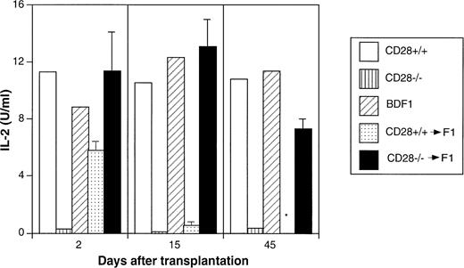
Profound immune deficiency appears rapidly after induction of the GVH reaction.24 25 To assess the effect of CD28 on T-cell function, IL-2 production was measured in the culture supernatants of splenocytes obtained 2, 15, and 45 days after transplantation and stimulated for 24 hours with ConA. Splenocytes from recipients transplanted with CD28+/+ showed a reduction in IL-2 production by day 2, lack of IL-2 production by day 15, and could not be tested on day 45 because the recipients were dead from GVHD. In contrast, splenocytes from recipients transplanted with CD28−/− cells produced IL-2 levels comparable with those of normal BDF1 splenocytes at all three time points (Fig 4). We conclude that CD28−/− donor T cells are unable to cause GVHD in this strain combination.
Immune deficiency does not develop in BDF1 recipients transplanted with CD28−/− donor cells. Splenocytes from CD28+/+ wild type, CD28−/− deficient, normal BDF1 mice, and BDF1 recipients of CD28+/+ or CD28−/− cells 2, 15, and 45 days after transplantation were cultured for 24 hours with ConA; and IL-2 in the supernatants was measured by using a sandwich enzyme-linked immunosorbent assay (ELISA) technique. IL-2 concentrations were calculated with reference to standard curves. Data represent the average +/− 1 SD of three mice per group analyzed separately for BDF1 recipients of CD28+/+ or CD28−/− cells. Data on CD28+/+, CD28−/− and normal BDF1 mice represent the study of one mouse at each time point. *Recipients of CD28+/+ donor cells died before day 45.
Immune deficiency does not develop in BDF1 recipients transplanted with CD28−/− donor cells. Splenocytes from CD28+/+ wild type, CD28−/− deficient, normal BDF1 mice, and BDF1 recipients of CD28+/+ or CD28−/− cells 2, 15, and 45 days after transplantation were cultured for 24 hours with ConA; and IL-2 in the supernatants was measured by using a sandwich enzyme-linked immunosorbent assay (ELISA) technique. IL-2 concentrations were calculated with reference to standard curves. Data represent the average +/− 1 SD of three mice per group analyzed separately for BDF1 recipients of CD28+/+ or CD28−/− cells. Data on CD28+/+, CD28−/− and normal BDF1 mice represent the study of one mouse at each time point. *Recipients of CD28+/+ donor cells died before day 45.
CD28 contributes to development of antihost cytotoxic effector cells in unirradiated recipients.
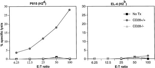
B cells of recipient origin were severely depleted after transplantation of CD28+/+ cells but remained at normal levels after transplantation of CD28−/− cells (Table 1). The loss of B cells in recipients of CD28+/+cells suggested the presence of antihost cytotoxic effectors. Spleen cells from recipients transplanted with CD28+/+ or CD28−/− cells were tested on day 15 after transplantation for antihost (H2d) activity in a 4-hour cytotoxicity assay against 51Cr-labeled targets without prior restimulation in vitro (Fig 5). Splenocytes from recipients transplanted with CD28+/+ cells showed cytotoxicity against recipient-type (H2d) targets but not against donor-type (H2b) targets (Fig 5). In contrast, splenocytes from normal BDF1 controls and from recipients transplanted with CD28−/− cells showed no activity against recipient-(H2d) or donor-(H2b) type targets. These results showed that CD28−/−donor T cells had a limited ability to generate cytotoxic effectors against recipient alloantigens, or the cytotoxic effectors could not be maintained.
Donor T cells lacking CD28 cannot generate cytotoxic effectors against the recipient. Fifteen days after transplantation, splenocytes from normal BDF1 mice and from BDF1 recipients of CD28+/+ or CD28−/− cells were assayed directly without in vitro restimulation. Three mice per group were analyzed separately, and the data represent the mean +/− 1 SD of three individual mice. The activity of anti-H2d or anti-H2b cytolytic effectors was measured in a 4-hour cytotoxicity assay against specific P815 (H2d) or control EL-4 (H2b) targets.
Donor T cells lacking CD28 cannot generate cytotoxic effectors against the recipient. Fifteen days after transplantation, splenocytes from normal BDF1 mice and from BDF1 recipients of CD28+/+ or CD28−/− cells were assayed directly without in vitro restimulation. Three mice per group were analyzed separately, and the data represent the mean +/− 1 SD of three individual mice. The activity of anti-H2d or anti-H2b cytolytic effectors was measured in a 4-hour cytotoxicity assay against specific P815 (H2d) or control EL-4 (H2b) targets.
Role of CD28 in prevention of rejection by donor CD8+cells.
In previous studies, we have shown that cytotoxic activity mediated by donor CD8+ cells is needed to prevent rejection of MHC class-I–disparate B6-Ly5.1 marrow in sublethally irradiated (550 cGy) bm1 recipients.22 In this model, recipients uniformly reject the marrow when the graft does not contain T cells and as few as 8.0 × 10⁵ CD28+/+ CD8+ cells are sufficient to prevent rejection in virtually all recipients (Table 2). CD28−/−CD8+ cells were impaired in their ability to prevent marrow graft rejection in this model. Rejection was not prevented when 8.0 × 10⁵ or 3.2 × 10⁶ CD8+cells from CD28−/− donors were added to the graft (Table 2, Experiment 1), but rejection was prevented when 1.0 × 10⁷ CD8+ cells from CD28−/− donors were added to the graft (Table2, Experiment 2). These results indicate that CD28−/− CD8+ cells have a limited ability to eliminate MHC class-I–disparate immune cells in vivo.
Ability of CD8+ Cells From CD28−/− Donors to Prevent Marrow Graft Rejection*
| Experiment . | CD8 Cells Added . | Percent Donor Marrow-Derived Cells . | |
|---|---|---|---|
| T Lymphocytes . | Granulocytes . | ||
| 1 | None | 1, 1, 0, 1 | 2, 3, 2, 3 |
| 8.0 × 10⁵ CD28+/+ | 69, 62, 79, 69 | 98, 94, 94, 95 | |
| 8.0 × 10⁵CD28−/− | 1, 0, 1, 0 | 1, 1, 1, 1 | |
| 3.2 × 10⁶ CD28−/− | 1, 0, 0, 1 | 2, 1, 1, 1 | |
| 2 | None | 1, 0, 1, 0, 0 | 3, 4, 2, 1, 1 |
| 8.0 × 10⁵ CD28+/+ | 65, 85, 73, 72 | 87, 95, 94, 96 | |
| 1.0 × 10⁷CD28−/− | 88, 91, 90, 46 | 96, 95, 97, 89 | |
*Groups of four or five irradiated (550 cGy) bm1 recipients were transplanted with 5.0 × 10⁶ T-cell–depleted B6-Ly5.1 marrow cells alone or together with the indicated number of CD8-enriched lymph nodes cells from CD28+/+ or CD28−/− B6 mice. The percent donor marrow-derived T cells and granulocytes in the peripheral blood on day 28 after transplantation was determined by two-color staining with CD3 and Ly5.1-specific antibodies. Data indicate results for individual recipients in two separate experiments. Data in experiment 2 were similar when results were analyzed on day 60 after transplantation (not shown).
Role of CD28 in GVHD mediated by CD4+ T cells in irradiated recipients.
We tested separately the role of CD28 on GVHD mediated by CD4+ cells against MHC class-II–incompatible (B6 × bm12)F1 recipients and CD8+ cells against MHC class-I–incompatible (B6 × bm1)F1 recipients. We selected these donor and recipient combinations because small numbers of purified CD4+ or CD8+ T cells from B6 mice respectively induce lethal GVHD in 100% of irradiated (B6 × bm12)F1 or (B6 × bm1)F1 recipients.26
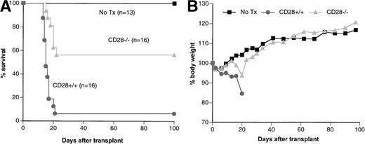
Irradiated (700 cGy) (B6 × bm12)F1 recipients were injected with 2.0 × 10⁶ CD4+ lymph node T cells from either CD28+/+ or CD28−/− B6 donors. Irradiated (B6 x bm12)F1 controls injected with PBS developed transient pancytopenia, but all recovered and survived longer than 100 days. Recipients of CD28+/+ cells became acutely ill with GVHD, characterized by progressive weight loss, ruffled fur, and hunched back, and 15 of 16 (94%) recipients died at a median of 15 days after transplant. In contrast, recipients of CD28−/− cells had less severe and delayed GVHD, and 7 of 16 (44%) mice died at a median of 20 days (Fig 6). Thus, the absence of CD28 on donor T cells was associated with increased recipient survival (P < .001). On day 15, recipients of CD28+/+ cells had a hematocrit of 15.7% ± 7.6%, significantly lower than the hematocrit of 27.8% ± 11.1% in recipients of CD28−/− cells (P < .001). Surviving recipients of CD28−/− cells eventually recovered from GVHD, and regained body weight to the level of irradiation controls (Fig 6). They also recovered T- and B-cell numbers and function comparable with those of irradiation controls (Table 3). To confirm engraftment of donor cells, we tested for the presence ofneo in recipients of CD28−/− cells and found that donor cells persisted beyond 100 days after transplant in all recipients tested (Fig 3B).
CD4+ CD28−/− donor T cells have a limited capacity to cause GVHD in irradiated (B6 × bm12)F1 recipients. (B6 × bm12)F1 mice were irradiated (700 cGy) and transplanted with CD28+/+ CD4+ cells or CD28−/− CD4+ cells from B6 donors. Irradiated (B6 × bm12)F1 mice were injected with PBS alone as control. (A) Survival curves; (B) Weight curves show the mean body weight for each group. Data were pooled from two replicate experiments.
CD4+ CD28−/− donor T cells have a limited capacity to cause GVHD in irradiated (B6 × bm12)F1 recipients. (B6 × bm12)F1 mice were irradiated (700 cGy) and transplanted with CD28+/+ CD4+ cells or CD28−/− CD4+ cells from B6 donors. Irradiated (B6 × bm12)F1 mice were injected with PBS alone as control. (A) Survival curves; (B) Weight curves show the mean body weight for each group. Data were pooled from two replicate experiments.
Role of CD28 in GVHD mediated by CD8+ T cells in irradiated hosts.
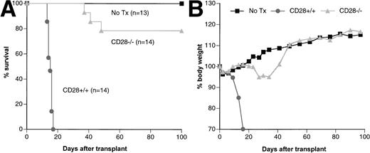
Irradiated (700 cGy) (B6 × bm1)F1 recipients were injected with 2.0 × 10⁶ CD8+ lymph node T cells from either CD28+/+ or CD28−/− B6 donors. Recipients of CD28+/+ cells became acutely ill with GVHD, characterized by progressive weight loss, ruffled fur, and hunched back, and all 14 recipients died at a median of 15 days after transplant. In contrast, recipients of CD28−/−cells had a delayed and less-severe GVHD, and only 3 of 12 (25%) recipients died at a median of 41 days (Fig7). On day 15, recipients of CD28+/+ cells had a hematocrit of 5.8% ± 2.6% compared with 26.3% + 4.9% in recipients of CD28−/− cells (P < .001). Surviving recipients transplanted with CD28−/− cells eventually recovered from GVHD and regained body weight to the level of irradiation controls (Fig 7). These recipients also recovered T and B cell number and function comparable to those of irradiation controls (Table 3). To assess engraftment of donor cells, we tested for the presence of neo in recipients of CD28−/− cells and found that donor cells persisted for at least 100 days after transplant in all recipients (Fig3C).
CD8+ CD28−/− donor T cells have a limited capacity to cause GVHD in irradiated (B6 × bm1)F1 recipients. (B6 × bm1)F1 mice were irradiated (700 cGy) and transplanted with CD28+/+ CD8+ cells or CD28−/− CD8+ cells from B6 donors. Irradiated (B6 × bm1)F1 mice were injected with PBS alone as control. (A) Survival curves; (B) Weight curves show the mean body weight for each group. Data were pooled from two replicate experiments.
CD8+ CD28−/− donor T cells have a limited capacity to cause GVHD in irradiated (B6 × bm1)F1 recipients. (B6 × bm1)F1 mice were irradiated (700 cGy) and transplanted with CD28+/+ CD8+ cells or CD28−/− CD8+ cells from B6 donors. Irradiated (B6 × bm1)F1 mice were injected with PBS alone as control. (A) Survival curves; (B) Weight curves show the mean body weight for each group. Data were pooled from two replicate experiments.
Recipients of CD28−/− Donor Cells Recovered Normal Numbers of Thymocytes and Normal Numbers and Function of Splenic T and B Cells*
| . | (B6 × bm12)F1 . | (B6 × bm1)F1 . | ||
|---|---|---|---|---|
| No Transplant . | CD28−/− . | No Transplant . | CD28−/− . | |
| Number of cells (×10−6)† | ||||
| Total thymocytes | 69 ± 34 | 72 ± 11 | 90 ± 25 | 97 ± 22 |
| CD4+CD8+ thymocytes | 57 ± 30 | 53 ± 14 | 74 ± 22 | 75 ± 18 |
| B splenocytes | 62 ± 12 | 52 ± 29 | 46 ± 14 | 46 ± 13 |
| T splenocytes | 30 ± 6 | 30 ± 13 | 23 ± 6 | 17 ± 6 |
| Thymidine uptake‡ | ||||
| LPS stimulation | 80 ± 20 | 69 ± 18 | 68 ± 27 | 47 ± 20 |
| ConA stimulation | 26 ± 13 | 24 ± 20 | 51 ± 16 | 48 ± 20 |
*Thymus and spleens were obtained from 100 to 105 days post-transplant. Data represent the average of six mice per group pooled from two separate experiments as shown in Figs 6 and 7.
Thymocytes were stained for CD4 and CD8 expression, and splenocytes were stained for CD3 and B220 expression. The absolute count of each population was calculated by the total number of cells times the percentage of the population.
B-cell proliferation was tested after 3 days in culture with LPS at 10 μg/mL, and T-cell proliferation was tested after 3 days in culture with ConA at 5 μg/mL. Data represent cpm × 10−3.
DISCUSSION
In this study we have compared the pathogenicity of CD28−/− and CD28+/+ donor T cells in GVHD induced by parental B6 grafts in unirradiated BDF1 recipients and in irradiated MHC class-I (bm1) or class-II (bm12) –incompatible recipients. In the absence of CD28, donor T cells have markedly reduced proliferative and cytolytic activity against the recipient and induce less-severe GVHD resulting in lower fatality. These findings indicate that CD28 plays an important role in the pathogenesis of GVHD but is not necessary for development of the disease.
One concern in this study was that donor engraftment could be compromised by the lack of CD28, especially in B6→BDF1 transplants. Hybrid resistance mediated by natural killer (NK) cells is particularly effective in this strain combination,26 and there was the possibility that hybrid resistance could not be overcome in the absence of donor CD28 despite transplantation of a large number of splenocytes. We have shown, however, that engraftment of CD28−/− donor T cells was evident at 2 and 15 days after transplantation by flow cytometry, and very low levels of engraftment were detected at 28 days after transplantation by the more sensitive PCR assay. In the B6→(B6 × bm12)F1 and B6→(B6 × bm1)F1 strain combinations, there is no hybrid resistance27 and graft rejection was not expected. Results of PCR assays showed that cells from CD28−/−donors persisted in the recipients beyond 100 days after transplantation. We found that CD28−/− cells have limited ability to prevent rejection of T-cell–depleted marrow cells in sublethally (550 cGy) irradiated bm1 recipients. However, CD28−/− CD8+ cells can overcome graft resistance by the host, if administered in sufficiently large numbers. Therefore, we conclude that CD28 on donor CD8+cells helps to eliminate MHC class-I–incompatible host-immune cells.
The role of CD28 on GVHD was apparent in all three strain combinations tested, although there were notable differences among them. GVHD after B6→BDF1 transplants appeared to be heavily dependent on CD28 costimulation, because CD28−/− donor T cells had marginal alloresponsiveness, lacked antihost cytotoxicity, and recipients of CD28−/− cells survived with no manifestations of the disease. On the other hand, GVHD after B6→(B6 × bm12)F1 and B6→(B6 × bm1)F1 transplants was less dependent on CD28 costimulation, because not all (B6 × bm12)F1 or (B6 × bm1)F1 recipients of CD28−/− CD4+ or CD8+cells survived. Irradiation of the recipients might account for this difference. (B6 × bm12)F1 and (B6 × bm1)F1 recipients received sublethal irradiation that caused marrow hypoplasia, and marrow failure was caused by GVHD. The release of proinflammatory cytokines, such as TNF-α and IL-1 induced by irradiation might also predispose towards the development of GVHD.28-31
Recently, Speiser et al21 found that CD28−/− and CD28+/+ spleen cells were equally capable of causing lethal acute GVHD, and concluded that CD28 costimulation is not necessary for the induction of lethal acute GVHD. The different results between their study and ours presented here and the studies of others in which B7 blocking agents prevented GVHD15-19 may reflect differences in genetic disparity between donor and recipient. Speiser et al21 transplanted cells from B6 mice into irradiated BALB/c recipients or vice versa under conditions in which recipients transplanted with wild-type T cells died early after transplantation with severe GVHD. We, and others,16,17 transplanted splenocytes from B6 mice into BDF1 recipients or purified T cells from B6 mice into (B6 × bm1)F1 or (B6 × bm12)F1, under conditions in which the GVHD induced by wild-type T cells was less severe. Blazar et al15 reported that CTLA-4Ig protected B10.BR/SgSnJ recipients from GVHD induced by B6 splenocytes under conditions in which the GVHD induced by wild-type T cells was less severe than the GVHD observed by Speiser et al.21 It is possible that costimulatory requirements of T cells may differ in response to distinct alloantigens and that high TCR affinity for alloantigen might overcome the requirements for CD28 costimulation, as shown in other systems.32-34
A defect in the expansion of CD28−/− cells appears to account for their limited ability to induce GVHD.35 Our data indicated that neither CD4 nor CD8 donor T cells were able to expand after transplantation in the absence of CD28 costimulation (Table 1). CD28−/− donor T cells had reduced proliferation and IL-2 production in response to recipient alloantigens and could not generate cytotoxic effectors against the host. These results are consistent with the observation by Hakim et al17 that CTLA-4Ig inhibited the expansion of CD4+ and CD8+ cells from B6 donors in BDF1 recipients, and the results of Blazar et al,18 who observed that anti-CD80 plus anti-CD86 MoAbs inhibited the expansion of CD4+ T cells from B6 donors in irradiated bm12 recipients. Therefore, the lack of CD28 costimulation appears to result consistently in a decreased T-cell expansion, IL-2 production, and CTL generation. Because we used CD28−/− T cells rather than B7 inhibitors that bind to both CD28 and CTLA-4, our data provide definitive evidence that CD28 contributes to T-cell activation and expansion after recognition of alloantigen in vivo. Because engagement of CTLA-4 can deliver a negative signal to T cells and facilitate peripheral T-cell tolerance,6-8 our findings provide a rationale to investigate whether alloantigen-specific tolerance can be achieved by selectively blocking CD28 costimulation while still allowing CTLA-4 engagement on donor T cells.
ACKNOWLEDGMENT
We thank Drs Craig B. Thompson and Patricia Noel for their generous gift of the CD28-/- mice; Dr Stanley Riddell for providing PCR primers specific for neo and β-actin; Drs Yoshiki Akatsuka and Ming-Tsen Lin for helpful technical advice; and Ms Alison Sell for assistance in preparing the manuscript.
Supported by Grants No. CA 18209, AI 40680, AI 33484, and HL 55257 from the Department of Health and Human Services of the National Institutes of Health, Bethesda, MD.
Address correspondence to Claudio Anasetti, MD, Fred Hutchinson Cancer Research Center, 1100 Fairview Ave N, D2-100, Seattle, WA 98109.
The publication costs of this article were defrayed in part by page charge payment. This article must therefore be hereby marked "advertisement" is accordance with 18 U.S.C. section 1734 solely to indicate this fact.


![Fig. 2. Spontaneous ex vivo proliferation and IL-2 production of donor T cells was reduced in the absence of CD28. Splenocytes were obtained from control BDF1 mice without transplant and from BDF1 recipients of CD28+/+ or CD28−/− cells 2 days after transplantation. Three mice per group were analyzed separately, and the data represent the mean +/− 1 SD of three individual mice. (A) Splenocytes were cultured at 2.0 × 10⁵/well for 4 hours in medium, and triplicate cultures were pulsed with [3H]TdR at the beginning of the culture. Results show the proliferative capacity of splenocytes early in GVHD. (B) Splenocytes were cultured at 5.0 × 10⁶/mL for 24 hours in medium alone, and supernates were assayed for IL-2 by the proliferation of CTLL-2 cells in the presence of anti–IL-4 MoAb.](https://ash.silverchair-cdn.com/ash/content_public/journal/blood/92/8/10.1182_blood.v92.8.2963/5/m_blod42013002x.jpeg?Expires=1764989895&Signature=iDUyTdgQHM295pNBu6kohP15WUPQLxERPvvdo8wywwIW-nRCiaHoyIZvPs3RUB6bXG90hegYQx9vFnbOLXG3s7XIXuUYH2b9joCJ~jPTfyitjU0tqI1qTtOHx6j8YkvpQqHi4DCLM81y3l9vVRlEn0tdgL8wulZVmXM7JEA-xjNYd89dqj2661x~9eM8ALPYoKoOFz7OmQEAYjsrUKu4Yfysg1wYN~9j6yxrIbMbYD8zBsVTTfTCGGgZ~6I3-vRdY8rxlo7C0Ewd7ncvhMj3oCfMZHOZx7C2jI1Yv-n9YjPOhpJ9Xtlzf-MJ0VfP-iQMSDkPoM6REtfRyhABESpQtQ__&Key-Pair-Id=APKAIE5G5CRDK6RD3PGA)


This feature is available to Subscribers Only
Sign In or Create an Account Close Modal