Abstract
We have previously identified a novel protein that mediates the attachment of erythroblasts to macrophages in vitro. This attachment promotes terminal maturation and enucleation of erythroblasts (Hanspal and Hanspal, Blood 84:3494, 1994). This protein is referred to here as Emp for erythroblast macrophageprotein. Two immunologically related isoforms of Emp with apparent molecular weights of 33 kD and 36 kD were detected in macrophage membranes. The complete amino acid sequence of the larger isoform of Emp was deduced from the nucleotide sequence of a full-length 2.0-kb cDNA that was isolated from a human macrophage cDNA library using affinity-purified anti-Emp antibodies. Of the 2,005 bp, 1,185 bp encode for 395 amino acids representing 43 kD (the sodium dodecyl sulfate-polyacrylamide gel electrophoresis [SDS-PAGE] molecular mass is 36 kD). Northern blot analysis of human macrophage poly(A) RNA detected a message for Emp of 2.1 kb. The deduced amino acid sequence contains a putative transmembrane domain near the N-terminus. To investigate the structure/function relationships of Emp, recombinant fusion proteins of full-length and truncated Emp were produced in bacteria, COS-7, and HeLa cells. Cell binding assays showed that the N-terminus is exposed on the cell surface. The recombinant Emp functions as a cell attachment molecule when expressed in heterologous cells. Furthermore, we showed that the demise of erythroblasts in the absence of Emp-mediated erythroblast-macrophage association is accompanied by apoptosis. We postulate that Emp-mediated contact between erythroblasts and macrophages promotes terminal maturation of erythroid cells by suppressing apoptosis.
© 1998 by The American Society of Hematology.
THE ASSOCIATION OF erythroblasts with macrophages plays a central role in the human bone marrow, where erythropoiesis occurs in distinct anatomic units called erythroblastic islands. These islands consist of a central macrophage surrounded by a ring of developing erythroblasts.1-4 Although erythroblastic islands have been well characterized morphologically, virtually nothing is known about the structural and functional basis of cellular contacts within erythroblastic islands. We have previously shown that physical contact between erythroblasts and macrophages promotes the terminal maturation of erythroblasts, leading to their enucleation in vitro. Importantly, we identified a novel protein now termed Emp (erythroblast macrophageprotein) that mediates the association of erythroblasts with macrophages.5
The molecular basis of erythroblast-macrophage interactions is poorly understood. In addition to the Emp-mediated association noted above, very late activation antigen-4 (VLA-4; or α4β1 integrin)-vascular cell adhesion molecule-1 (VCAM-1) has been reported to mediate the contact of erythroblasts with macrophages.6 This was evidenced by the finding that central macrophages in erythroblastic islands isolated from anemic mouse spleens express VCAM-1, whereas the surrounding erythroblasts express α4β1 integrin. Moreover, monoclonal antibodies against α4 integrin and VCAM-1 disrupted erythroblastic islands, suggesting that these molecules play an important role in erythroblast-macrophage interactions. Whereas VCAM-1 is expressed by central macrophages of erythroblastic islands but not by other macrophages, such as those of peritoneal origin,6 VLA-4 is distributed on a variety of hematopoietic cells, including stem cells, lymphocytes, erythroblasts, monocytes, and immature granulocytes.7-9 Thus, if VLA-4 and VCAM-1 were the only molecules involved in erythroblast-macrophage interactions, several types of hematopoietic cell islands could be formed. However, in vivo, only erythroid cells are found in association with bone marrow macrophages. This observation suggests that other molecules, such as Emp, may also be required for the formation of erythroblastic islands in vivo.
In contrast to the limited understanding of erythroblast-macrophage cell interaction, there is considerable data on the adhesion of hematopoietic progenitor cells to bone marrow stromal cells. Such adhesive interactions promote the localization of hematopoietic progenitor cells to the bone marrow and support their proliferation and differentiation.10,11 In the bone marrow, the cell-cell and cell-extracellular matrix (ECM) interactions involve multiple adhesive molecules. Cell-cell interactions among hematopoietic progenitors in the bone marrow mainly involve cell adhesion molecules of the Ig superfamily. However, recently, a member of the cadherin family of cell-cell adhesion receptors was also shown to be involved in the hematopoietic system. E-cadherin, which is mainly expressed by cells of the epithelial origin, was detected in human erythroblasts and was shown to be involved in the differentiation of erythroid cells.12 The interactions of hematopoietic progenitor cells with ECM, on the other hand, are largely mediated by integrins, whose expression is developmentally regulated.13
We have previously shown that the Emp-mediated association of macrophages with erythroblasts promotes terminal maturation of erythroblasts followed by their enucleation.5 We report here the complete amino acid sequence of the larger isoform of Emp translated from the nucleotide sequence of human macrophage cDNA. The apparent molecular weight, cellular localization, and binding specificities distinguish Emp from other known adhesive molecules present in hematopoietic tissues. Also, we show that Emp-mediated attachment of erythroblasts to macrophages prevents apoptosis of erythroblasts. Based on this observation, we propose that, in the absence of this attachment, apoptosis contributes to the inability of erythroid cells to fully mature. The availability of the primary structure of Emp will permit further characterization of its physiological functions in both erythroid and nonerythroid cells.
MATERIALS AND METHODS
Production of anti-Emp antibodies.
Polyclonal antibodies were raised against gel-purified Emp isolated from human macrophage membranes. The antibodies were affinity-purified on nitrocellulose membrane containing immobilized Emp, as described by Olmsted,14 and were tested against macrophage membranes separated by sodium dodecyl sulfate (SDS) gels containing 18% polyacrylamide by Western blotting. The immunoreactive bands were detected by an enhanced chemiluminescence (ECL) system (Amersham, Arlington Heights, IL).
Cloning of Emp cDNA.
For immunoscreening, a cDNA library constructed from the human monocytic cell line, U937, was used. U937 cells express high levels of Emp as tested by Western blotting using affinity-purified anti-Emp antibodies (data not shown). A U937 cDNA library made in the expression vector λgt11 (Clonetech, Palo Alto, CA) was used to infect overnight cultures of Escherichia coli strain Y1090 at a density of approximately 5 × 10⁴ plaque-forming units/180-mm diameter plate. The infected cells, after appropriate dilutions, were then placed on 1.5% LB agar plates with the top 0.7% agar and incubated for 3.5 hours at 42°C. Protein expression was induced by overlaying the plate with a nitrocellulose membrane that had been saturated with 10 mmol/L isopropyl β-D-thiogalactoside (IPTG). Incubation was continued for another 3.5 hours at 37°C. Nitrocellulose membranes were blocked with 5% nonfat dried milk in phosphate-buffered saline (PBS) containing 3 mmol/L sodium azide for 1 hour. Incubation with affinity purified anti-Emp IgG prepared from rabbit antiserum was performed for 1 hour. This was followed by incubation with [125I] protein A. Positive plaques were visualized by autoradiography. Positive recombinant phage plaques were then purified to 100% homogeneity by three cycles of rescreening, and the cDNA inserts were subcloned into pGEM7Z (Promega, Madison, WI) for further analysis.
A 1.6-kb positively reacting partial cDNA clone was obtained by immunoscreening. This clone covered almost the entire coding region, with the exception of the very amino terminus of Emp. The amino terminus was completed by 5′ RACE (rapid amplification of cDNA ends) using the Marathon cDNA amplification method (Clonetech). First-strand synthesis of 1.0 μg of U937 Poly(A) RNA was performed using a modified lock-docking oligo (dT) primer that consists of two degenerate nucleotide positions at the 3′ end.15 After second-strand synthesis, the cDNA pool was blunt-end ligated to the cDNA adaptors. The first polymerase chain reaction (PCR) was performed with a 27-mer sense primer (AP1) specific for the adaptor and an Emp-specific antisense primer (5′-TCCCAGCTGGTG-TAGTCGGTAGTT-3′ nucleotides 744 to 721; see Fig 3). The second round of PCR was performed with a nested 23-mer sense primer (AP2) and an Emp-specific antisense primer (5′-GAAGGC-CAGCATGCCCATGG-3′ nucleotides 623 to 604; see Fig 3). The conditions for both PCRs were 95°C for 30 seconds, 60°C for 30 seconds, and 68°C for 5 minutes. The nucleotide sequences of the collinear primers, AP1 and AP2, are described in the RACE protocol (Clonetech). The amplified product was subcloned in a TA cloning vector system (Invitrogen, San Diego, CA) and sequenced by the dideoxynucleotide chain-termination method.16 Both strands of the cDNA clones were sequenced.
Generation of peptide-specific antibodies.
A peptide (p1) corresponding to residues 15-26 (YETLNKRFRAAQ) of human Emp was synthesized. Antibodies to the peptide were raised in rabbits, and the antiserum was tested for immunospecificity for Emp by Western blots of macrophage membranes. Peptide-specific antibodies were obtained by affinity purification of antiserum on cyanogen bromide immobilized peptide. Antibodies were eluted with 0.1 mol/L glycine, pH 2.5, immediately neutralized with Tris, and dialyzed against PBS.
Northern blot analysis.
Poly(A) RNA was isolated by using the FastTrack kit from Invitrogen. Five micrograms of poly(A) RNA was electrophoresed on a 1% agarose gel containing 6.6% formaldehyde. RNA was then transferred to nitrocellulose and hybridized with a full-length [32P]-labeled Emp cDNA probe overnight at 42°C in 50% formaldehyde, 4× SSC, 0.5 mg/mL salmon testes DNA (denatured), 1× Denhardt’s solution, 0.25 mg/mL yeast t-RNA, 10% dextran sulphate, and 1% SDS. This was followed by washes in 0.1× SSC, 0.1% SDS at room temperature for 5 minutes, at 52°C for 20 minutes, and finally in 0.1× SSC, 0.5% SDS at 60°C for 30 minutes. The filter was air-dried briefly and subjected to autoradiography using Kodak x-ray films (Eastman Kodak, Rochester, NY).
Construction and expression of GST-Emp fusion protein.
Human recombinant Emp was expressed in bacteria. Two constructs (designated GST-Emp-1 and GST-Emp-2) were prepared in the pGEX-2T vector (Pharmacia, Uppsala, Sweden). The GST-Emp-1 construct contained the entire coding region of Emp that was amplified by the PCR using the following primers containing EcoRI adaptors: (sense) 5′-AAGAATTCCGGTGGAGTCGGCGGCTCAG-3′, nucleotides 20 to 38; and (antisense) 5′-CCGAATTCCAGAGTA TTTATCGTTGCTCA-3′, nucleotides 1210 to 1229. The sense primer was designed upstream of the initiator methionine. Thus, the GST-Emp-1 construct encodes full-length Emp with an additional eight amino acids upstream of the initiator methionine. The GST-Emp-2 construct contained cDNA insert encoding sequence between amino acids 98 and 395. The insert was amplified by PCR using the following sense primer containingEcoRI adaptor: 5′-AAGAAT TCATAGCAGCGACCAGCCCGCG-3′ nucleotides 336 to 356, together with the same antisense primer used for the GST-Emp-1 construct. After digestion with EcoRI, the PCR-amplified products were ligated into the pGEX-2T vector. All constructs were sequenced from both directions to ascertain sequence fidelity and proper orientation. Transformants containing Emp cDNA inserts were grown in the presence of IPTG to induce expression of the fusion proteins. Expression of fusion proteins in transformants was determined by SDS-polyacrylamide gel electrophoresis (SDS-PAGE) of total cell lysates, followed by the cell attachment assay. A parallel control was included that contained only the GST protein.
The cell attachment assay.
The attachment of erythroblasts to Emp was studied by the cell attachment assay as described previously.5 Briefly, bacterial cell lysates expressing full-length or truncated Emp were separated by SDS-PAGE and transferred to a nitrocellulose membrane using the standard transfer procedure.17 The nitrocellulose membrane was blocked with 1% bovine serum albumin (BSA) in PBS for 1 hour at 22°C and incubated with [125I]-labeled erythroblasts for 2 hours at 4°C. The membrane was washed 4 to 5 times with PBS, air-dried, and exposed to Kodak x-ray film for autoradiography.
Expression of Emp in mammalian cells.
A eukaryotic expression vector pMT3, containing a hemagglutinin (HA) tag, was used. The vector pMT3 and monoclonal antibodies against the HA tag were generously provided by Dr K. Andrabi (Harvard Medical School, Boston, MA). Two constructs of human Emp cDNA were made: one containing nucleotides 21 to 1229 (designated Emp-1) encoding full-length Emp (residues 1-395), whereas the other containing nucleotides 336 to 1229 (designated Emp-2) encoding truncated Emp from residues 114 to 395. The inserts were PCR amplified using the following primers containing Nsi I adaptors: (sense for Emp-1) 5′-TGCATGCAT CGGTGGAGT CGGCGGCT CA GT-3′; (sense for Emp-2) 5′-TGCATGCATCCCATAG CAG CGAC CAGCCCGC-3′; and (antisense for both Emp-1 and Emp-2) 5′-GGCATGCATCAGAG TAT TTATCGTTGCTCA-3′. After digestion with Nsi I, the PCR-amplified products were ligated into the unique Pst1 site of pMT3. cDNAs were inserted to produce an in-frame fusion with the HA tag at the C-terminus, thus expressing a fusion protein containing the HA tag and some vector sequence, thereby adding 35 amino acids (3.9 kD) to the molecular mass of Emp. All constructs were sequenced from both directions to ascertain sequence fidelity and proper orientation. The plasmids were purified on Qiagen columns (Qiagen Inc, Chatsworth, CA) and transiently transfected into COS-7 cells by diethyl aminoethyl (DEAE)-dextran method.18
Three days after the transfection, COS-7 cells were harvested, metabolically labeled with [35S] methionine, and immunoprecipitated with monoclonal antibodies against the HA tag. For immunoprecipitation, metabolically labeled cells were treated with diisopropyl fluorophosphate (Sigma Chemical Co, St Louis, MO) and lysed in 0.5 mL of immunoprecipitation buffer (10 mmol/L Tris-HCl, pH 7.5, 150 mmol/L NaCl, 5 mmol/L EDTA, 1% NP-40, 0.5% sodium deoxycholate, 2 mg/mL BSA, 0.2 mmol/L TLCK, 0.2 mmol/L TPCK, and 2 mmol/L phenylmethyl sulfonyl fluoride [PMSF]). The samples were centrifuged at 800g for 5 minutes to remove nuclei. The supernatants were diluted 10-fold with the immunoprecipitation buffer. Ten microliters of monoclonal antibody against the HA tag was added to each and the samples were incubated overnight at 4°C with gentle shaking. Thereafter, 100 μL of protein A-Sepharose (Pharmacia; 50 mg/mL of buffer A [10 mmol/L Tris-HCl, pH 7.5, 130 mmol/L NaCl, 5 mmol/L EDTA, and 1% NP-40]) was added and the samples incubated for another 4 hours at 4°C. The protein A-Sepharose beads were then collected and washed three times with buffer A. The final pellet was resuspended in 70 μL SDS sample buffer and boiled for 2 minutes. Beads were removed by centrifugation and the supernatant was directly loaded on SDS-polyacrylamide gels according to the buffer system of Laemmli.19
To determine the cellular orientation of the expressed Emp cDNA, transfected COS-7 cells were surface labeled with [125I] using the glucose oxidase-lactoperoxidase method,20followed by immunoprecipitation with monoclonal antibodies against the HA tag as described above.
Stable transfections.
To study the attachment of erythroblasts to Emp cDNA-transfected cells, a stably transfected HeLa cell line was generated. HeLa cells were cotransfected with Emp cDNA constructs (either Emp-1 or Emp-2) and pHook vector (Invitrogen) (containing the neomycin resistance gene under the direction of a cytomegalovirus [CMV] promoter) by DEAE-dextran method. Three days after transfection, cells were harvested and replated in the selective medium containing 200 μg/mL of G418. Resistant cells were further propagated in selective medium to establish a cell line. Human erythroblasts cultured by the two-phase liquid culture system5 were mixed with transfected or nontransfected HeLa cells and grown in complete culture medium in the absence of G418 for 3 to 4 days. On day 3 or 4, floating cells were removed and the adherent cells were washed two times with PBS. Cells adhering on the petri dishes were fixed and stained with Wright-Giemsa without detaching from the dishes and examined by bright-field microscopy.
In vitro maturation of erythroid progenitors.
Erythroblasts were generated by in vitro culture of peripheral blood-derived mononuclear cells using a two-phase liquid culture system as described previously.5 This system supports the proliferation and maturation of largely erythroid progenitors. At the end of the culture, approximately 90% of the cells are late erythroblasts and approximately 10% of the remaining cells are macrophages. For experiments involving macrophage-depleted cultures, macrophages were removed using monoclonal antibodies MO2 against macrophage surface antigen as described earlier.5 Briefly, macrophages were removed on day 4 or 5 of the second phase of culture, when clusters of proerythroblasts and macrophages started to appear. The mixture of cells at a concentration of about 107/mL was incubated with 1:20 rat antihuman MO2 antibodies at 4°C for 45 minutes. After three washings in cold Iscove’s modified Dulbecco’s medium (IMDM), the cells were resuspended in IMDM containing 1:40 fluorescein isothiocyanate (FITC)-conjugated rabbit antirat IgG and incubated for a further 45 minutes. The cells were then washed, resuspended in IMDM, and incubated with sheep anti-FITC antibody attached to magnetic particles for 30 minutes at 4°C. MO2-positive cells were removed by magnetic separation using a Bio-Mag separator (Advanced Magnetic Inc, Cambridge, MA). The control cultures, ie, macrophage-containing cultures, were treated identically but were not exposed to MO2 monoclonal antibodies. Cell viability, as tested by trypan blue exclusion staining, was found to be greater than 95% in both the control and macrophage-depleted cultures before and after the magnetic separation procedure. The cultures were continued for 7 to 8 days after the removal of macrophages (a total of 12 days in the second phase of culture).
In situ detection of apoptosis.
Erythroblasts cultured in the presence and absence of macrophages were analyzed for DNA fragmentation, characteristic of apoptosis, using the TUNEL (terminal deoxynucleotidyltransferase-mediated dUTP nick end labeling) reaction. The latter was performed using the peroxidase-conjugated in situ cell death kit (Boehringer Mannheim, Indianapolis, IN) according to the manufacturer’s instructions.
RESULTS
Detection of two immunologically related Emp isoforms in macrophage membranes.
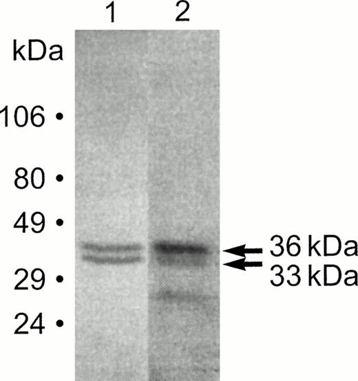
Anti-Emp antibodies detected two isoforms of Emp in the macrophage membranes (Fig 1, lane 1). These isoforms migrate on SDS gels with apparent molecular weights of 33 kD and 36 kD and are referred to as Emp-33 and Emp-36, respectively. The cell attachment assay involving incubation of radiolabeled erythroblasts with immobilized macrophage membrane proteins showed that both isoforms bind to radiolabeled erythroblasts (Fig 1, lane 2). Occasionally, we have observed a lower band of approximately 26 kD that also appears to bind to radiolabeled erythroblasts. Because this lower band is not always detected, it is likely that it represents a degradation product of Emp. Previously,5 the two isoforms were not easily discernible because of the diffuse appearance of protein bands on low polyacrylamide concentration gels. Western blot analysis using anti-Emp antibodies detected Emp-33 and Emp-36 from erythroblast membranes as well (data not shown).
Detection of Emp isoforms in macrophage membranes. Macrophage membrane proteins were separated by 18% SDS-PAGE and transferred to a nitrocellulose (NC) membrane. The NC membrane was probed with anti-Emp antibodies (lane 1) and immunoreactive bands were detected with an enhanced chemiluminescence system, or the NC membrane was incubated with radioiodinated erythroblasts (lane 2) and processed for autoradiography. Molecular weight standards are shown on the left.
Detection of Emp isoforms in macrophage membranes. Macrophage membrane proteins were separated by 18% SDS-PAGE and transferred to a nitrocellulose (NC) membrane. The NC membrane was probed with anti-Emp antibodies (lane 1) and immunoreactive bands were detected with an enhanced chemiluminescence system, or the NC membrane was incubated with radioiodinated erythroblasts (lane 2) and processed for autoradiography. Molecular weight standards are shown on the left.
Cloning and sequencing of the Emp cDNA.
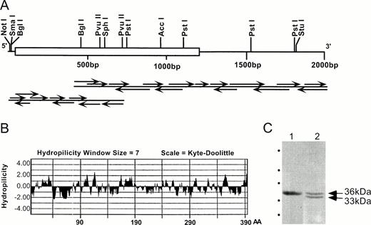
As a first step to elucidate the mechanism by which the Emp-mediated attachment of erythroblasts to macrophages promotes erythroid cell maturation, studies were initiated to determine the primary structure of Emp. A human macrophage (U937) library constructed in bacteriophage expression vector λgt11 was used for expression cloning. The library was screened with affinity-purified anti-Emp antibodies using standard techniques. A 1.6-kb cDNA clone was obtained by immunoscreening. This clone contained almost the entire coding region of Emp with the exception of its very amino terminus. The amino terminal sequence was obtained by 5′ RACE, which yielded a 0.7-kb overlapping cDNA fragment. Both cDNA clones were subcloned and sequenced (Fig 2).
Organization of human Emp cDNA. (A) The restriction map was obtained from two overlapping clones isolated from a human U937 λgt11 cDNA library. The central block (nucleotides 45-1229) represents the coding region. Both strands were sequenced, and arrows indicate the sequencing strategy. (B) Hydrophilicity plot of the deduced amino acid sequence of Emp. Positive values indicate hydrophilicity, whereas negative values indicate hydrophobicity. AA, amino acids. (C) Immunochemical identification of the Emp sequence. Macrophage membrane proteins were separated by 18% SDS-PAGE, transferred to NC membrane, and probed with affinity purified anti-p1 antibodies (lane 1) or affinity-purified antibodies against native Emp (lane 2). Anti-p1 was raised against the synthetic peptide corresponding to residues 15-26. The position of the molecular weight standards (same as those used in Fig 1) is shown on the left.
Organization of human Emp cDNA. (A) The restriction map was obtained from two overlapping clones isolated from a human U937 λgt11 cDNA library. The central block (nucleotides 45-1229) represents the coding region. Both strands were sequenced, and arrows indicate the sequencing strategy. (B) Hydrophilicity plot of the deduced amino acid sequence of Emp. Positive values indicate hydrophilicity, whereas negative values indicate hydrophobicity. AA, amino acids. (C) Immunochemical identification of the Emp sequence. Macrophage membrane proteins were separated by 18% SDS-PAGE, transferred to NC membrane, and probed with affinity purified anti-p1 antibodies (lane 1) or affinity-purified antibodies against native Emp (lane 2). Anti-p1 was raised against the synthetic peptide corresponding to residues 15-26. The position of the molecular weight standards (same as those used in Fig 1) is shown on the left.
Together, these two clones gave a sequence of 2,005 bps that represented the complete sequence containing a translation initiation codon at nucleotide 45 and a stop codon at nucleotide 1229 (Fig 3). An open reading frame of 1,185 bp predicted a protein containing 395 amino acids with a calculated molecular mass of 43 kD (although the protein expressed in E coli and COS-7 cells migrates with an apparent molecular weight of ∼36 kD; see below) and an isoelectric point of 9.86. The stop codon is followed by an untranslated region of 776 bp with a consensus polyadenylation sequence (AATAAA) 10 bp before the poly A tail. The hydrophilicity plot shows a hydrophobic segment near the N-terminus between amino acids 56 and 80 (Fig 2). Although the hydrophobicity index is relatively low, this segment is followed immediately by highly charged residues, consistent with the stop transfer signals found following most membrane spanning domains.21
Nucleotide and deduced amino acid sequence of human Emp cDNA. The putative membrane spanning domain is underlined, the putative PTB binding motif is dotted underlined, and the polyadenylation consensus sequence (AATAAA) is doubly underlined.
Nucleotide and deduced amino acid sequence of human Emp cDNA. The putative membrane spanning domain is underlined, the putative PTB binding motif is dotted underlined, and the polyadenylation consensus sequence (AATAAA) is doubly underlined.
The predicted amino acid sequence of Emp contains a consensus sequence, N-D-K-Y at the C-terminus (Fig 3), which, if tyrosine phosphorylated, could serve as a binding site for phosphotyrosine binding (PTB) domains. The PTB domains are structurally different from SH2 domains and specifically recruit tyrosine-phosphorylated proteins into signaling complexes by binding to phosphotyrosine within a sequence motif N-X-X-Y (where X represents any amino acid).22,23 The recognition sequence of the PTB domain is specified by residues N-terminal of the tyrosine and especially the −3 and the −5 residues, which correspond respectively to an asparagine and to a hydrophobic residue.22,24-27 Emp also contains tyrosine residues that could participate in other cell signaling events. For example, tyrosine 127 (which is part of a YXN sequence), if phosphorylated, could possibly be recognized directly by the SH2 domain of GRB2.28,29 Similarly, tyrosine 312 of Emp could potentially bind to the SH2 domain of phospholipase Cγ.30
Database searches did not show any protein that is highly homologous over the entire coding sequence of Emp. However, short segments of Emp were found to have homology with the δ chain of sheep T-cell surface glycoprotein CD3. The segment of Emp between amino acids 236 and 264 is 41% identical, and the segment between amino acids 67 and 96 is 33% identical with the δ chain of T-cell receptor. The CD3/T-cell receptor complex has been shown to prevent apoptosis of immature T cells in thymic cultures.31
Peptide-specific antibody detects native protein in macrophage membranes.
To confirm the authenticity of cloned Emp cDNA, we have raised antibodies against a selected domain of recombinant Emp. Based on the deduced amino acid sequence, a peptide (p1) corresponding to residues 15-26 was synthesized. Rabbits were immunized with the peptide p1, and antibodies were purified on a p1 affinity matrix. In immunoblots of macrophage membranes, affinity-purified anti-p1 antibodies detected a single 36-kD band that comigrates with the larger isoform of Emp (Fig2C). These results show that the full-length recombinant Emp corresponds to the native 36-kD Emp isoform from macrophages. Also, this result suggests that the smaller isoform of Emp lacks the peptide p1.
Emp is widely distributed in nonerythroid cells.
To determine whether Emp is expressed in various cell types and tissues, Northern blot analysis was performed. Analysis of poly(A) RNA from U937 cells, the human monocytic cell line used for cDNA cloning, showed a message size of 2.1 kb for Emp, which is in agreement with the 2.0-kb nucleotide sequence of the cDNA clones. After induction with 12-O-tetradecanoylphorbol-13-acetate (TPA), which results in their differentiation into adherent macrophage-like cells,32 33the U937 cells showed a 50% decrease in the message for Emp (Fig 4A). A transcript of the same size was also observed using poly(A) RNA isolated from human heart, brain, placenta, lung, liver, skeletal muscle, kidney, and pancreas (Fig 4B). To address the concern regarding possible contamination of tissues with peripheral blood, Northern blot analysis was repeated with poly(A) RNA isolated from various cell lines. As shown in Fig 4C, Emp transcripts were also present in K562, COS-7, HeLa, and Jurkat cell lines. These results show that Emp transcripts are widely distributed in nonerythroid cells and tissues.
Northern blot analysis. (A) Five micrograms of Poly(A) RNA from uninduced (1) and induced (2) U937 cells. (B) Northern blot containing 20 μg of total RNA from human tissues was obtained from Clonetech. (C) Five micrograms of Poly(A) RNA from different cell lines. Each blot was hybridized with a full-length [32P]-labeled Emp cDNA probe. Blot A was stripped and reprobed with human β-actin cDNA. The size of the markers in kilobases is shown.
Northern blot analysis. (A) Five micrograms of Poly(A) RNA from uninduced (1) and induced (2) U937 cells. (B) Northern blot containing 20 μg of total RNA from human tissues was obtained from Clonetech. (C) Five micrograms of Poly(A) RNA from different cell lines. Each blot was hybridized with a full-length [32P]-labeled Emp cDNA probe. Blot A was stripped and reprobed with human β-actin cDNA. The size of the markers in kilobases is shown.
Emp expressed in E coli behaves as an erythroblast binding protein.
To determine if recombinant Emp binds to erythroblasts, GST-fusion protein was expressed in bacteria. Two constructs were prepared in the pGEX-2T vector: one containing the entire coding sequence (GST-Emp-1) and the other containing cDNA sequence from which 97 amino acids from the N-terminus were deleted (GST-Emp-2). These constructs were expressed as GST fusion proteins in E coli. Expression of fusion proteins in the transformants was determined by SDS-PAGE of total cell lysates, followed by the cell attachment assay. As shown in Fig 5A, the GST-Emp-1 construct produced a 65-kD product (indicated by an asterisk in lane 1) that contains full-length Emp (36 kD). The GST-Emp-2 construct was expressed as a 58-kD fusion protein (indicated by an asterisk in lane 2) containing GST and a truncated Emp (29 kD). Our cell attachment assay, which involves incubation of immobilized recombinant proteins with radiolabeled erythroblasts, showed a strong binding of erythroblasts to the 65-kD (full-length) product and only a weak binding to the 58-kD (truncated) product. Quantification of cell binding, as determined by scanning the autoradiogram, showed a 20-fold greater attachment of erythroblasts to the 65-kD product than to the 58-kD product under identical conditions. These results show that the recombinant Emp behaves identically to native protein in the cell attachment assay and that the cell binding domain of Emp is contained within the 97 amino acid region at the N-terminus. Affinity-purified anti-Emp antibodies detected both 65-kD and 58-kD products by Western blotting (Fig 5A).
Expression of recombinant Emp. (A) Full-length (Emp-1) and truncated (Emp-2) human Emp were expressed as glutathione S-transferase (GST) fusion proteins in bacteria. Cell lysates of bacteria transformed with GST-Emp-1 and -2 constructs (lanes 1 and 2, respectively) were separated by SDS-PAGE and stained directly with Coomassie blue or transferred to nitrocellulose, probed with radiolabeled erythroblasts followed by autoradiography, or probed with affinity-purified anti-Emp antibody and immunoreactive bands were detected by ECL. (B) Full-length (Emp-1) and truncated (Emp-2) human Emp were expressed in COS-7 cells as fusion proteins containing HA tag at the C-terminus. Transfected and nontransfected COS-7 cells were either metabolically labeled with [35S]methionine or surface labeled with [125I], followed by immunoprecipitation with anti-HA tag antibodies. Autoradiographs of the immunoprecipitates are shown. The position of the molecular weight standards is indicated on the left.
Expression of recombinant Emp. (A) Full-length (Emp-1) and truncated (Emp-2) human Emp were expressed as glutathione S-transferase (GST) fusion proteins in bacteria. Cell lysates of bacteria transformed with GST-Emp-1 and -2 constructs (lanes 1 and 2, respectively) were separated by SDS-PAGE and stained directly with Coomassie blue or transferred to nitrocellulose, probed with radiolabeled erythroblasts followed by autoradiography, or probed with affinity-purified anti-Emp antibody and immunoreactive bands were detected by ECL. (B) Full-length (Emp-1) and truncated (Emp-2) human Emp were expressed in COS-7 cells as fusion proteins containing HA tag at the C-terminus. Transfected and nontransfected COS-7 cells were either metabolically labeled with [35S]methionine or surface labeled with [125I], followed by immunoprecipitation with anti-HA tag antibodies. Autoradiographs of the immunoprecipitates are shown. The position of the molecular weight standards is indicated on the left.
The N-terminus of Emp is extracellular.
To confirm the topology of Emp in the plasma membrane, an epitope-tagged protein was produced in mammalian cells. Two constructs of human Emp cDNA were made in a eukaryotic expression vector pMT3 containing an HA tag: one containing the entire coding sequence (Emp-1) and another containing truncated Emp (113 amino acids deleted from the N-terminus) (Emp-2). The cDNA fragments were inserted in frame to produce a fusion with the HA tag at the C-terminus. These two constructs were transiently transfected into COS-7 cells by the DEAE-dextran method. Three days after transfection, COS-7 cells were harvested, metabolically labeled with [35S] methionine, and immunoprecipitated with monoclonal antibodies against the HA tag. A 41-kD protein was immunoprecipitated from COS-7 cells transfected with the Emp-1 construct, and a 32-kD protein was immunoprecipitated from COS-7 cells transfected with the Emp-2 construct (Fig 5B). As described in Materials and Methods, recombinant Emp is expressed as a fusion protein containing the HA tag and some vector sequence, thereby adding 35 amino acids (3.9 kD) to the molecular mass of Emp. Thus, the SDS-PAGE molecular mass of full-length recombinant Emp expressed in COS-7 cells is 37.1 kD and that of the truncated protein is 28.1 kD.
Next, COS-7 cells transfected with the Emp constructs were surface labeled with [125I], followed by immunoprecipitation with monoclonal antibodies against the HA tag. As shown in Fig 5B, a radiolabeled product of 41 kD was immunoprecipitated from COS-7 cells transfected with the Emp-1 plasmid containing the entire coding region of Emp cDNA. However, immunoprecipitations under identical conditions from COS-7 cells transfected with the Emp-2 plasmid lacking 113 amino acids from the N-terminus did not precipitate a radiolabeled 32-kD product. These results suggest that the N-terminus, which is encoded only by the Emp-1 construct, must be exposed on the cell surface because it was radiolabeled by surface iodinating agents, whereas the C-terminus carrying the HA tag must be intracellular and hence inaccessible to the surface labeling reagents. This observation is further supported by the fact that the recombinant GST-Emp-1 fusion protein showed a strong attachment with erythroblasts (Fig 5A).
Recombinant Emp expressed in HeLa cells mediates attachment of erythroblasts.
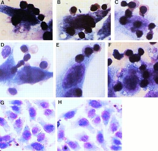
Because transient transfections allow only a small number of cells to acquire DNA, it was difficult to assess the attachment of erythroblasts to Emp-transfected COS-7 cells. Therefore, a stably transfected cell line was generated using HeLa cells. We have previously shown that erythroblasts do not bind to nontransfected HeLa cells.5The latter were cotransfected with Emp cDNA constructs (either Emp-1 or Emp-2) and pHook vector (Invitrogen). pHook vector contains CMV promoter for high-level transcription and a neomycin resistance gene for the selection of stable cell lines. To demonstrate that the transfected HeLa cells support the attachment of erythroblasts in culture, human erythroblasts were isolated by the two-phase liquid cultures of peripheral blood mononuclear cells as described previously.5 Erythroblasts at the proerythroblast/basophilic normoblast stage harvested on day 7 or 8 of the second phase were mixed with transfected or nontransfected HeLa cells, and cultures were continued in petri dishes for 3 to 4 days at 37°C in a 5% CO2 incubator. On day 3 or 4, floating cells were removed. The adherent cells were washed, fixed, stained with Wright-Giemsa without detaching from the dishes, and examined by bright-field microscopy. As shown in Fig6A through F, erythroblasts specifically attached to HeLa cells that had been transfected with the Emp-1 plasmid containing the entire coding sequence of the Emp cDNA insert in the sense orientation. No attachment was observed with HeLa cells transfected with the plasmid containing a truncated Emp cDNA or to nontransfected HeLa cells (Fig 6G and H). Hence, the transfection of full-length cDNA of Emp can produce erythroblast binding characteristics in HeLa cells. These results indicate that the cDNA encodes an authentic Emp. Furthermore, these results provide evidence that the Emp cDNA encodes a novel cell adhesion molecule and that the N-terminal of Emp is extracellular and is involved in cell:cell contact.
Attachment of erythroblasts to transfected and nontransfected HeLa cells. Early erythroblasts were cultured with HeLa cells transfected with the full-length (Emp-1) plasmid (A through F), with the truncated (Emp-2) plasmid (H), or with nontransfected HeLa cells (G) for 3 to 4 days. On day 3 or 4, floating cells were removed and the adherent cells were washed two times to remove any trapped nonadherent cells. Cells adhering on the petri dishes were fixed and stained with Wright-Giemsa without detaching from the dishes and examined by bright-field microscopy. Original magnification: (A through F) × 100; (G and H) × 40.
Attachment of erythroblasts to transfected and nontransfected HeLa cells. Early erythroblasts were cultured with HeLa cells transfected with the full-length (Emp-1) plasmid (A through F), with the truncated (Emp-2) plasmid (H), or with nontransfected HeLa cells (G) for 3 to 4 days. On day 3 or 4, floating cells were removed and the adherent cells were washed two times to remove any trapped nonadherent cells. Cells adhering on the petri dishes were fixed and stained with Wright-Giemsa without detaching from the dishes and examined by bright-field microscopy. Original magnification: (A through F) × 100; (G and H) × 40.
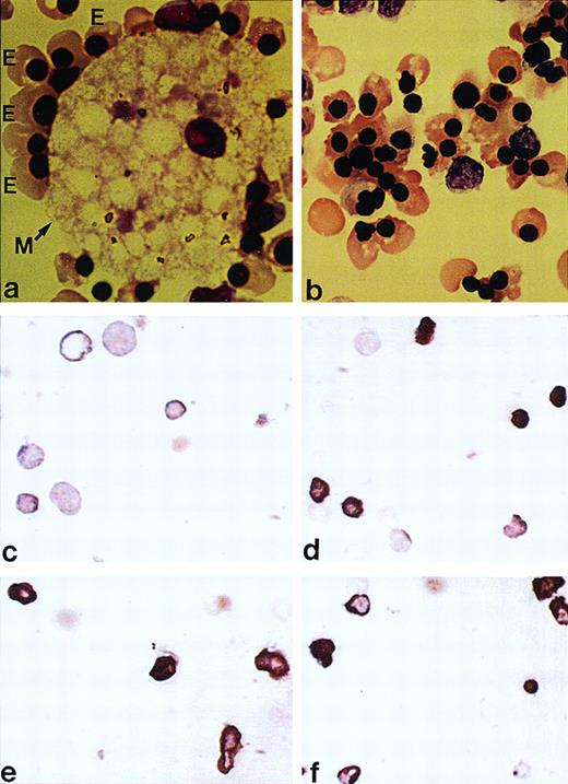
In situ analysis for DNA fragmentation. (Top panel) Wright-Giemsa–stained cytocentrifuged preparations of cells obtained on day 12 of the second phase of (a) macrophage-containing and (b) macrophage-depleted cultures. An erythroblastic island consisting of a central macrophage (M) surrounded by a ring of late erythroblasts (E) is shown in (a). (Middle and bottom panels) Erythroblasts harvested on day 12 of the second phase of each culture were examined by the TUNEL method, (c) macrophage-containing culture, (d) macrophage-depleted culture, (e) macrophage-containing culture in the presence of anti-Emp peptide (p1) antibodies, and (f) macrophage-depleted culture supplemented with macrophage conditioned medium. Apoptotic cells are identified by more dense staining.
In situ analysis for DNA fragmentation. (Top panel) Wright-Giemsa–stained cytocentrifuged preparations of cells obtained on day 12 of the second phase of (a) macrophage-containing and (b) macrophage-depleted cultures. An erythroblastic island consisting of a central macrophage (M) surrounded by a ring of late erythroblasts (E) is shown in (a). (Middle and bottom panels) Erythroblasts harvested on day 12 of the second phase of each culture were examined by the TUNEL method, (c) macrophage-containing culture, (d) macrophage-depleted culture, (e) macrophage-containing culture in the presence of anti-Emp peptide (p1) antibodies, and (f) macrophage-depleted culture supplemented with macrophage conditioned medium. Apoptotic cells are identified by more dense staining.
Emp-mediated association inhibits apoptosis of erythroblasts.
We have previously shown that the association of erythroblasts with macrophages mediated by Emp promotes erythroid maturation leading to their enucleation. In the absence of this association, erythroid cells mature to the late erythroblast stage but they do not enucleate and ultimately die. To determine whether the demise of erythroblasts in the absence of Emp-mediated erythroblast-macrophage association is due to apoptosis, erythroblasts cultured in the presence and absence of macrophages were analyzed for DNA fragmentation in situ using the TUNEL reaction.
Erythroblasts isolated on day 12 of the second phase of macrophage-containing as well as macrophage-depleted cultures are typically at the late stage of erythroid maturation as assessed by Wright-Giemsa staining of cytocentrifuged cell preparations (Fig 7a and b). However, if the cultures are continued for another 3 to 4 days, erythroblasts in macrophage-containing cultures undergo enucleation, whereas those in macrophage-depleted cultures die. For the following studies, we examined erythroblasts on day 12 of the second phase. Erythroblasts were subjected to TUNEL assay for in situ detection of apoptosis. As shown in Fig 7d, erythroblasts cultured in absence of macrophages stained intensely by the TUNEL method that detects in situ endonucleolytic cleavage characteristic of apoptosis. Quantification of the TUNEL reaction showed that 66% erythroblasts in macrophage-depleted cultures versus 10% in macrophage-containing cultures were apoptotic (Table 1). To determine if the magnetic separation procedure for macrophage removal induced any apoptosis of erythroblasts, TUNEL assay was performed on erythroblasts harvested from cultures that underwent mock macrophage removal. No apoptosis was observed in these erythroblasts (data not shown).
Percentage of Apoptosis in Erythroblasts Cultured in the Presence or Absence of Macrophages
| Culture . | % Apoptosis . |
|---|---|
| +Macrophages | 10 (n = 156) |
| −Macrophages | 66 (n = 236) |
| +Macrophages + anti-Emp Ab | 59 (n = 172) |
| −Macrophages + Macrophage CM | 40 (n = 170) |
| Culture . | % Apoptosis . |
|---|---|
| +Macrophages | 10 (n = 156) |
| −Macrophages | 66 (n = 236) |
| +Macrophages + anti-Emp Ab | 59 (n = 172) |
| −Macrophages + Macrophage CM | 40 (n = 170) |
Quantification of the TUNEL reaction. Erythroblasts harvested on day 12 of the second phase of each culture were examined by the TUNEL method. The percentage of intensely stained cells representing apopotic cells was determined in duplicate. The data represented the mean value of two experiments with very similar results. n represents the total number of cells counted.
The results noted above suggest that macrophages inhibit apoptosis of erythroblasts. To determine whether the effect of macrophages is through Emp-mediated contact with erythroblasts, this contact was specifically interfered by adding affinity-purified antibodies against the peptide, p1, in the extracellular domain of Emp to the macrophage-containing cultures. Antibodies (20 μg/mL) were added on day 4 of the second phase and cultures were continued up to day 12. Erythroblasts from control and antibody-containing cultures were subjected to TUNEL assay. As seen in Fig 7e and Table 1, 59% to 60% erythroblasts cultured in the presence of anti-Emp antibody were apoptotic, suggesting that Emp-mediated cell:cell contact inhibits apoptosis of erythroblasts. Similar results were obtained when the monovalent Fab fragment of anti-p1 IgG was used (data not shown). Apoptosis was also observed in erythroblasts when macrophage-depleted cultures were reconstituted with macrophage conditioned medium (Fig7f): 10% macrophage conditioned medium was added to cultures on day 4 of the second phase after macrophage removal and the cultures were continued up to day 12. These results further document the role of erythroblast-macrophage contact in inhibiting apoptosis.
DISCUSSION
In this study, we have characterized Emp, a novel protein present on the surface of erythroblasts and macrophages. Emp mediates erythroblast-erythroblast and erythroblast-macrophage interactions in vitro, suggesting a role of Emp in homophilic interactions that involve binding to homotypic and heterotypic cells.5 Two isoforms of Emp with apparent molecular weights of 33 kD and 36 kD were detected in macrophage membranes. Using cell attachment assay, we have shown that both isoforms of Emp bind to radiolabeled erythroblasts. This report describes the complete amino acid sequence of the larger isoform of human macrophage Emp, which will facilitate assignment of structural and functional domains within the protein.
The complete amino acid sequence of Emp was deduced from the nucleotide sequence of a full-length cDNA that was isolated by screening a human macrophage cDNA library using affinity-purified anti-Emp antibodies. To demonstrate that the predicted sequence encodes an authentic Emp, antibodies were raised against a peptide p1, corresponding to residues 15-26 in the amino terminus of recombinant Emp. Affinity-purified anti-p1 antibodies detected the larger isoform of Emp in macrophage membranes, suggesting that recombinant Emp corresponds to the 36-kD isoform native to macrophage membranes.
Northern blot analysis showed the presence of a single 2.0-kb species of Emp mRNA in U937 cells, a human macrophage cell line (Fig 4). Upon induction with phorbol ester TPA, the U937 cells differentiate into adherent macrophage-like cells. Our observation that the Emp mRNA is downregulated twofold upon induction of U937 cells with TPA (Fig 4A) is consistent with the fact that adherent macrophages do not participate in the formation of erythroblastic islands. Moreover, the presence of Emp transcripts in various human tissues and cell lines suggests that Emp or its homologues are widely distributed and may be involved in other cell:cell interactions.
The expression of full-length recombinant Emp in heterologous cells and erythroblast attachment assays provide conclusive evidence that Emp is a cell attachment molecule. A 20-fold reduction in the erythroblast attachment to the truncated Emp lacking 97 amino acids from the N-terminus shows that the cell binding domain of Emp is located at the N-terminus within 97 residues. Recombinant Emp was also expressed in COS-7 cells as a fusion protein with an HA tag at the C-terminus. Surface labeling of transfected cells provided further evidence that the N-terminus is exposed on the cell surface. Furthermore, full-length and truncated (113 amino acids from the N-terminus deleted) Emp cDNA fragments were stably transfected into HeLa cells. Incubation of transfected cells with human erythroblasts confirmed that the N-terminal extracellular domain is involved in cell:cell contact. In summary, the deduced primary structure of Emp consists of a relatively small amino terminal extracellular domain involved in cell-cell contact, a single transmembrane domain, and a large cytoplasmic domain. The N-terminal extracellular domain is the likely site for homophilic binding interactions between homotypic and heterotypic cells. The C-terminal cytoplasmic domain of Emp contains several tyrosine residues that, when phosphorylated, could be recognized by well-characterized protein recognition modules such as PTB or SH2 domains.22-30 Through these interactions, Emp may be recruited into signaling complexes and may provide mechanism to connect with signal transduction pathways.
The results presented here provide the convincing evidence that Emp is an integral membrane protein with the N-terminus on the extracellular side and the C-terminus on the cytoplasmic side. But, because it lacks an amino-terminal cleavable signal sequence, the mechanism by which the amino terminus of Emp is translocated across the endoplasmic reticulum is less clear. Nevertheless, it is believed that the membrane insertion of proteins without a cleavable signal sequence is assisted by internal hydrophobic sequences that are inserted in the membrane by the same mechanism that operates for cleavable signal sequences, except that no postinsertional proteolysis occurs.21 34-36 We believe that a similar mechanism is applicable for Emp’s insertion in the membrane.
Most adhesion proteins, for example, cadherins, integrins, and Ig superfamily, consist of a relatively large extracellular domain, a single membrane spanning domain, and a short cytoplasmic domain.37 In contrast, Emp appears to contain a relatively small extracellular domain at the N-terminus and a large cytoplasmic domain at the C-terminus. Thus, Emp may belong to a subfamily of novel adhesion proteins displaying large cytoplasmic domains or Emp may be a component of a large complex of which all the components are required for proper functioning.
Two other macrophage receptors that have been proposed to be involved in adhesion to erythroid cells include erythroblast receptor, EbR,38 and a lectin-like sheep erythrocyte receptor, SER.39,40 EbR mediates reversible divalent cation-dependent binding of hematopoietic cells to murine fetal liver macrophages. In contrast, Emp, which is present on the surface of both macrophages and erythroblasts, does not require divalent cations to mediate cellular contact.5 SER, a 185-kD plasma membrane glycoprotein, mediates binding of unopsonized sheep erythrocytes via recognition of sialylated glycoconjugates and may interact with sialylated ligands on bone marrow cells. Using monoclonal antibodies, Crocker et al41 have shown that SER is diffusely localized at the contact zones between macrophages and erythroblasts within erythroblastic islands. The apparent molecular weight of Emp distinguishes it from SER. These observations indicate that Emp is a novel molecule that is involved in the attachment of macrophages to erythroid cells.
In the absence of Emp-mediated interaction of erythroblasts with macrophages, erythroid cells mature to the late erythroblast stage but fail to enucleate and undergo apoptosis. This observation is of particular interest because portions of Emp are homologous to the δ chain of the sheep T-cell surface glycoprotein CD3 and because antibodies to CD3/T-cell receptor complex have been shown to induce cell death in immature T cells by apoptosis. It is also of relevance to note that the interaction of follicular dendritic cell–B-cell clusters mediated by VLA-4–VCAM-1 interactions inhibits apoptosis of germinal center B cells.42 Similarly, the central macrophage in erythroblastic islands may inhibit apoptosis of erythroblasts through the VLA-4–VCAM-1 interaction or through interactions involving Emp. The data presented here show that the disruption of erythroblast-macrophage contact by anti-Emp antibodies causes apoptosis, thus suggesting that one of the functions of the Emp-mediated cell-cell contact is to prevent apoptosis of erythroblasts. Numerous erythroid-expressed genes have been linked to regulation of apoptosis. For example, erythropoietin regulates survival of erythroid progenitors by preventing apoptosis.43Recently, GATA-1, an erythroid transcription factor, has been shown to support the viability of erythroid precursors by suppressing apoptosis.44
In summary, the molecular cloning of Emp describes a novel protein involved in erythroblast-macrophage interaction. This interaction promotes the terminal maturation of erythroid cells leading to their enucleation by suppressing apoptosis. To elucidate the mechanism by which Emp mediates erythroblast-macrophage contact and promotes terminal maturation and enucleation of erythroid cells, it will be essential to identify the interactions of Emp with other cellular components, including cytoskeletal and signaling proteins. These interactions are likely to show a new mechanism of cellular adhesion that may mediate cell:cell contacts in an hematopoietic microenvironment.
ACKNOWLEDGMENT
The authors thank Dr Athar Chishti for his advice and encouragement during the course of these studies, Jennifer Wu for the preparation of the peptide p1 and anti-p1 antibodies, Dr Eugenia Cifuentes for preparing the Fab fragment of anti-p1, and Donna-Marie Mironchuk for the artwork.
Supported in part by a grant from the American Cancer Society to M.H. and a grant from the National Institutes of Health (HL 27215) to the late Dr Jiri Palek.
Presented in part in abstract form at the 38th Annual Meeting of the American Society of Hematology, Orlando, FL, 1996.
Genbank accession no. AFO84928.
Address reprint requests to Manjit Hanspal, PhD, Department of Biomedical Research, St Elizabeth’s Medical Center, 736 Cambridge St, Boston, MA 02135; e-mail: mbh@tiac.net.
The publication costs of this article were defrayed in part by page charge payment. This article must therefore be hereby marked "advertisement" is accordance with 18 U.S.C. section 1734 solely to indicate this fact.


![Fig. 4. Northern blot analysis. (A) Five micrograms of Poly(A) RNA from uninduced (1) and induced (2) U937 cells. (B) Northern blot containing 20 μg of total RNA from human tissues was obtained from Clonetech. (C) Five micrograms of Poly(A) RNA from different cell lines. Each blot was hybridized with a full-length [32P]-labeled Emp cDNA probe. Blot A was stripped and reprobed with human β-actin cDNA. The size of the markers in kilobases is shown.](https://ash.silverchair-cdn.com/ash/content_public/journal/blood/92/8/10.1182_blood.v92.8.2940/5/m_blod42031004w.jpeg?Expires=1765113373&Signature=xUL1-JTBVax09iJ1aWmITAL6c0SBGBxhumKvvqTXciiubj51twwaJtTzHRnsC2j3Api~2m4Bt9v5JcrgE7J6UWTvajtCWqC65r8xOUhnbvX-O7HsjdL~JaJ3w-7go36dho7b2JQEqYk8OfsmCOIPEeH1uX2PlE26dTFz9cV3tA8f7r-mPF1LJX1d9D2yVzKdVzaAO2zvFBKuViinHykObSlkExjR8Nx43OXD~y4PiMZ3qSdNLOJVH4GQBj0y~FjbjRqWrZeRKpHULATRuDQtaqzgSi~cSnL1usFYMONHGW~3f-L~yEHvKo367zysgk2dwPR03iij9Ens9Tncw~R4BA__&Key-Pair-Id=APKAIE5G5CRDK6RD3PGA)
![Fig. 5. Expression of recombinant Emp. (A) Full-length (Emp-1) and truncated (Emp-2) human Emp were expressed as glutathione S-transferase (GST) fusion proteins in bacteria. Cell lysates of bacteria transformed with GST-Emp-1 and -2 constructs (lanes 1 and 2, respectively) were separated by SDS-PAGE and stained directly with Coomassie blue or transferred to nitrocellulose, probed with radiolabeled erythroblasts followed by autoradiography, or probed with affinity-purified anti-Emp antibody and immunoreactive bands were detected by ECL. (B) Full-length (Emp-1) and truncated (Emp-2) human Emp were expressed in COS-7 cells as fusion proteins containing HA tag at the C-terminus. Transfected and nontransfected COS-7 cells were either metabolically labeled with [35S]methionine or surface labeled with [125I], followed by immunoprecipitation with anti-HA tag antibodies. Autoradiographs of the immunoprecipitates are shown. The position of the molecular weight standards is indicated on the left.](https://ash.silverchair-cdn.com/ash/content_public/journal/blood/92/8/10.1182_blood.v92.8.2940/5/m_blod42031005w.jpeg?Expires=1765113373&Signature=06apAJXo6KAooluK7YkrLQzzAgxdg2e3AvL3zcotObNGyrbmKVcu8e3KN8Mmv4mBe788FHeWi9fSrIyJgco6nE1V-BmNBI9rzTH0D1RU7ERX8FoEGikPUtdAtkf8WQxKda9nSiv8uUxDRySbk11IdMmSkgqJKkhPjKTefKC1Crak6Rv87jebRZNoo36h0JTvmee2TIIZWIi1qoxQ0od7R8SvsHk8YGjpxevdAPJ~XD44aiHQFgqgrFNZV6RTvuclq8G8p3erm2yFm7h3PFQmRp6ajNz0HKKBaIn673cH7J8b4y2LS7XQYT4wWj2NeXmgEApm7W9SeWjucmpyZOz~AQ__&Key-Pair-Id=APKAIE5G5CRDK6RD3PGA)


This feature is available to Subscribers Only
Sign In or Create an Account Close Modal