Abstract
Mast cells contain a lot of mast cell-specific proteases. We have reported that the expression of mouse mast cell protease 6 (MMCP-6) is remarkably reduced in both cultured mast cells (CMCs) and skin mast cells of mi/mi mutant mice. In the present study, we found that the expression of MMCP-5 was reduced in CMCs but not in skin mast cells of mi/mi mice, and we compared the regulation mechanisms of MMCP-5 with those of MMCP-6. The mi locus encodes a member of the basic-helix-loop-helix-leucine zipper (bHLH-Zip) protein family of transcription factors (hereafter called MITF ). The consensus sequence recognized and bound by bHLH-Zip transcription factors is CANNTG. The overexpression of the normal (+) MITF but not of mi-MITF normalized the poor expression of the MMCP-5 gene in mi/mi CMCs, indicating the involvement of +-MITF in transactivation of the MMCP-5 gene. Although +-MITF directly bound CANNTG motifs in the promoter region of the MMCP-6 gene and transactivated it, the binding of +-MITF to the CAGTTG motif in the promoter region of the MMCP-5 gene was not detectable. The +-MITF appeared to regulate the transactivation of the MMCP-5 gene indirectly. Moreover, addition of stem cell factor to the medium normalized the expression of the MMCP-5 but not of the MMCP-6 gene in mi/mi CMCs. Despite the significant reduction of both MMCP-5 and MMCP-6 expressions in mi/mi CMCs, their regulation mechanisms appeared to be different.
THE MI LOCUS OF MICE on chromosome 6 encodes a member of the basic-helix-loop-helix-leucine zipper (bHLH-Zip) protein family of transcription factors (hereafter called mi-transcription factor [MITF ]).1,2 The MITF encoded by the mutant mi locus deletes 1 of 4 consecutive arginines in the basic domain (hereafter mi-MITF ).1,3,4 The mi/mi mice show microphthalmia, depletion of pigment in both hair and eyes, osteopetrosis, and a decrease in the number of mast cells.5-9 In addition to the decrease in number, the phenotype of mast cells was abnormal in mi/mi mice.10-13 In the skin of normal (+/+) mice, most mast cells were stained with berberine sulfate that bound heparin proteoglycan,14-17 and half of them expressed the mouse mast cell protease 6 (MMCP-6) gene.11 In contrast, only a few mast cells were berberine sulfate-positive and MMCP-6 mRNA-positive in the skin of mi/mi mice.11 The expression of the MMCP-6 gene was remarkably reduced in cultured mast cells (CMCs) derived from the spleen of mi/mi mice as well.10 13
We previously showed that normal (+) MITF (hereafter called +-MITF ) directly regulated the expression of the MMCP-6 and c-kit genes.18-20 The defect in the transactivation ability of mi-MITF caused the decrease of the MMCP-6 gene expression in mi/mi mice. The decrease of the mast cell number in tissues of mi/mi mice was attributed to the low expression of the c-kit gene, which encodes the receptor for stem cell factor (SCF ).9,10,18 We also showed that the deficient transactivation ability of the mi-MITF was caused by two different mechanisms: the loss of DNA binding and the impaired nuclear localization.19 21
Mast cells of mice contain various proteases besides MMCP-6. The cDNAs and genes that encode mast cell carboxypeptidase A (MC-CPA) and six of the seven mouse mast cell-specific serine proteases (MMCP) have been cloned and sequenced.22-29 The MMCP-1, MMCP-2, MMCP-4, and MMCP-5 are predicted to be chymases, whereas the MMCP-6 and MMCP-7 to be tryptases from the deduced amino acid sequences. The MC-CPA is an exopeptidase that prefers C-terminal aliphatic amino acids. The MMCP-1, MMCP-2, MMCP-4, and MMCP-5 genes reside on the chromosome 14 and link with a gene complex encoding blood cell proteases, such as the neutrophil cathepsin G and four granzymes specific to cytotoxic T lymphocytes.30-33 On the other hand, the MC-CPA gene resides on the chromosome 3, and the MMCP-6 and MMCP-7 genes reside on the chromosome 17.29 The expression of MMCP-6 was strongly regulated by the +-MITF, but that of MC-CPA was not influenced by the +-MITF.20 In the present study, we studied the effect of the +-MITF on the expression of MMCP-5 and found that the +-MITF was involved in the transactivation of the MMCP-5 gene. However, the mechanism of the MMCP-5 regulation by the +-MITF appeared to be different from that of MMCP-6.
MATERIALS AND METHODS
Mice. The original stock of C57BL/6-mi/+ (mi/+) mice was purchased from the Jackson Laboratory (Bar Harbor, ME) and were maintained in our laboratory by consecutive backcrosses to our own inbred C57BL/6 colony (more than 15 generations at the time of the present experiment). Female and male mi/+ mice were crossed together, and the resulting mi/mi mice were selected by their white coat color.5,6 The original stock of VGA-9-tg/tg mice, in which the mouse vasopressin-Escherichia coli β-galactosidase transgene was integrated at the 5′ flanking region of the mi (MITF ) gene, were kindly given by H. Arnheiter (National Institutes of Health, Bethesda, MD). The integrated transgene was maintained by repeated backcrosses to our own inbred C57BL/6 colony (more than 5 generations at the time of the present experiment). Female and male tg/+ mice were crossed together, and the resulting tg/tg mice were selected by their white coat color.1
Cells. Pokeweed mitogen-stimulated spleen cell conditioned medium (PWM-SCM) was prepared according to the method described by Nakahata et al.34 Mice of mi/mi or tg/tg genotype and control C57BL/6-+/+ (+/+) mice were used at 2 to 3 weeks of age to obtain CMCs. Mice were killed by decapitation after ether anesthesia and spleens were removed. Spleen cells derived from mi/mi, tg/tg, or +/+ mice were suspended as described previously and cultured in α-minimal essential medium (α-MEM; ICN Biomedicals, Costa Mesa, CA) supplemented with 10% PWM-SCM and 10% fetal calf serum (FCS; Nippon Bio-supp Center, Tokyo, Japan). Half of the medium was replaced every 7 days. More than 95% of cells were CMCs 4 weeks after the initiation of the culture.16 A helper virus-free packaging cell line (ψ2) was maintained in Dulbecco's modification of Eagle's medium (DMEM; ICN Biomedicals) supplemented with 10% FCS.35 Mouse mastocytoma cell line, FMA/3, was maintained in α-MEM supplemented with 10% FCS.36 When various amounts of recombinant mouse SCF (rmSCF; a generous gift of Kirin Brewery Co Ltd, Tokyo, Japan) were added, mi/mi and +/+ CMCs were cultured in α-MEM containing 10% PWM-SCM and 10% FCS.
Construction of retrovirus and its infection. Bluescript KS(−) plasmids (pBS; Stratagene, La Jolla, CA) containing the whole coding regions of +-MITF or mi-MITF (hereafter called pBS-+-MITF and pBS-mi-MITF, respectively) had been constructed in our laboratory.19 A retroviral vector pM5Gneo,37 a derivative of myeloproliferative sarcoma virus vector, was a kind gift from Dr W. Ostertag (Universität Hamburg, Hamburg, Germany). The purified Sma I-HincII fragment from pBS-+-MITF or pBS-mi-MITF was introduced into the blunted EcoRI site of pM5Gneo. The resulting plasmids (hereafter called pM5Gneo-+-MITF and pM5Gneo-mi-MITF, respectively) or the pM5Gneo vector were transfected into the packaging cell line (ψ2) using the calcium phosphate method,38 and neomycin-resistant ψ2 cell clones were selected by culturing in DMEM containing 10% FCS and G418 (0.8 mg/mL; GIBCO BRL, Grand Island, NY). For the gene transfer, spleen cells obtained from mi/mi mice were incubated on irradiated (30 Gy) subconfluent monolayer of virus-producing ψ2 cells for 72 hours in α-MEM supplemented with 10% PWM-SCM and 10% FCS. Neomycin-resistant CMCs were obtained by continuing the culture in α-MEM containing 10% PWM-SCM, 10% FCS, and G418 (0.8 mg/mL) for 4 weeks. FMA/3 cells were infected with the retrovirus containing pM5Gneo-+-MITF, pM5Gneo-mi-MITF, or pM5Gneo plasmid by culturing on the virus-producing ψ2 cells. Neomycin-resistant FMA/3 cells were obtained by continuing the culture in α-MEM containing 10% FCS and G418 (0.8 mg/mL) for 4 weeks.
Northern blot analysis. Total RNAs were prepared using the lithium chloride-urea method.39 The fragments of MMCP-5,25 MC-CPA,23 MMCP-6,26 and β-actin40 cDNAs were used as probes after being labeled with [32P]-α-dCTP (DuPont/NEN Research Products, Boston, MA; 10 mCi/mL) by random oligonucleotide priming. After hybridization at 42°C, blots were washed to a final stringency of 0.2× SSC (1× SSC is 150 mmol/L NaCl, 15 mmol/L trisodium citrate, pH 7.4) at 50°C and subjected to autoradiography.
Reverse transcriptase modification of polymerase chain reaction (RT-PCR). Total RNA (5.0 μg) obtained from +/+ or mi/mi CMCs was reverse transcribed in 20 μL of the reaction mixture containing 20 U of avian myeloblastosis virus reverse transcriptase (Boehringer Mannheim GmbH Biochemica, Mannheim, Germany) and random hexamer. One microliter of each reaction product was amplified in 25 μL of PCR mixture containing 0.125 U of Taq DNA polymerase (Takara Shuzou, Kyoto, Japan) and 12.5 pmol each of sense and antisense primers by 30 cycles of 1 minute of denaturation at 94°C, 2 minutes of annealing at 55°C, and 2 minutes of synthesis at 72°C. Ten microliters of the PCR products was electrophoresed in 1.0% agarose gel containing ethidium bromide. The following oligonucleotide primers were used: MMCP-5: sense primer, 5′-CTACATGGCCTATCTGGAAA (130 through 149), antisense primer, 5′-TCCAGTTCCAGATTTCCTCA (795 through 814); MMCP-6: sense primer, 5′-TGTCCCTGAGCATTCCTGAGACCTA (1509 through 1533), antisense primer, 5′-TACTTTAATGAGCCACAGAAGAGAG (1738 through 1762); MC-CPA: sense primer, 5′-GTGATGACTGCTGCACACTG (194 through 213), antisense primer, 5′-CTTGAAGAGTCTGACTCAGG (769 through 788). The numbers represent the nucleotide numbers on the complementary strands of each cDNA sequence.23,25 26
In situ hybridization. Skin pieces were removed from the back of 20-day-old mice and smoothed onto a piece of the filter paper to keep them flat. The skin pieces were fixed in 4% paraformaldehyde in 0.1 mol/L phosphate buffer (PB; pH 7.4), and embedded in paraffin. CMCs were collected, washed with phosphate-buffered saline, and immobilized by agarose.41 The pieces of agarose-containing CMCs were fixed with 4% paraformaldehyde in 0.1 mol/L PB overnight and then embedded in paraffin. The preparation of MMCP-5, MMCP-6, and MC-CPA probes and the technique of in situ hybridization were described previously.11 42 The number of MMCP-5 mRNA-positive cells and that of Alcian blue-positive cells were counted in the serial sections, and the proportion of MMCP-5 mRNA-positive cells to Alcian blue-positive cells was calculated. The proportion of MMCP-6 mRNA-positive cells and that of MC-CPA mRNA-positive cells were also obtained.
Isolation of the promoter region of the MMCP-5 gene. The isolation of the promoter region of MMCP-5 gene was performed with the Mouse Promoter Finder Kit (Clonetech Laboratories, Palo Alto, CA) according to the manufacturer's instructions. The isolated promoter region was cloned into pBS and sequenced.
Construction of reporter plasmids. The luciferase gene subcloned into pSP72 (pSPLuc) was generously provided by Dr K. Nakajima (Osaka University Medical School, Osaka, Japan).43 To construct reporter plasmids, a DNA fragment containing a promoter region and the first exon (noncoding region) of the MMCP-5 gene (−2212 to +30) was cloned into the upstream of luciferase gene in pSPLuc. The deletion of the MMCP-5 promoter was produced by using the appropriate restriction enzyme. The mutation was introduced by PCR with mismatch primers. Deleted or mutated products were verified by sequencing.
Transfection of reporter plasmids and luciferase assay. Ten micrograms of reporter plasmid and 1 μg of pSTneo were transfected to 107 FMA/3 cells with electroporation (BioRad, Hercules, CA; 975 μF, 350 V). Neomycin-resistant cells were obtained by continuing the culture in α-MEM containing 10% FCS and G418 (0.8 mg/mL) for 4 weeks. The genomic DNA was extracted from neomycin-resistant cells, and the copy number of the luciferase gene was examined with the Southern blot analysis. For the assay of the luciferase activity, the cells were lysed with 0.1 mol/L potassium phosphate buffer (pH 7.4) containing 1% Triton X-100. Soluble extracts were then assayed for luciferase activity with a luminometer LB96P (Berthold GmbH, Wildbad, Germany). The activity was normalized by the copy number of the integrated luciferase gene and total protein concentration. When the reporter plasmids were transfected to FMA/3 cells infected by retrovirus containing pM5Gneo-+-MITF, pM5Gneo-mi-MITF, or pM5Gneo, the hygromycin-resistant gene was cotransfected. The hygromycin-resistant cells were selected by culturing in α-MEM containing 10% FCS and hygromycin (2 mg/mL) for 4 weeks.
Electrophoretic gel mobility shift assay (EGMSA). The production and purification of glutathione-S-transferase (GST)-+-MITF fusion protein was described previously.19 The nuclear extract from FMA/3 cells overexpressing +-MITF or mi-MITF was obtained by the method described by Schreiber et al.44 The whole cell extract was obtained by the method described by Pagano et al.45 Oligonucleotides were labeled with α-[32P]-dCTP by filling 5′-overhangs and used as probes of EGMSA. DNA-binding assays were performed in a 20 μL reaction mixture containing 10 mmol/L Tris-HCl (pH 8.0), 1 mmol/L ethylenediaminetetraacetic acid (EDTA), 75 mmol/L KCl, 1 mmol/L dithiothreitol, 4% Ficoll type 400, 50 ng of poly (dI-dC), 25 ng of labeled DNA probe, and 3.5 μg of GST-+-MITF fusion protein, nuclear extract, or whole cell extract. After incubation at room temperature for 15 minutes, the reaction mixture was subjected to electrophoresis at 14 V/cm at 4°C on a 5% polyacrylamide gel in 0.25× TBE buffer (1× TBE is 90 mmol/L Tris-HCl, 64.6 mmol/L boric acid, and 2.5 mmol/L EDTA, pH 8.3). The polyacrylamide gels were dried on Whatman 3MM chromatography paper (Whatman, Maidstone, UK) and subjected to autoradiography.
RESULTS
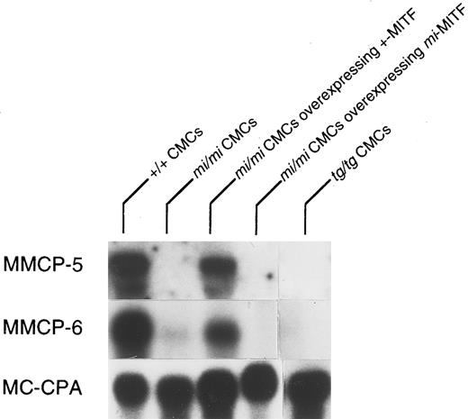
Impaired MMCP-5 expression in mi/mi CMCs but not in mi/mi skin mast cells. The expression of the MMCP-5 gene was examined by Northern blot using total RNA extracted from +/+, mi/mi, and tg/tg CMCs. The mRNA expression of the MMCP-5 gene was easily detectable in +/+ CMCs but hardly detectable in mi/mi and in tg/tg CMCs (Fig 1). As reported previously, the expression of MMCP-6 gene showed the similar expression pattern to that of the MMCP-5 gene,20 and the expression of the MC-CPA gene did not decrease in mi/mi and tg/tg CMCs (Fig 1).10 We introduced the cDNA encoding +-MITF or mi-MITF to mi/mi CMCs. The magnitude of mRNA expression of +-MITF was comparable to that of mi-MITF, and the expression level of introduced +- or mi-MITF was much greater than that of endogenous mi-MITF. We examined the MMCP-5 mRNA expression in mi/mi CMCs overexpressing +-MITF and mi/mi CMCs overexpressing mi-MITF (Fig 1). The expression of the MMCP-5 mRNA recovered to the nearly normal level in mi/mi CMCs overexpressing +-MITF but not in mi/mi CMCs overexpressing mi-MITF. The expression of the MMCP-6 mRNA showed the similar pattern to that of the MMCP-5 mRNA. The introduction of +- or mi-MITF cDNA did not affect the mRNA expression of the MC-CPA, as reported previously.20
Reduced expression of MMCP-5 mRNA in mi/mi and tg/tg CMCs and normalization of the MMCP-5 expression in mi/mi CMCs by the introduction of the +-MITF cDNA but not of mi-MITF cDNA. The blot was hybridized with 32P-labeled cDNA probe of MMCP-5, MMCP-6, or MC-CPA.
Reduced expression of MMCP-5 mRNA in mi/mi and tg/tg CMCs and normalization of the MMCP-5 expression in mi/mi CMCs by the introduction of the +-MITF cDNA but not of mi-MITF cDNA. The blot was hybridized with 32P-labeled cDNA probe of MMCP-5, MMCP-6, or MC-CPA.
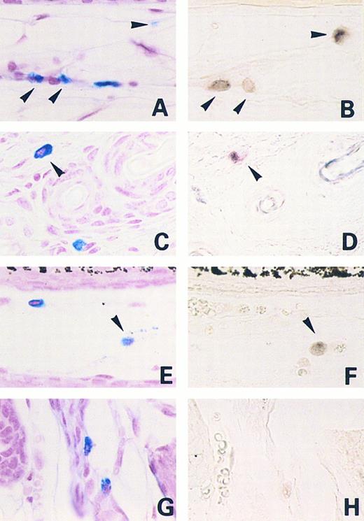
We next examined the expression of the MMCP-5 gene in skin mast cells of mi/mi mice by in situ hybridization. Approximately half of the skin mast cells expressed the MMCP-5 mRNA in both +/+ and mi/mi mice (Table 1 and Fig 2). Because mast cells in the skin were considered to be supported by SCF synthesized by fibroblasts,17 rmSCF was added to the culture medium of mi/mi and +/+ CMCs. The CMCs of mi/mi genotype do not proliferate in the medium containing rmSCF alone.10 Thus, we added PWM-SCM to the culture medium as well. After the incubation with 10 or 50 ng/mL of rmSCF for 2 weeks, we examined the proportion of mi/mi CMCs expressing the MMCP-5, MMCP-6, or MC-CPA mRNA by in situ hybridization (Table 2). The proportion of mi/mi CMCs expressing the MMCP-5 mRNA increased significantly even in the presence of 10 ng/mL rmSCF (Table 2). In contrast, the proportion of mi/mi CMCs expressing the MMCP-6 or MC-CPA mRNA was not influenced by the addition of rmSCF. The proportion of +/+ CMCs expressing the MMCP-5, MMCP-6, or MC-CPA mRNA was not affected by the addition of rmSCF to the culture medium (Table 2).
Proportion of MMCP-5-mRNA Expressing Mast Cells in the Skin of +/+ and mi/mi Mice Shown by In Situ Hybridization
| Genotype of Mice . | No. of Mice . | No. of . | No. of . | Proportion of . |
|---|---|---|---|---|
| . | . | Alcian Blue+ Cells . | MMCP-5 mRNA+ . | MMCP-5 mRNA+ to . |
| . | . | per cm of Skin* . | Cells per cm of Skin* . | Alcian Blue+ Cells (%)* . |
| +/+ | 5 | 228 ± 9 | 127 ± 6 | 56 ± 3 |
| mi/mi | 5 | 47 ± 13† | 27 ± 9† | 57 ± 4 |
Mean ± SE.
P < .01 by t-test when compared with the value of +/+ mice.
Mast cells expressing MMCP-5 or MMCP-6 mRNA in the skin of +/+ and mi/mi mice. Serial sections from the skin of a +/+ mouse (A and B, E and H) and a mi/mi mouse (C and D, G and H). (A, C, E, and G) Stained with alcian blue and nuclear fast red; (B and D) in situ hybridization with MMCP-5 probe; (F and H) in situ hybridization with MMCP-6 probe. Arrowheads show identical cells in each set of serial sections. Original magnification × 400.
Mast cells expressing MMCP-5 or MMCP-6 mRNA in the skin of +/+ and mi/mi mice. Serial sections from the skin of a +/+ mouse (A and B, E and H) and a mi/mi mouse (C and D, G and H). (A, C, E, and G) Stained with alcian blue and nuclear fast red; (B and D) in situ hybridization with MMCP-5 probe; (F and H) in situ hybridization with MMCP-6 probe. Arrowheads show identical cells in each set of serial sections. Original magnification × 400.
Effect of rmSCF on Proportions of CMCs Expressing MMCP-5, MMCP-6, or MC-CPA mRNA in +/+ or mi/mi CMCs Shown by In Situ Hybridization
| Genotype of CMCs . | Concentration of rmSCF . | Proportion of . | Proportion of . | Proportion of MC-CPA mRNA+ to Alcian Blue+ Cells (%)† . |
|---|---|---|---|---|
| . | (ng/mL)* . | MMCP-5 mRNA+ to Alcian Blue+ Cells (%)† . | MMCP-6 mRNA+ to Alcian Blue+ Cells (%)† . | . |
| +/+ | 0 | 45 ± 2 | 44 ± 3 | 92 ± 5 |
| mi/mi | 0 | 4 ± 3‡ | 3 ± 1‡ | 86 ± 4 |
| +/+ | 10 | 54 ± 7 | 52 ± 6 | 93 ± 3 |
| mi/mi | 10 | 41 ± 4ρ | 5 ± 4‡ | 85 ± 5 |
| +/+ | 50 | 52 ± 6 | 53 ± 7 | 86 ± 7 |
| mi/mi | 50 | 38 ± 4ρ | 5 ± 2‡ | 82 ± 6 |
+/+ or mi/mi CMCs were incubated with 10% PWM-SCM and various concentrations of rmSCF for 2 weeks.
Mean ± SE of five independent experiments.
P < .01 by t-test when compared with the value of +/+ CMCs, which were cultured without rmSCF.
ρ P < .01 by t-test when compared with the value of mi/mi CMCs, which were cultured without rmSCF.
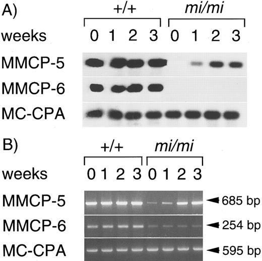
We examined the effect of duration of rmSCF exposure on induction of the MMCP-5 mRNA expression by Northern blotting and RT-PCR technique. The MMCP-5 mRNA expression was not detectable in mi/mi CMCs that were cultured with 10% PWM-SCM alone by the Northern blotting, but a weak band was detectable by RT-PCR (Fig 3). One week after initiation of the culture with 10% PWM-SCM and 10 ng/mL rmSCF, the message became detectable even by Northern blotting. The level of MMCP-5 expression increased until 2 weeks after the addition of rmSCF. The expression of MMCP-5 mRNA in +/+ CMCs was not influenced by the addition of rmSCF. The expression of MMCP-6 mRNA was not detectable in mi/mi CMCs by the Northern blotting, but weak band was detectable by RT-PCR. In contrast to MMCP-5, the magnitude of MMCP-6 expression was not influenced by the addition of rmSCF (Fig 3).
Effect of the addition of rmSCF on the expression of MMCP-5, MMCP-6, or MC-CPA gene in +/+ and mi/mi CMCs. (A) Northern blotting. Total RNA was obtained from +/+ or mi/mi CMCs cultured in the presence of 10% PWM-SCM and 10 ng/mL SCF for 0, 1, 2, or 3 weeks. The blot was hybridized with 32P-labeled cDNA probes for MMCP-5, MMCP-6, and MC-CPA. (B) RT-PCR analysis for the expression of MMCP-5, MMCP-6, or MC-CPA gene. The same RNAs were used as those used in Northern blotting. PCR products were electrophoresed in 1.0% agarose gel containing ethidium bromide.
Effect of the addition of rmSCF on the expression of MMCP-5, MMCP-6, or MC-CPA gene in +/+ and mi/mi CMCs. (A) Northern blotting. Total RNA was obtained from +/+ or mi/mi CMCs cultured in the presence of 10% PWM-SCM and 10 ng/mL SCF for 0, 1, 2, or 3 weeks. The blot was hybridized with 32P-labeled cDNA probes for MMCP-5, MMCP-6, and MC-CPA. (B) RT-PCR analysis for the expression of MMCP-5, MMCP-6, or MC-CPA gene. The same RNAs were used as those used in Northern blotting. PCR products were electrophoresed in 1.0% agarose gel containing ethidium bromide.
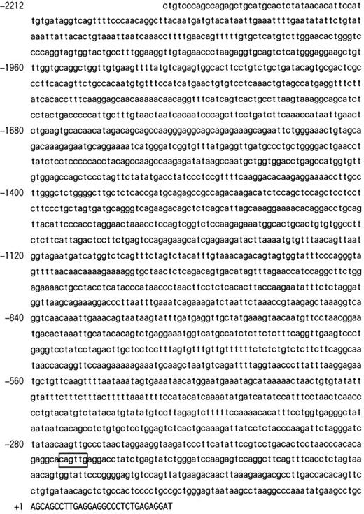
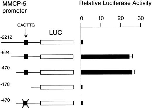
The analysis of the promoter region of MMCP-5 gene. The result that the introduction of +-MITF cDNA normalized the impaired MMCP-5 expression in mi/mi CMCs indicated the involvement of +-MITF in the regulation of the MMCP-5 gene expression. To examine the regulation mechanism by the +-MITF, we cloned 2212 bases of the 5′-upstream region of the MMCP-5 gene (Fig 4). The reporter plasmid that contained the luciferase gene under the control of the MMCP-5 promoter starting from nt −2212 (+1 shows the transcription initiation site) was constructed. We also constructed the deleted reporter plasmids containing the MMCP-5 promoter starting from nt −924, −470, or −178 to examine the elements that mediate the transactivation. The reporter plasmids were transfected into the FMA/3 mastocytoma cells, which expressed the +-MITF and MMCP-5 mRNAs. When the reporter plasmid containing the MMCP-5 promoter starting from nt −2212 was introduced, the FMA/3 cells showed the low luciferase activity. On the other hand, when the reporter plasmid containing the MMCP-5 promoter starting from nt −924 was introduced, the luciferase activity increased approximately 30-fold (Fig 5). The reporter plasmid starting from nt −470 also showed a comparable level of the luciferase activity in FMA/3 cells. However, when the reporter plasmid starting from nt −178 was introduced, the luciferase gene was not transactivated (Fig 5). There was only one CANNTG motif in the MMCP-5 promoter region starting from nt −470, ie, CAGTTG between nt −204 and nt −199. The CANNTG motif is recognized by the bHLH-Zip protein family of transcription factors, including MITF.46 We mutated the CAGTTG motif to CTGTAG in the reporter plasmid starting from nt −470. The mutation abolished the transactivation ability of the MMCP-5 promoter in FMA/3 cells (Fig 5).
The nucleotide sequence of 5′ flanking region of the MMCP-5 gene. The CANNTG motif was boxed. A part of the first exon is shown by capitals, and the 5′ flanking region is shown by lower case. The transcription initiation site was numbered as +1.
The nucleotide sequence of 5′ flanking region of the MMCP-5 gene. The CANNTG motif was boxed. A part of the first exon is shown by capitals, and the 5′ flanking region is shown by lower case. The transcription initiation site was numbered as +1.
Luciferase activity in FMA/3 cells under the control of the normal, deleted, or mutated MMCP-5 promoter. The value of the luciferase activity was divided by the value obtained with the reporter plasmid starting at nt −178 and is shown as the relative luciferase activity. The data represent the mean ± standard error (SE) of three experiments. In some cases, the SE was too small to be shown by the bars.
Luciferase activity in FMA/3 cells under the control of the normal, deleted, or mutated MMCP-5 promoter. The value of the luciferase activity was divided by the value obtained with the reporter plasmid starting at nt −178 and is shown as the relative luciferase activity. The data represent the mean ± standard error (SE) of three experiments. In some cases, the SE was too small to be shown by the bars.
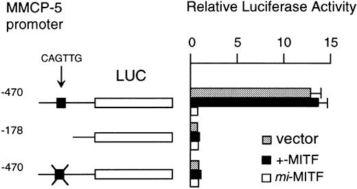
Effect of overexpression of mi-MITF on the MMCP-5 promoter. The luciferase gene under the control of the MMCP-5 promoter was transfected into FMA/3 cells overexpressing +- or mi-MITF. Because there is a possibility that the transcriptional activity in the FMA/3 cells infected with the retrovirus is different from that of the original FMA/3 cells, the FMA/3 cells infected with the retrovirus containing the vector alone were used as a control. When the reporter plasmid starting from nt −470 was transfected, the high luciferase activity was observed in the FMA/3 cells overexpressing the +-MITF (Fig 6). The value was comparable to the value observed in FMA/3 cells containing the retroviral vector alone (Fig 6). In contrast, when the same plasmid was transfected into FMA/3 cells overexpressing the mi-MITF, luciferase activity was hardly detectable (Fig 6). The deletion to nt −178 or the mutation at the CAGTTG motif to CTGTAG resulted in the abolishment of the luciferase activity in all three types of FMA/3 cells.
The effect of overexpression of +- or mi-MITF on the luciferase activity in FMA/3 cells. The luciferase gene under the control of the normal, deleted, or mutated MMCP-5 promoter was transfected to the FMA/3 cells overexpressing +- or mi-MITF or to the FMA/3 cells containing the retroviral vector (pM5Gneo) alone. The data represent the mean ± SE of three experiments. In some cases, the SE was too small to be shown by the bars.
The effect of overexpression of +- or mi-MITF on the luciferase activity in FMA/3 cells. The luciferase gene under the control of the normal, deleted, or mutated MMCP-5 promoter was transfected to the FMA/3 cells overexpressing +- or mi-MITF or to the FMA/3 cells containing the retroviral vector (pM5Gneo) alone. The data represent the mean ± SE of three experiments. In some cases, the SE was too small to be shown by the bars.
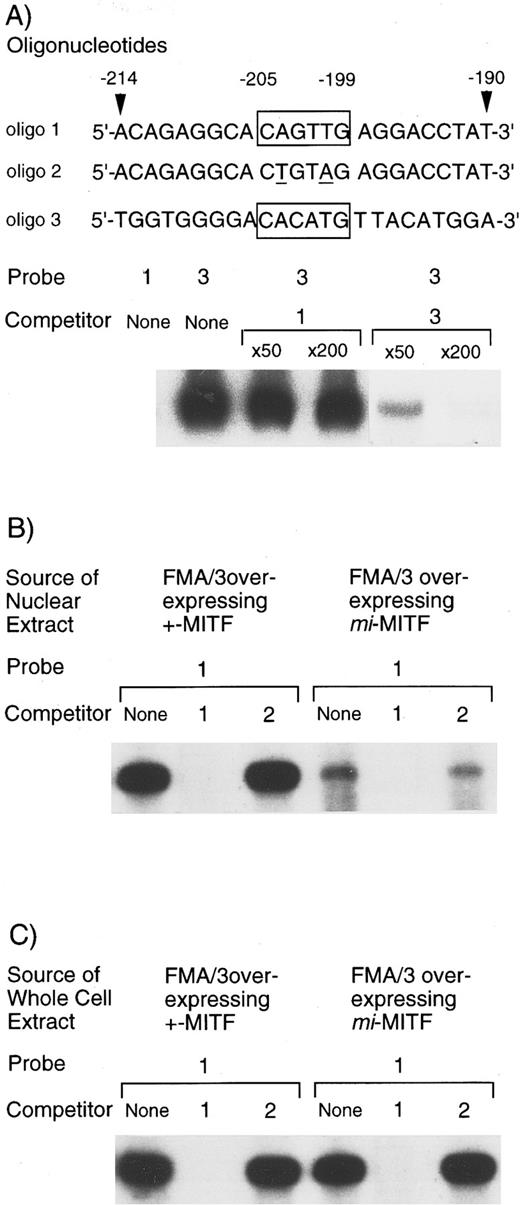
EGMSA with the CAGTTG motif as a probe. The transfection assay using the deleted or mutated reporter plasmids suggested a possibility that the CAGTTG motif mediated the transactivation ability of the +-MITF. First, we examined the binding of +-MITF to the oligonucleotide containing the CAGTTG motif. The purified GST-+-MITF fusion protein did not bind the oligonucleotide (Fig 7A). Moreover, the binding of GST-+-MITF fusion protein to the CACATG motif in the MMCP-6 promoter was not competed with the excess amount of the nonlabeled oligonucleotide containing the CAGTTG motif.
EGMSA using GST-+-MITF fusion protein or using the nuclear extract obtained from FMA/3 cells overexpressing +-MITF or mi-MITF. (A) Nonbinding of GST-+-MITF to the CAGTTG motif in the MMCP-5 promoter. The labeled 5′-ACAGAGGCACAGTTGAGGACCTAT oligonucleotide in the MMCP-5 promoter (oligo 1) or the labeled 5′-TGGTGGGGACACATGTTACATGGA oligonucleotide in the MMCP-6 promoter (oligo 3) was used as a probe (each hexameric motif is boxed in the figure). Competition for the binding of GST-+-MITF to the labeled oligo 3 was also examined. The excess amount of nonlabeled oligo 1 or oligo 3 was added. The binding was inhibited by nonlabeled oligo 3 but not by nonlabeled oligo 1. (B) EGMSA using nuclear extract obtained from FMA/3 cells overexpressing +-MITF or mi-MITF. Oligo 1 was used as a probe. The excess amount of nonlabeled oligo 1 or the nonlabeled oligonucleotide mutated at the CAGTTG motif (to CTGTAG, oligo 2) was added. The binding was inhibited by nonlabeled oligo 1 but not by nonlabeled oligo 2. (C) EGMSA using whole cell extract obtained from FMA/3 cells overexpressing +-MITF or mi-MITF. Oligo 1 was used as a probe. The excess amount of nonlabeled oligo 1 or oligo 2 was added.
EGMSA using GST-+-MITF fusion protein or using the nuclear extract obtained from FMA/3 cells overexpressing +-MITF or mi-MITF. (A) Nonbinding of GST-+-MITF to the CAGTTG motif in the MMCP-5 promoter. The labeled 5′-ACAGAGGCACAGTTGAGGACCTAT oligonucleotide in the MMCP-5 promoter (oligo 1) or the labeled 5′-TGGTGGGGACACATGTTACATGGA oligonucleotide in the MMCP-6 promoter (oligo 3) was used as a probe (each hexameric motif is boxed in the figure). Competition for the binding of GST-+-MITF to the labeled oligo 3 was also examined. The excess amount of nonlabeled oligo 1 or oligo 3 was added. The binding was inhibited by nonlabeled oligo 3 but not by nonlabeled oligo 1. (B) EGMSA using nuclear extract obtained from FMA/3 cells overexpressing +-MITF or mi-MITF. Oligo 1 was used as a probe. The excess amount of nonlabeled oligo 1 or the nonlabeled oligonucleotide mutated at the CAGTTG motif (to CTGTAG, oligo 2) was added. The binding was inhibited by nonlabeled oligo 1 but not by nonlabeled oligo 2. (C) EGMSA using whole cell extract obtained from FMA/3 cells overexpressing +-MITF or mi-MITF. Oligo 1 was used as a probe. The excess amount of nonlabeled oligo 1 or oligo 2 was added.
We then examined whether the nuclear extract from FMA/3 cells overexpressing +- or mi-MITF contained proteins that bound the oligonucleotide with the CAGTTG motif. The protein-DNA complex was made when the oligonucleotide containing the CAGTTG motif was incubated with the nuclear extract from FMA/3 cells overexpressing either +-MITF or mi-MITF. However, the amount of the protein-DNA complex was significantly greater in the nuclear extract from the FMA/3 cells overexpressing +-MITF than in the nuclear extract from the FMA/3 cells overexpressing mi-MITF (Fig 7B). The amount of the protein-DNA complex was reduced by the addition of the excess amount of the nonlabeled oligonucleotide containing the CAGTTG motif, whereas the amount of the complex was not reduced by the addition of the excess amount of the oligonucleotide whose CAGTTG motif was mutated to CTGTAG.
We then performed a similar experiment using the whole cell extract instead of the nuclear extract. The whole cell extract was obtained from FMA/3 cells overexpressing +- or mi-MITF. The amount of protein-DNA complex was compared. Despite the remarkable difference in the amount of nuclear protein-DNA complex between FMA/3 cells overexpressing +-MITF and FMA/3 cells overexpressing mi-MITF, the amount was comparable between the whole cell extract obtained from these two types of FMA/3 cells (Fig 7C). The binding was specific because the addition of the excess amount of nonlabeled oligonucleotide containing CAGTTG abolished the binding (Fig 7C).
We examined the possibility that the protein that bound the oligonucleotide with the CAGTTG motif might be MITF, USF1, USF2, MYC, or FOS using specific antibodies. Any antibodies tested did not interfere with the formation of the protein-DNA complex (data not shown).
DISCUSSION
The expression of MMCP-5 was deficient in mi/mi CMCs and tg/tg CMCs. The poor expression of MMCP-5 mRNA in mi/mi CMCs is reminiscent of the poor expression of MMCP-6.20 However, the character of MMCP-5 as the protease is different from that of MMCP-6; MMCP-5 is a chymase, whereas the MMCP-6 is a tryptase.22 The protease mRNA expression phenotype of +/+ CMCs is influenced by various growth factors.29,47,48 Interleukin-4 (IL-4) inhibits the SCF-induced expression of the MMCP-4 gene in +/+ CMCs and the IL-10–induced expression of MMCP-1 and MMCP-2 genes in +/+ CMCs.47 Although IL-4 and IL-3 do not influence the MMCP-5 expression,47,48 Eklund et al47 reported that SCF increased the amount of MMCP-5 mRNA in +/+ CMCs. This is inconsistent with the present result that the addition of SCF did not remarkably influence the MMCP-5 mRNA expression in +/+ CMCs. The discrepancy may be attributable to the different concentrations of SCF and different mouse strains used. Eklund et al47 used 200 ng/mL of rmSCF and CMCs derived from BALB/c strain, whereas we used 10 ng/mL of rmSCF and CMCs from C57BL/6 strain. Further studies are necessary to clarify the discrepancy. Even if SCF potentiates the MMCP-5 mRNA expression in +/+ CMCs, we consider that the phenomenon is more apparent in mi/mi CMCs than in +/+ CMCs. The overexpression of +-MITF but not of mi-MITF in mi/mi CMCs normalized the transcription of the MMCP-5 gene. This apparently indicated that the +-MITF played an important role for the expression of the MMCP-5 gene.
Xia et al49 reported that MMCP-2 is posttranscriptionally regulated by IL-10. They indicated that IL-10 induces expression of a trans-acting factor(s) that stabilizes the MMCP-2 mRNA or facilitates its processing. As a result, the amount of MMCP-2 mRNA is greater in the presence of IL-10 than in its absence. In the present study, we indicated that SCF increased the amount of MMCP-5 mRNA. There is a possibility that MMCP-5 may be posttranscriptionally regulated by SCF. In contrast to MMCP-5, the addition of SCF did not increase the amount of MMCP-6 mRNA. This suggested that the regulational mechanism of MMCP-5 expression was different from that of MMCP-6. Further analysis such as nuclear run-on assay will clarify the transcriptional or posttranscriptional regulation of MMCP-5 expression.
The transfection of the reporter plasmid starting from nt −470 of the MMCP-5 promoter into the FMA/3 cells resulted in the high luciferase activity. This also indicated that the +-MITF was strongly involved in the transactivation of the MMCP-5 gene, because FMA/3 cells expressed +-MITF. The +-MITF induces the expression of various mast cell-specific genes18-20 and melanocyte-specific genes.50-53 The reporter activity increases when the +-MITF cDNA was coexpressed with the reporter gene under the control of the promoter of the MMCP-6,19,20 c-kit,18 tyrosinase,50 or tyrosinase related protein 1 (TRP-1) gene.51,52 The stable transfection of the +-MITF cDNA converted NIH/3T3 fibroblasts to the cells with melanocyte characteristics that expressed the tyrosinase and TRP1 genes.53 However, little is known about the molecular mechanism of the transactivation by the +-MITF, except for the cases of the c-kit18 and the MMCP-6 genes.20 We previously reported that the MMCP-6 expression was transactivated by the binding of +-MITF to the CANNTG motifs in the promoter region.19 20 Because the MMCP-5 and MMCP-6 genes showed the similar expression pattern in mi/mi or tg/tg CMCs or in mi/mi CMCs overexpressing +-MITF, we speculated that the +-MITF might transactivate the MMCP-5 gene by the direct binding, as observed in the case of the MMCP-6 gene. Only one CANNTG motif was present in the 2212 bases of the MMCP-5 promoter region, ie, the CAGTTG motif between nt −204 and −199. The reporter plasmid whose CAGTTG motif was mutated to CTGTAG lacked the luciferase activity completely in FMA/3 cells. This indicated the significance of the CAGTTG motif for the expression of the MMCP-5 gene.
However, EGMSA showed that the GST-+-MITF fusion protein did not bind the CAGTTG motif between −204 and −199 of the MMCP-5 promoter. We examined whether proteins that bound the CAGTTG motif were present in the nuclear extract from the FMA/3 cells overexpressing +-MITF. At least a protein that bound the CAGTTG motif was detectable. The addition of the anti-MITF antibody did not influence the formation of the protein-DNA complex. This suggested that the protein that bound the CAGTTG motif in the nuclear extract did not form a complex with the +-MITF. Many examples of indirect interactions between transcription factors and the cis-elements have been reported.54-56 The Id proteins that lack the DNA binding domain bind bHLH-Zip transcription factors such as myoD and E2A and inhibit the binding of these bHLH-Zip proteins to DNA.54 The I-κB proteins form complexes with the active form of NF-κB proteins and sequester them in the cytoplasm, keeping them in the latent form.55 56 These transcription factors do not bind DNA itself, but they function by associating with other intermediate proteins. There is a possibility that the +-MITF may indirectly interact with the CAGTTG motif through the CAGTTG-binding protein detected in the nuclear extract.
The amount of the CAGTTG-binding protein was smaller in the nuclear extract of the FMA/3 cells overexpressing mi-MITF than in the nuclear extract of the FMA/3 cells overexpressing +-MITF (Fig 7B). This may be consistent with another result that the transactivation of the luciferase gene under the control of the MMCP-5 promoter was suppressed by the overexpression of mi-MITF in FMA/3 cells (Fig 6). The amount of the CAGTTG-binding protein in the nucleus may be regulated by the following two mechanisms. (1) The +-MITF may regulate the transcription of the gene encoding the CAGTTG-binding protein. (2) The +-MITF may regulate the nuclear transport of the CAGTTG-binding protein. The second mechanism is consistent with our previous finding that the mi-MITF inhibited the nuclear transport of +-MITF.21 In fact, the amount of the CAGTTG-binding protein was comparable between the whole cell extract from FMA/3 cells overexpressing +-MITF and that of FMA/3 cells overexpressing mi-MITF (Fig 7C). On the other hand, the amount of the CAGTTG-binding protein was apparently larger in the nuclear extract of FMA/3 cells overexpressing +-MITF than in the nuclear extract of FMA/3 cells overexpressing mi-MITF. Taken together, it was suggested that the CAGTTG-binding protein remained in the cytoplasm of FMA/3 cells overexpressing mi-MITF. The +-MITF appeared to be necessary for the nuclear transport of the CAGTTG-binding protein. In other words, the overexpression of the mi-MITF may inhibit the nuclear transport of the CAGTTG-binding protein. Recently Nechushtan et al57 reported that +-MITF was associated with USF2. We examined whether the anti-USF2 antibody bound CAGTTG-binding protein, but no binding was observed. The identification of the CAGTTG-binding protein might clarify the molecular mechanism of the transactivation of the MMCP-5 gene through the +-MITF. The present result at least indicates that the transactivation mechanism of the MMCP-5 gene is different from that of the MMCP-6 gene.
The addition of SCF to the culture medium significantly increased the proportion of mi/mi CMCs that expressed MMCP-5 mRNA. The expression of c-kit gene was decreased in mi/mi CMCs, but it was not completely abolished.18 SCF appeared to be able to stimulate mi/mi CMCs through the moderately expressed c-kit receptors. Eklund et al58 reported that CMCs derived from W/Wv mice, in which the function of c-kit receptor was severely impaired, expressed nearly normal levels of MMCP-5 mRNA. The MMCP-5 gene expression appeared to be regulated by at least two mechanisms: the c-kit-mediated signal transduction and the MITF-mediated one. When one of the two mechanisms functions normally, the MMCP-5 gene may be expressed. In mi/mi CMCs, the former was moderately impaired, and the latter was severely impaired. This may result in the abolishment of the MMCP-5 expression in mi/mi CMCs. The addition of SCF may make the former mechanism functional and increase the MMCP-5 expression in mi/mi CMCs.
In contrast to mi/mi CMCs, mi/mi skin mast cells expressed MMCP-5 mRNA. SCF synthesized by fibroblasts may induce the MMCP-5 expression in mi/mi skin mast cells. The different pattern of MMCP-5 expression between mi/mi CMCs and mi/mi skin mast cells may be attributable to the different concentration of SCF surrounding mast cells. The addition of SCF did not increase the MMCP-6 mRNA-expressing CMCs in mi/mi CMCs, and the skin mast cells in mi/mi mice did not express the MMCP-6 gene.11 These results indicated that SCF did not induce the MMCP-6 gene expression in mi/mi mast cells. The regulatory mechanisms of the MMCP-5 gene expression are apparently different from those of the MMCP-6 gene expression. The MMCP-5 gene is a good model to investigate the regulation of gene expression that is influenced by both transcription factors and growth factors.
ACKNOWLEDGMENT
The authors thank Dr S. Nomura (Osaka University) and Dr M. Yamamoto (Tsukuba University, Tsukuba, Japan) for valuable discussions, Dr H. Arnheiter for VGA9-tg/tg mice, Dr K. Nakajima for pSPLuc, and Kirin Brewery Co Ltd for rmSCF.
Supported by grants from the Ministry of Education, Science and Culture.
Address reprint requests to Yukihiko Kitamura, MD, Department of Pathology, Osaka University Medical School, Yamada-oka 2-2, Suita 565, Japan.

This feature is available to Subscribers Only
Sign In or Create an Account Close Modal