POLYMORPHONUCLEAR leukocytes were discovered by Paul Ehrlich,1 when fixation and staining techniques made it possible to identify the lobulated nucleus and the granules that have given name to these cells and allowed for their classification as eosinophils, basophils, and neutrophils. Neutrophilic granulation refers to staining by a mixture of basic and acid, ie, neutral, dyes,1 whereas the term specific granules was used by Paul Ehrlich to distinguish true granules from artifacts with a granular appearance.2 It was later realized that two types of granules could be distinguished in neutrophils on the basis of their affinity for dye: azurophil granules, which take up the basic dye azure A at the promyelocytic stage, due to their content of acid mucopolysaccharide,3and specific granules, which do not. A clear distinction between these two types of granules was established when the peroxidase staining method for electron microscopy was adopted to identify myeloperoxidase (MPO), which is present only in azurophil granules.4,5 This distinction was corroborated by the development of subcellular fractionation techniques for separation of the granules.6-9 It has become dogma that these two types of granules are fundamentally different. Specific granules have been characterized as secretory granules that play important roles in initiating the inflammatory response,10 whereas azurophil granules are often viewed as lysosomes that are particularly active in the digestion of phagocytosed material.11-14 

This simplistic view of granules has been challenged by results from subcellular fractionation experiments in which granule proteins were found in more than two peaks on density gradients.9,13,15,16 In addition, the existence of a tertiary granule type, identified by its late appearance during myeloid maturation, was indicated by electron microscopy.3 During the last 15 years, high-resolution subcellular fractionation techniques, alone or in combination with immune-electron microscopy and flow cytometry, have shown a bewildering heterogeneity of the neutrophil's granules17-22 and have furthermore identified an additional regulated exocytotic storage organelle, the secretory vesicle.23-26 

A novel aspect of the physiology of granules was unraveled by the discovery that these regulated storage organelles (granules and secretory vesicles) are not just simple bags of proteolytic or bactericidal proteins that are kept in store until liberated either to the outside of the cell or to the phagocytic vacuole, but are also important reservoirs of membrane proteins that become incorporated into the surface membrane of the neutrophils when these organelles fuse with the plasma membrane and exocytose their content.17 27 In this way, granules and secretory vesicles may fundamentally change the ability of the neutrophil to interact with its environment.

Realizing this, a number of important questions immediately appear. Why this heterogeneity? Does it provide the neutrophil with the ability to differentially exocytose (or, in case of membrane proteins, translocate) proteins stored in different granules, and if so, how is this differential exocytosis controlled? Or, is the reason for the granule heterogeneity simply that some proteins must be segregated to survive inside the neutrophil, or for the neutrophil to survive, much as two-component glue has to be stored in two separate tubes, although both will be used at the same time? Whether one or the other reason applies, both raise the question of how the neutrophil can target proteins into different granule subsets.

The purpose of this review is to present the heterogeneity of granules and discuss the functional importance of this and, furthermore, to address how the neutrophil controls the structure and mobilization of these different granules.

Granules start to form at the stage of neutrophil maturation marked by transition from myeloblast to promyelocyte.4,5 From here on, formation of granule proteins continues even up to the stage of segmented cells.28 

In general, granules are believed to be formed by aggregation of immature transport vesicles that bud off from the trans-Golgi network (TGN), in which sorting takes place between constitutively secreted proteins and proteins that are routed into the regulated secretory pathway, ie, go to granules.29-31 The original study by Bainton et al4,5 showed that such vesicles bud off from cis-Golgi to form storage granules at the promyelocyte stage, but from the trans-Golgi at the myelocyte stage, where specific granules are formed. This implies that the sorting apparatus (if existing) is localized in the cis-Golgi in promyelocytes and moves to the TGN in more mature cells. Unlikely as this may seem, it agrees with the finding that MPO, a major protein of azurophil granules, does not contain complex carbohydrate side chains,32,33 but it is contradicted by the finding that several other azurophil granule proteins, including elastase, cathepsin G, and proteinase 3, acquire complex oligosaccharide side chains.34,35 Refining of carbohydrate side chains from simple to complex is a feature of the intermediate- and trans-Golgi stacks.36 

Several regulatory steps will be required if granules are formed by fusion from smaller unit transport vesicles that bud off from the Golgi and undergo homotypic fusion until they achieve the size of a granule. It must be secured that such transport vesicles do not fuse with the plasma membrane, as do transport vesicles that mediate constitutive secretion. Furthermore, it must be secured that such transport vesicles do not fuse with already formed granules, because this would lead to mixing of granule proteins destined for different types of granules.

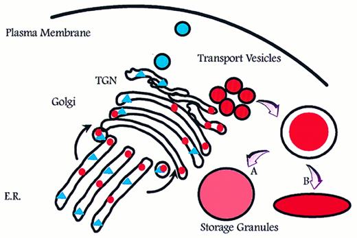
Aggregation of smaller unit vesicles to form mature granules creates a surface to volume problem because the surface area of the final granule is the sum of the surface area of the transport vesicles that fused to create the mature granule, and the volume of the mature granule is the sum of the volume of cargo carried by the transport vesicles (Fig 1). Azurophil granules are generally described as spherical or football shaped, whereas specific granules are known to adopt more irregular and elongated forms.5 This might be a reflection of volume adjustment in azurophil granules, which are known to proteolytically process a significant part of the proteins that are targeted to these.32,37,38 In this respect, azurophil granules resemble lysosomes.39 In contrast, no processing and therefore no increase in osmotic activity due to proteolysis has been observed in specific granules, with one possible exception.40 

Fig. 1.

Intracellular routing of newly synthesized proteins. Newly synthesized proteins are sorted into pathways for constitutive (triangles) and regulated (circles) exocytosis. If formation of storage granules occurs by homotypic aggregation of transport vesicles, a surface to volume problem will arise that can be solved only by either adjusting (expanding) the volume to fit the surface of a spherical particle (A) or by adjusting the shape so that the surface fits the volume (B). E.R., endoplasmic reticulum; TGN, trans-Golgi network.

Close modal

The question of whether granules of neutrophils are formed by fusion of unit vesicles has not been addressed, but patch-clamp capacitance studies that accurately quantitate the size of granules that are incorporated into the plasma membrane have been performed on horse eosinophils and rat basophils. These studies have shown that individual granules differ in size by the size of unit vesicles. This indicates that granules are formed by homotypic fusion of a finite but slightly varying number of smaller unit vesicles.41 42 

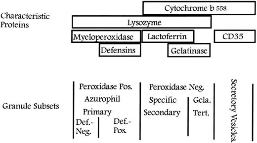
Granules may be classified on the basis of their size, morphology, or electron density or with reference to a given protein.5 The initial classification into two major types of granules was based on the content of MPO.4 However, the granules can be further subdivided on the basis of other intragranular proteins,21,43,44 as observed in Fig 2. It should be emphasized that, according to the targeting-by-timing hypothesis that will be discussed later, classification of granules is arbitrary, because granules form a continuum from azurophil granules to gelatinase granules, sharing some proteins, eg, lysozyme,45 whereas other proteins can be chosen to serve as specific markers of one particular subset, eg, MPO, lactoferrin, and gelatinase. A primary question is whether classification of granules is physiologically meaningful. A secondary question is whether it is of use in clinical hematology, eg, for classification of myeloproliferative or myelodysplastic disorders.

Fig. 2.

Classification of granules in neutrophils. Peroxidase-positive (azurophilic or primary) granules are characterized by the content of MPO and may be further divided based on their content of defensins into large, defensin-rich granules and the smaller defensin-poor granules. The peroxidase-negative granules may be divided into specific (secondary) granules and gelatinase (tertiary) granules on the basis of their relative content of lactoferrin and gelatinase. All granules contain lysozyme. Secretory vesicles share some of their membrane proteins with peroxidase-negative granules, whereas others are unique to secretory vesicles. Def., defensins; Gela., gelatinase; Tert., tertiary.

Close modal

It is well established that major differences exist between the different granule subsets regarding the extent to which these are mobilized both in vitro and in vivo.46-50 Gelatinase granules (identified by gelatinase) are mobilized more readily than specific granules (identified by lactoferrin),16,51,52 which again are exocytosed more readily than azurophil granules (identified by MPO). This hierarchy applies both when neutrophils isolated from peripheral blood are stimulated with various secretagogues and when exudate neutrophils collected in a skin window chamber are analyzed and further stimulated.48,50 It therefore makes sense from a functional point of view to classify the neutrophil granules into peroxidase-positive (or azurophilic or primary) granules, defined by their content of MPO, and to further subdivide the peroxidase-negative granules into specific (or secondary) granules, defined by their content of lactoferrin, and gelatinase (or tertiary) granules, defined by their high concentration of gelatinase20 21 (Fig 2). A number of proteins have been identified in these granules. Table 1 gives the content of the different types of granules. The localization of some of these proteins has been determined by electron microscopy, some by subcellular fractionation, and some by mobilization, assuming that proteins that are mobilized together also localize together.

Table 1.

Content of Human Neutrophil Granules and Secretory Vesicles

Azurophil GranulesSpecific GranulesGelatinase GranulesSecretory Vesicles
 
Membrane 
CD6359 289  
CD68290  
V-type H+-ATPase291  Membrane 
CD11b64,104 
CD15 antigens305  
CD66306  
CD67306  
Cytochrome b55817,52,105 
fMLP-R131,307 308  
Fibronectin-R309 
G-proteinα-subunit310 311  
Laminin-R309 
NB 1 antigen312  
19-kD protein313  
155-kD protein314  
Rap1, Rap2315,316 
SCAMP170  
Thrombospondin-R317 
TNF-R318  
Urokinase-type plasminogen activator-R319 
VAMP-2170  
Vitronectin-R309 Membrane 
CD11b64,109,327-330 
Cytochrome b55820 
Diacylglycerol-deacylating enzyme331  
fMLP-R109 131  
SCAMP170  
Urokinase-type plasminogen activator-R319 
VAMP-2170  
V-type H+-ATPase291  Membrane 
Alkaline phosphatase23-26 66  
CR165 
Cytochrome b55862,333 
CD11b62,64 
CD14334  
CD1663 334* 
fMLP-R131  
SCAMP170  
Urokinase-type plasminogen activator-R319 
V-type H+-ATPase291  
VAMP-2170  
CD10, CD13, CD45335* 
C1q-receptor336* 
DAF60* 
Matrix 
Acid β-glycerophosphatase11  
Acid mucopolysaccharide292  
α1-Antitrypsin293  
α-Mannosidase11  
Azurocidin/CAP37/heparin binding protein294-296  
Bactericidal permeability increasing protein297  
β-Glycerophosphatase7  
β-Glucuronidase7 298  
Cathepsins11  
Defensins43 299  
Elastase300  
Lysozyme7,8 298  
Myeloperoxidase301  
N-Acetyl-β-glucosaminidase7  
 
Proteinase-3302 
Sialidase303  
Ubiquitin-protein304  Matrix 
β2-Microglobulin320  
Collagenase321  
Gelatinase21 322  
hCAP-18117  
Histaminase323  
Heparanase324  
Lactoferrin301  
Lysozyme7,8 298  
NGAL126  
Urokinase-type plasminogen activator319 325  
Sialidase303  
SGP2840  
Vitamin B12-binding protein326  Matrix 
Acetyltransferase332  
β2-Microglobulin20  
Gelatinase16,21 107  
Lysozyme45  Matrix 
Plasma proteins24 25 (including tetranectin) 
*

This localization is based on kinetics of upregulation in response to stimulation with inflammatory mediators, but has not yet been demonstrated by subcellular localization by immunocytochemistry.

When considering exocytosis of granules, it should be kept in mind that exocytosis of granule content is a consequence of fusion of granule membrane with the plasma membrane and incorporation of granule membrane into the plasma membrane. In many cells, this membrane is rapidly retrieved for re-use (adipocytes, neurons),53-57 but in neutrophils, the membrane of mobilized granules largely remains part of the plasma membrane.58-61 In this way, membrane proteins located to the membrane of granules translocate to the surface membrane and furnish the cell with new receptors and other functional proteins.17 

The observation that the β2 -integrin Mac-1 (αmβ2 , CD11b/CD18) became incorporated into the plasma membrane without corresponding exocytosis of granule content led to the discovery of the most rapidly mobilizable intracellular structure in neutrophils, the secretory vesicle.23,24,26,58 Secretory vesicles are important because of their membrane, which is particularly rich in receptors.62-65 The only known intravesicular content of secretory vesicles is plasma. Albumin therefore currently serves as marker of these vesicles.25 This indicates that secretory vesicles are endocytic in origin. It should be noted that secretory vesicles are not part of a constitutive endocytosis/exocytosis organelle. Once mobilized, secretory vesicles are not reformed, either in vitro or in vivo.66-70 

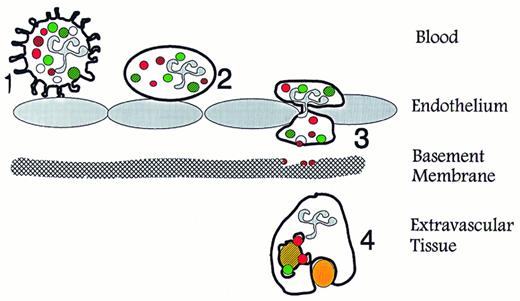
When combining the known content of the different granules and secretory vesicles with their order of mobilization, it becomes clear that these serve different and significant functions (Fig 3). Our view on secretory vesicles is that these are mobilized when the neutrophil establishes the primary rolling contact with activated endothelium, which is mediated primarily by selectins and their ligands, eg, PSGL-1.71-74 The mobilization of secretory vesicles may be mediated by signaling through the selectins75,76 or by inflammatory mediators liberated from the endothelium.77,78 This view is in agreement with the observation that secretory vesicles have been completely mobilized in neutrophils that are collected in plasma in a skin window chamber.50 Thus, mobilization of secretory vesicles transforms the neutrophil to a β2 -integrin presenting cell79,80 and in this way changes the cell from a generally passive cell, well suited for circulation, to a highly responsive cell, primed for migration into tissues.81-93 

Fig. 3.

Specific functions of neutrophil granules and secretory vesicles in relation to diapedesis and phagocytosis. (1) Primary contact between endothelium and circulating neutrophils is established via selectins and their ligands, which causes the neutrophils to roll along the activated endothelium. This contact may transduce signals in the neutrophils that mobilize secretory vesicles. (2) Integration of the membrane of secretory vesicles and its associated CD11b/CD18 enhances the potential of the neutrophil for firm adhesion to endothelium. (3) Exocytosis of gelatinase from gelatinase granules may help degradation of type IV collagen in basement membranes. (4) Mobilization of specific granules to the surface membrane may enhances the phagocytic potential of the neutrophils by providing CR3 (CD11b/CD18). Fusion of azurophil and specific granules with the phagosome creates conditions for oxygen-dependent and -independent bactericidal activity. (Modified and reprinted with permission.200 ).

Close modal
Fig. 4.

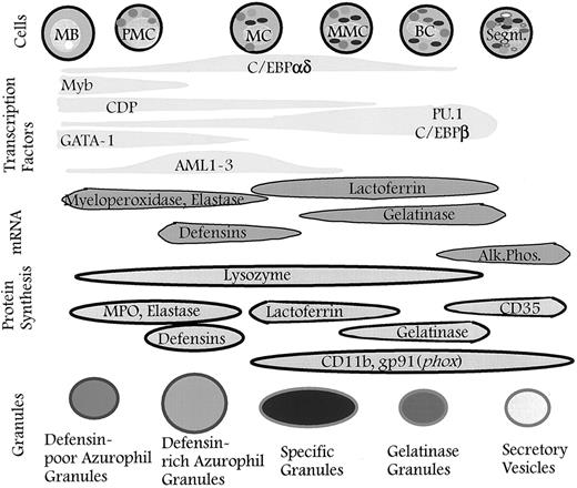
Granules defined by timing of biosynthesis of their characteristic proteins. The granules formed at any given stage of maturation of neutrophil precursors will be composed of the granule proteins synthesized at that time. The different subsets of granules identified are the result of differences in the biosynthetic windows of the various granule proteins during maturation and not the result of specific sorting between individual granule subsets. When the formation of granules ceases, secretory vesicles will form337 (this point has not yet been proven). The control of biosynthesis is exerted by transcription factors that control the expression of genes for the various granule proteins. It cannot be ruled out that posttranscriptional control occurs so that biosynthesis of proteins is not a precise reflection of the corresponding mRNA levels. MB, myeloblast; PMC, promyelocyte; MC, myelocyte; MMC, metamyelocyte; BC, band cell; Segm., segmented cell.

Close modal

Type IV collagen, a major constituent of basement membranes, and type V collagen of interstitial tissues, are substrates of gelatinase.94-96 It is likely that exocytosis of gelatinase from gelatinase granules is essential for migration of neutrophils through basement membranes.97,98 Although evidence for disruption of the basement membrane has not been obtained by in vivo experiment,99 in vitro experiments support the need for gelatinase activity in migration of neutrophils through Matrigel and amnion membranes,100 but the appropriate gene knock-out experiment has not been performed to address this. A human gene knock-out is provided by the rare condition, specific granule deficiency,101,102 although the functional defects in this condition, which include inability of neutrophils to infiltrate into tissues, may be caused by the lack of expression of other granule proteins than gelatinase.103 

Although subject to much previous debate,104-109 the membrane of specific granules has not definitively been shown to be different from that of gelatinase granules, except by quantitative measures (total area of specific granules is larger than the total area of gelatinase granules).20,21 The membranes of specific-and gelatinase granules relate to phagocytosis and intracellular killing, because these are the main stores of Mac-1 (αmβ2 , CD11b/CD18) and of the flavocytochrome b558 (gp91phox/p22phox ), an essential component of the NADPH oxidase.20,104 105 

With respect to intragranular proteins, specific granules are dominated by lactoferrin,8,110 the function of which is still unknown. Of the constituents listed in Table 1, the zymogen collagenase should be mentioned because this, like gelatinase of gelatinase granules, most likely is important for the ability of the neutrophil to make its way through tissues.111-113 Azurophil granules are characterized by their content of hydrolytic and bactericidal proteins such as elastase, bactericidal permeability-increasing protein, defensins, and MPO.5,7,13 Azurophil granules have been further subdivided on the basis of several proteins.114 Defensins are the dominating protein in a major subset of azurophil granules.43 Three novel proteins of specific granules that may turn out to contribute significantly to the function of neutrophils have recently been identified. These are NGAL,115,116 hCAP-18,117-119 and SGP28.40 

NGAL.NGAL is a member of the lipocalin family of proteins, which were recently reviewed by Flower.120 Lipocalins, the archetype of which is retinol-binding protein, are 25- to 30-kD proteins that have a three-dimensional structure121 that has been likened to a coffee filter.122 Lipocalins are known to bind small lipophilic substances (eg, retinol). NGAL, so named because of its ability to complex with gelatinase (neutrophil gelatinase associated lipocalin),115,116 is the human homologue of the mouse protein 24p3 that was identified in SV40 transformed kidney cells.123,124 A closely homologous protein was recently identified as the protein whose expression is increased the most in neu-transformed rat mammary carcinomas. This protein was termed neu-related lipocalin.125 NGAL is normally found only in the specific granules of human neutrophils,126,127 but its production is significantly induced in colon epithelial cells during inflammation (Crohn's disease, ulcerative colitis, appendicitis, and diverticulitis), as evidenced both by immune-histochemistry and by in situ hybridization.128 The synthesis of NGAL has been shown to be induced in peripheral blood neutrophils treated with granulocyte-macrophage colony-stimulating factor (GM-CSF ).129 Two studies on the subcellular localization of the fMLP receptor by photo-affinity labeling identified NGAL as a 25-kD fMLP-binding protein of specific granules.130 131 Our hypothesis is that NGAL may participate in regulating the inflammatory response by binding small lipophilic inflammatory mediators such as fMLP, platelet activating factor (PAF ), leukotriene B4 (LTB4 ), and lipopolysaccharide (LPS).

hCAP-18.hCAP-18, also named FALL-39, was recently identified independently by three groups.117-119 It is the only human member of the cathelicidin family of bactericidal peptides132 (see Zanetti et al133 and Levy134 for recent reviews). These peptides were first discovered in neutrophils of ruminants.135 Cathelicidins are proteins that share a high level of homology in the N-terminal 14-kD part of the protein that is homologous to cathelin, a protein purified from porcine neutrophils,136 whereas the C-terminal parts of cathelicidins are highly diverse, ranging from 12135 to 100 amino acids.137 The C-termini are highly cationic. Some are extremely rich in proline and arginine,135,138,139 whereas others, including the C-terminal 39 aminoacids of hCAP-18, form an amphipatic α-helix.119 Indeed, we discovered hCAP-18 as a protein highly enriched in the Triton-phase during Triton X-114 phase separation of proteins from specific granules.117 

Cathelicidins are stored as intact proteins in specific granules (or large granules of ruminant neutrophils), but the C-termini may be cleaved off when the proteins are exposed to elastase140,141 during concomitant degranulation of specific and azurophil granules, and the bactericidal activity of the C-terminal peptides is unleashed.118,141 The C-terminal peptides are not only microbicidal, but may also be toxic to eucaryotic cells.142 

SGP28.SGP28 is a glycoprotein of specific granules with a molecular weight of 28 kD, hence its name.40 It is homologous to two other proteins: Tpx-1, which is a human testis specific protein,143 and sperm-coating glycoproteins of rat epididymis.144 These proteins constitute a family of cystein-rich proteins termed CRISPs (cysteine-rich secretory proteins).145 SGP28 was independently cloned from a salivary gland cDNA library and termed CRISP-3 and was found expressed (Northern blotting) also in pancreas and epithelial cells of the large bowel.145 Part of SGP28 shows high amino acid sequence homology to plant proteins known as pathogenesis-related proteins. There are proteins that are believed to be important for resistance to infections caused by both virus, bacteria, and fungi.146 

The generation of reactive oxygen metabolites that is essential for proper microbicidal activity of neutrophils is dependent on components of both peroxidase-negative granules (which harbor the flavocytochrome b558 , an essential component of the NADPH oxidase) and of azurophil granules (which contain MPO that transforms the relatively innocuous product of the NADPH oxidase, H2O2 , to hypochlorous acid147 ). As mentioned above, proteases from azurophil granules may activate the cathelicidin present in specific granules by proteolytically removing the inhibitory (and protective) N-terminal cathelin-like part of the protein. Likewise, gelatinase and collagenases may be converted from their proform to active proteases by elastase liberated from azurophil granules97 or by reactive oxygen metabolites generated by the NADPH oxidase.112 148 

As alluded to above, secretory vesicles and the different kinds of granules are mobilized in a hierarchy, which seems adjusted to the different roles these organelles play during the journey of the neutrophil from the circulation to the inflammatory focus. Because all granules and secretory vesicles appear randomly distributed throughout the cytosol in the circulating cell21,25,62,63 and localized towards the lamellipod in the (fMLP-) activated cell,70 this hierarchy must rely on mechanisms that discriminate between the different granule subsets. However, hierarchic mobilization excludes independent mobilization, ie, mobilization of specific granules without concomitant and more extensive mobilization of gelatinase granules. This indicates that differences in the mobilization of the granule subsets are due to quantitative differences and not to qualitative differences in the machinery that controls exocytosis of the individual granule subsets.

Elevation of intracellular Ca2+ is known to elicit exocytosis of storage granules in a variety of cells,47,149-152 but the molecular mechanism by which this occurs is unknown. Differences in sensitivity toward intracellular Ca2+ as a signal to elicit mobilization were observed among the different granule subsets49,51,153 and secretory vesicles, in correspondence with the hierarchy of their mobilization. It has been shown that a few cytosolic proteins translocate to granules in a Ca2+-dependent way (annexins I, II, III, IV, VI, and XI).154-160 No clear-cut qualitative differences were observed between binding of different annexins to the different granule subsets, although annexin III was suggested to bind preferentially to specific granules.156 Furthermore, the Ca2+ concentrations that elicited binding of annexins were found to differ among the different granule subsets.155 A non-annexin Ca2+-binding protein, later identified as grancalcin,161 was found to be translocated selectively to secretory vesicles.154 However, the role, if any, of these proteins in regulation of exocytosis is unclear.

Recently, the studies of intracellular transport (from ER to Golgi and within Golgi) have been extended to exocytosis, and a unifying hypothesis called the SNAP/SNARE-hypothesis has been forwarded according to which specific targeting of granules and vesicles is provided by specific interaction of v-SNAREs (present on the membrane of donor organelles) with their cognate t-SNAREs (present on the target membrane). According to this hypothesis, fusion is mediated by the combined action of the cytosolic factors NSF (N-ethyl maleimide sensitive factor) and the attachment proteins for NSF, the SNAPs (soluble NSF-attachment proteins), in further combination with Ca2+, synaptotagmin, and other membrane proteins.30,31,162,163 The experimental evidence for the relevance of this mechanism to control exocytosis is derived mainly from studies on neuronal tissue.164-166 One of the strongest indications to support this hypothesis is the observation that the neurotoxins botulinus and tetanus toxins specifically cleave SNARE proteins.167-169 

In neutrophils, the t-SNARE protein, syntaxin-4, is found exclusively in the plasma membrane, whereas the cognate v-SNARE, VAMP-2, is present in the membrane of the mobilizable granules and secretory vesicles, with the highest density on the membrane of secretory vesicles and gelatinase granules followed by specific granules. VAMP-2 could not be detected on azurophil granules.170 This could indicate that exocytosis of the neutrophil granules and of secretory vesicles is stochastic, ie, that the likelihood that any given granule will be exocytosed, is determined entirely by the density of fusion-proteins in the membrane of that granule (as recently observed in sea urchin eggs171 ) and that no qualitative differences exist among fusion proteins associated with the different types of granules.

The signal transduction pathways that lead to granule exocytosis are not completely known. Involvement of G-proteins is indicated by the ability of the nonhydrolyzable GTP-γ-S to induce exocytosis in permeabilized neutrophils172,173 and by localization of distinct G-proteins to granules.174 Patch clamp capacitance measurements provide a powerful tool to analyze granule exocytosis. Using this technique, incorporation of granule membrane into the plasma membrane is quantitated as increase in the electrical capacitance of the plasma membrane.175 This technique has documented the role of GTP and Ca2+ in control of granule exocytosis and holds promise for further delineation of the molecular mechanism of exocytosis.176 

The differences among different granule subsets in their ability to become exocytosed have been shown to extent also to orientation of exocytosis. Whereas peroxidase-negative granule are often characterized as secretory granules that mainly mediate extracellular release of their proteins,177,178 peroxidase-positive granules have been characterized as specialized lysosomal structures4,11 that do not participate significantly in extracellular release, but form a digestive organelle for phagocytosed particles.178 179 

The notion that azurophil granules are specialized lysosomes has recently been challenged by the observation that the membrane of azurophil granules is devoid of the characteristic lysosomal membrane proteins (LAMPs),180,181 which instead are found in multilamellar and multivesicular bodies in the neutrophil, probably defining these organelles as the true lysosomal structures of neutrophils.181 182 

The mechanism for controlling the protein profile of the individual granule subsets, including the proteins that determine their later exocytosis, must be highly efficient to account for the differences in granule composition and for the hierarchy in exocytosis presented above. Two essentially unrelated steps are involved in targeting of proteins to granules. The first step is sorting between transport vesicles that mediate constitutive exocytosis of secretory proteins and insertion of membrane proteins into the plasma membrane and vesicles that will form storage granules capable of undergoing regulated exocytosis. The second step is sorting between different subsets of storage granules.

Sorting between regulated and constitutive exocytotic pathways.Very little is known with respect to mechanisms that sort proteins into the constitutive or the regulated exocytotic pathways in general, let alone in myeloid cells. Sorting of insulin and neuropeptides is (partly) controlled by pH and Ca2+-mediated aggregation,183-185 without involvement of carrier proteins or membrane attachments.186 The well-known targeting of mannose 6-phosphate containing glycoproteins to lysosomes via the cation-dependent and cation-independent mannose 6-phosphate receptors187 has not been shown to be important for targeting of proteins to granules in neutrophils. Although MPO is decorated with mannose 6-phosphate that is recognized by the cation-independent mannose 6-phosphate receptor,33,181 this is not important for sorting to azurophil granules.33,188 In addition, azurophil granules contain proteins that do not have the mannose 6-phosphate label (eg, lysozyme189 and defensins190 ). Recently, MPO was found associated with calreticulin as a chaperone,191 but the significance of this for sorting is unknown. No common primary protein structure has been identified in proteins that are retained versus those that are constitutively secreted. N-terminal hydrophobic domains have been suggested as a sorting signal in some neuropeptides.192 

An essential question is whether retention signals are common for all cells, ie, is a protein that in neutrophils is retained in granules also retained if transfected to other cell types? If so, is this dependent on whether the cells have the capacity for formation of granules? This has been studied with defensins, the constituents of azurophil granules, as probe. It has been observed that defensins can localize to granules in nonmyeloid cells, indicating that sorting to storage granules occurs by mechanisms common to all cells.193 This is also supported by transfection of other azurophil granule proteins to other myeloid cells.194,195 Yet, to prove the point, it is necessary to show active sorting between two proteins synthesized in the same cell, but handled differently by the sorting machinery. Because granules are so abundant in granulocytes and appear to be formed rapidly, at least as evaluated in eosinophils,196 there may be little need for a sorting mechanism if the bulk of cargo will go to granules anyway. This question must be addressed by examining whether all proteins that are routed through the Golgi go to granules with the same efficiency or whether some are more efficiently retained than others.

No studies have been presented to address the route of transport of membrane proteins that eventually localize to granules in granulocytes. In neuroendocrine cells, the targeting of peptidylglycyl α-amidating monooxygenase seems to be mediated by two targeting signals: a carboxyterminal cytoplasmic domain that retains the protein in the TGN (as opposed to being routed to the plasma membrane) and a lumenal domain that is essential for routing to secretory granules.197 P-selectin, as opposed to E-selectin, is targeted to its intracellular localization in endothelial cells by specific signals present in the cytosolic carboxy-terminal tail.198 199 Such have not been reported regarding the localization of flavocytochrome b558 and CD11b/CD18, proteins that are localized to the membrane of peroxidase-negative granules. Thus, it has not been addressed whether there is need for specialized targeting or whether sufficient amounts of the membrane proteins going through the Golgi will go passively to the granules at the time of their formation.

Sorting between individual granule subsets.In view of the considerable heterogeneity of neutrophil granules, sorting of proteins into different granules would require a very complex sorting mechanism if it was dependent on sorting information present in the individual proteins. We have forwarded the hypothesis that there is no specific sorting of proteins to individual granule subsets and that all granule proteins that are synthesized at the same time will localize to the same granules.28,200,201 This implies that the differences in protein content that define the different subsets of granules result from differences in the biosynthetic window of the various granule proteins in relation to maturation, as depicted in Fig 4. This extends the original observation of Dorothy F. Bainton that peroxidase-positive granules are formed at the promyelocyte stage, whereas peroxidase-negative granules are formed at the myelocyte stage.5 We were able to show that the differences among peroxidase-negative granules with regard to their content of gelatinase and lactoferrin could be explained by differences in the time of biosynthesis of these proteins, ie, lactoferrin synthesis occurred in cells at the myelocyte/metamyelocyte stage, whereas maximal gelatinase synthesis occurred at the metamyelocyte/band cell stage.28 

Although the available information regarding the localization of proteins and their time of biosynthesis is in agreement with this hypothesis,37,202-204 formal proof that no targeting is required for sorting into individual granule subsets was achieved by our analysis of the granule protein NGAL. NGAL is synthesized at the same stage as lactoferrin in normal neutrophil precursors28 and colocalizes with lactoferrin in specific granules.126 When NGAL was transfected to HL-60 cells under control of a constitutively active cytomegalovirus promoter, NGAL became synthesized at the same time as the endogenous granule protein MPO. NGAL was efficiently retained in granules of these HL-60 cells and colocalized with MPO in azurophil granules.201 However, NGAL, so targeted to azurophil granules by changing its timing of expression, was slowly degraded in the proteolytic milieu of the azurophil granules or their precursors.201 This illustrates that, in addition to the need for sequential mobilization of different granule proteins, the need for segregating proteins that cannot coexist may be another reason for the development of neutrophil granule heterogeneity.

The degradation of NGAL in azurophil granules or their precursors is in line with a number of observations that point to the azurophil granule as the place of terminal processing of many of its proteins. A variety of proteins localized to azurophil granules (MPO,32 defensins,37 cathepsin G, and elastase38 ) are proteolytically modified to their mature form after exit from the TGN. This is supported by the observation that defensins and MPO are not proteolytically modified when targeted to storage granules of cells of nonmyeloid origin.193,205 Signals present in the N-terminus of defensins have been shown to be important for correct targeting to granules.206 This most likely illustrates that the N-terminal pro-piece neutralizes the highly cationic C-terminus190 and prevents the protein from becoming stuck to membranes en route through the ER and Golgi and underscores the importance of final processing occurring in azurophil granules and not before. The fact that pro-segments are described for a variety of azurophil granule proteins, but not for granule proteins localized to peroxidase-negative granules, may be a matter of semantics. A variety of the specific granule proteins are only active after N-terminal trimming of a pro-piece has occurred (cathelicidin, gelatinase, and collagenase, as previously discussed), but this only takes place after exposure to azurophil granule proteins or to external proteases.97,113,140,141,207 Thus, it is possible that pro-segments play a role for correct transport through the biosynthetic machinery, but maybe not for specific sorting, as was recently suggested.208 As mentioned in the legend and text to Fig 1, the proteolytic processing in granules may even affect the shape of the granules by generating osmotically active substances.

HL-60 cells share with other human promyelocytic cell lines, eg, NB4 cells, the inability to express endogenous proteins normally localized to specific granules, despite the fact that the gene structure, including the 5′-untranslated regions assumed to control transcription of these genes, are intact, where investigated.209-211 Yet, these cells retain the ability to express proteins normally localized in the membrane of specific granules when driven into maturation by retinoic acid or dimethyl sulfoxide (DMSO).204,212 We therefore investigated the fate of the transfected specific granule protein NGAL in HL-60 cells when these were forced to maturation by retinoic acid and DMSO and had stopped the synthesis of endogenous azurophil granule proteins.213 In these cells, newly synthesized NGAL was not retained, but was routed to the extracellular medium, and the endogenous specific granule membrane protein, the flavocytochrome b558 was localized to the plasma membrane and not to granules. This is in contrast to the undifferentiated transfected cells, in which NGAL was localized to azurophil granules, as previously discussed. This illustrates that a shift of routing from the regulated secretory pathway to the constitutive secretory pathway takes place, when HL-60 cells mature. This indicates that the ability to maintain a regulated secretory pathway is dependent on continued synthesis of proteins that are needed for formation of regulated storage granules. This is consistent with the observation that formation of transport vesicles that mediate constitutive secretion continues after cycloheximide treatment, whereas formation of transport vesicles that mediate regulated secretion is blocked.214 It has recently been observed that transfection of fibroblasts with synaptotagmin is able to transform constitutive secretion to regulated secretion.215 It appears, therefore, that the difference between regulated and constitutive secretion may rely on a few proteins and is not due to fundamental differences in the budding and targeting mechanisms of the TGN. The proteins that are needed for formation of regulated storage granules have not been identified in myeloid cells.

The rare condition termed specific granule deficiency is characterized by defects in expression of defensins and of matrix proteins of specific granules, but preservation of the ability to form some membrane proteins that are normally localized to specific granules.103 These membrane proteins colocalize with the plasma membrane in specific granule deficient neutrophils216 and, in this respect, these cells resemble differentiated HL-60 cells.

If, as we believe, targeting of proteins to granules is entirely dependent on the time of biosynthesis, then control mechanisms must exist to secure that vesicles, budding off the TGN and carrying newly synthesized proteins, do not fuse with already formed granules. One such mechanism could be that granules bud off from Golgi at their final size with no further homotypic aggregation, although the observations that have been made on the size of granules do not support this, as previously mentioned. On the other hand, all cells that contain regulated storage granules must have a mechanism to control the size of the granules, otherwise the cells would continue to put all proteins into an ever-growing granule. The targeting-by-timing hypothesis therefore does not invoke further control mechanisms than are already needed in all cells that form granules. An indication that factors exist that limit the size of storage vesicles is given by the observation that such factors seem to be absent from cells of the promyelocytic mouse cell line 32D cl3, which accumulate granule proteins into large intracellular sacs,206 and by the large granules of neutrophils, eosinophils, and other cells from patients with the Chédiak-Higashi syndrome,22,217,218 now shown to be defective in Lyst protein.219 

The control of granule protein biosynthesis can be on either the transcriptional or the translational level. Whereas the transcriptional control is exerted by transcription factors, translational control is more elusive.220 221 Although growth factors may exert translational control, translational control has not been shown to be relevant for expression of myeloid granule proteins.

The stage of maturation of neutrophil precursors at which biosynthesis of granule proteins starts is in agreement with the start of transcription of the relevant genes in the cases in which this has been investigated (MPO,202,211,222-224 defensins,211,224-228 lactoferrin,211,224,229,230 vitamin B12 -binding protein,211,224 CD11b,204 and alkaline phosphatase202 ), although no distinct temporal difference between gelatinase and lactoferrin mRNA expression was observed during maturation in a mouse myeloid cell.231 It is not quite clear whether there is a complete correlation between stop of protein biosynthesis and downregulation of mRNA. This indicates that start of protein synthesis is controlled by transcription, whereas the termination of protein synthesis, at least for some granule proteins, may be under translational control.

Of the largely myeloid-specific granule proteins listed in Table 1, the following have been cloned at the genomic level and their 5′-upstream or flanking sequences have been characterized: MPO,232 elastase,233,234 cathepsin G,233,235,236 proteinase 3,237 lysozyme,238 defensin,239 lactoferrin,210 gp91phox,240-243  p22phox,244 CD11b,245-247 CD18,248-250 vitamin B12 -binding protein,251 hCAP-18/FALL-39,132 NGAL (Cowland and Borregaard, EMBL database accession no. X99133), gelatinase,252,253 and alkaline phosphatase.254 Putative binding sites for a variety of transcription factors have been identified in these promoter regions, but only a few promoter regions have been characterized by functional analysis (expression and gel shift analysis, and experiments of nature [CGD states241,255]). These include MPO,256,257 cathepsin G,236 lysozyme,238 elastase,234,258 CD11b,246,247,259,260 CD18,248-250 gp91242,243,261  and gelatinase.253 

GATA-1.The transcription factor GATA-1 is upregulated in the very early stages of commitment of the hematopoietic progenitor cells as a result of induction of cell division.262 This results in a gradual increase in the concentration of GATA-1 over a period of 1 to 2 days,263 followed by an abrupt downregulation and the disappearance of GATA-1 within 4 to 5 days from cells committed to the myelomonocytic lineage.262 A GATA-1 site capable of binding the GATA-1 factor is found in the genes for lactoferrin and for the Mac-1 subunit CD11b, but it has not been shown that these sites are of any importance for the control of expression of these genes.210 247 

AML (PEBP2/CBP).The AML (PEBP2/CBP) transcription factors are a family of heterodimeric proteins consisting of a common β-subunit and a specific DNA-binding α-subunit, of which three different types are known in humans.264 This is further varied by different splice variants of the individual α-unit transcripts.264,265 A functionally important PEBP2/CBP site has been identified in the promoters of the human GM-CSF266 and in the mouse MPO and elastase genes.267,268 PEBP2/CBP is upregulated in the murine 32D cl3 cells upon induction of differentiation (days 1 to 3) and downregulated upon exit from the promyelocytic stage (days 5 to 6).267 Besides the regulatory effects of upregulation and downregulation of the AML (PEBP2/CBP) factors and the need for co-operativity with other transcription factors for optimal function, a positive and negative regulatory effect of two differently spliced AML-1 transcripts has been observed on differentiation of murine 32D cl3 cells.265 

c-Myb.c-Myb is found to be highly expressed in immature hematopoietic cells and completely absent from mature granulocytes.269-271 Overexpression of c-myb causes continued proliferation of progenitor cells and blocks differentiation of the cells.272 In humans, only genes expressed at early stages of granulocyte differentiation, such as elastase213,267 and MPO,268 have been shown to carry a functional important Myb site. Potential c-Myb sites have also been identified in the early expressed proteinase-3 and azurocidin genes. Co-operativity with C/EBP has been shown for the min-1 gene273,274 and with both C/EBP and PU.1 for the elastase gene.213 

PU.1.PU.1 is specific for the hematopoietic system and highly expressed in B-lymphocytic, granulocytic, and monocytic cells.275,276 PU.1 is present at all stages of granulocyte differentiation.276,277 Its expression increases gradually up to the myelocytic stage of differentiation and then remains constant.276,277 This is reflected by the observation that many genes contain a functionally important PU.1 site, both some with an early onset of transcription (elastase,234 PU.1,278 GM-CSF receptor,279 and G-CSF receptor280 ) and some with a later onset of transcription during granulocytic differentiation (CD11b,247,281 CD18,249,282 and FcγRI283,284). Based on its pattern of expression, it can be concluded that PU.1 is likely to confer hematopoietic specificity to gene expression, but by itself is unable to exert a granulocytic- or stage-specific expression of the target genes. The latter has in some instances been shown to be achieved by co-operativety with other lineage- and/or stage-restricted transcription factors, as in the case of the elastase213 and GM-CSF receptor promoters.279 

C/EBPα.The C/EBP family of transcription factors contains at least 6 members, of which three (C/EBPα, -β, and -δ) are restricted to the myeloid cells within the hematopoietic lineages.285 C/EBPα and C/EBPδ expression is intense in immature myeloid cells and increases until the onset of the promyelocytic stage of differentiation and then declines rapidly, whereas the expression of C/EBPβ is low in immature cells, but increases steadily during the course of granulocytic maturation to reach a maximum in the mature granulocytic cell.285 A number of genes have been shown to carry a functionally important C/EBP site such as the elastase,213min-1,273 274  G-CSF receptor,286 and GM-CSF receptor genes.279 Co-operativity with c-Myb has been shown for the elastase,213 GM-CSF,279 and min-1 promoters.273,274 For the elastase promoter, additional co-operativity has also been observed with PU.1.213 

CCAAT displacement protein (CDP).CDP is a repressor of the ubiquitous CCAAT-binding transcription factor CP1. CDP has been shown to be involved in the regulation of the gp91phox gene240,242 and to play a regulatory role in the transcriptional regulation of proteins localized to the matrix of specific granules.287 These granule proteins are all expressed at the myelocytic stage of neutrophil development. This may indicate that de-repression of gene expression, due to downregulation of CDP over the course of granulocytic differentiation, is important for the stage-specific expression of some granule proteins.

It is likely that it is the appearance (activation) or disappearance (suppression) of one or more lineage- and developmental-specific transcription factors that, alone or in combination, determines the developmental expression pattern of neutrophil-specific genes in concert with ubiquitous transcription factors (Fig 4). No single transcription factor has been shown to confer myeloid-specific expression of genes, but PU.1, which is specific for hematopoietic cells, should be able to determine myeloid-specific gene expression in combination with the C/EBPs, which are found in many different cell types but, within the hematopoietic lineages, only in myeloid cells. Early myeloid expression can then be achieved by the requirement of c-Myb and/or a PEBP2/CBP binding for gene expression. Late granulocytic gene expression can be achieved by the disappearance of one or more repressors, such as CDP, or the appearance of a yet unidentified positive regulatory protein during differentiation. Another possibility is that the C/EBP sites of some of the genes expressed at the early stages of granulocytic differentiation have a high affinity for C/EBPα and a low affinity for C/EBPβ and that the opposite is the case for genes expressed at later stages. The expression patterns of these granulocyte-specific genes may then simply reflect the expression patterns of C/EBPα and C/EBPβ. Although no difference in specificity towards the recognized DNA core sequence has been ascribed for the two C/EBP transcription factors,288 a difference in binding affinity and specificity might be achieved by subtle differences in the surrounding sequences. Activation of transcription factors as a result of ligand binding to the GM-CSF and G-CSF receptors may also contribute to the regulation of late gene expression.

The granules of human neutrophils should be viewed as a continuum that reflects the differences in the time of biosynthesis of the various proteins that characterize the granules. With the growing knowledge about the stages of neutrophil maturation at which the different granule proteins are synthesized and about transcription factors involved in controlling this process, the subsets of neutrophil granules that may now be identified not only provide the basis for understanding essential parts of neutrophil physiology and pathophysiology, but also give a more detailed framework on which to put the defects in neutrophil maturation that are characteristic of myeloproliferative and myelodysplastic disorders.

The authors express their sincere appreciation of the collaboration with the previous and present staff at the Granulocyte Research Laboratory who contributed with much of the work that forms the basis of this review (Charlotte Horn, Ole Bjerrum, Lars Kjeldsen, Henrik Sengeløv, Karsten Lollike, Ole Sørensen, Sanne Christiansen, Veronique Le Cabec, Tomas Bratt, and Kristina Arnljots). We also thank Dr John K. Spitznagel for helpful information.

Address reprint requests to Niels Borregaard, MD, PhD, Rigshospitalet L-4042, 9 Blegdamsvej, DK-2100 Copenhagen, Denmark.

1
Ehrlich P, Lazarus A: Histology of the Blood. Normal and Pathological. (Myers W [ed and trans]: Cambridge, MA, Cambridge, 1900), reprinted in Himmelweit F (ed): The Collected Papers of Paul Ehrlich. Vol 1. Histology, Biochemistry, and Pathology. New York, NY, Pergamon, 1956, p 181
2
Ehrlich P: Über die specifischen granulationen des blutes. Archiv für anatomie und physiologie, Physiologische abteilung 571 Supplementum. Leipzig, Germany, Veit und comp, 1879
3
Spicer
 
SS
Hardin
 
JH
Ultrastructure, cytochemistry, and function of neutrophil leukocyte granules.
Lab Invest
20
1969
488
4
Bainton
 
DF
Farquhar
 
MG
Origin of granules in polymorphonuclear leukocytes.
J Cell Biol
28
1966
277
5
Bainton
 
DF
Ullyot
 
JL
Farquhar
 
M
The development of neutrophilic polymorphonuclear leukocytes in human bone marrow.
J Exp Med
143
1971
907
6
Baggiolini
 
M
Hirsch
 
JG
de Duve
 
C
Resolution of granules from rabbit heterophil leukocytes into distinct populations by zonal sedimentation.
J Cell Biol
40
1969
529
7
Bretz
 
U
Baggiolini
 
M
Biochemical and morphological characterization of azurophil and specific granules of human neutrophilic polymorphonuclear leukocytes.
J Cell Biol
63
1974
251
8
Leffell
 
MS
Spitznagel
 
JK
Association of lactoferrin with lysozyme in granules of human polymorphonuclear leukocytes.
Infect Immun
6
1972
761
9
West
 
BC
Rosenthal
 
AS
Gelb
 
NA
Kimball
 
HR
Separation and characterization of human neutrophil granules.
Am J Pathol
77
1974
41
10
Gallin
 
JI
Neutrophil specific granules: A fuse that ignites the inflammatory response.
Clin Res
32
1984
320
11
Baggiolini
 
M
The enzymes of the granules of polymorphonuclear leukocytes and their functions.
Enzyme
12
1972
132
12
Spitznagel
 
JK
Chi
 
MS
Cationic proteins and antibacterial properties of infected tissues and leukocytes.
Am J Pathol
43
1963
697
13
Welsh
 
IRH
Spitznagel
 
JK
Distribution of lysosomal enzymes, cationic proteins, and bactericidal substances in subcellular fractions of human polymorphonuclear leukocytes.
Infect Immun
4
1971
97
14
Cohn
 
ZA
Hirsch
 
JG
The isolation and properties of the specific cytoplasmic granules of rabbit polymorphonuclear leukocytes.
J Exp Med
112
1960
983
15
Spitznagel
 
JK
Dalldorf
 
FG
Leffell
 
MS
Folds
 
JD
Welsh
 
IRH
Cooney
 
MH
Martin
 
LE
Character of azurophil and specific granules purified from human polymorphonuclear leukocytes.
Lab Invest
30
1974
774
16
Dewald
 
B
Bretz
 
U
Baggiolini
 
M
Release of gelatinase from a novel secretory compartment of human neutrophils.
J Clin Invest
70
1982
518
17
Borregaard
 
N
Heiple
 
JM
Simons
 
ER
Clark
 
RA
Subcellular localization of the b-cytochrome component of the human neutrophil microbicidal oxidase: Translocation during activation.
J Cell Biol
97
1983
52
18
Rice
 
WG
Kinkade
 
JM
Parmley
 
RT
High resolution of heterogeneity among human neutrophil granules: Physical, biochemical, and ultrastructural properties of isolated fractions.
Blood
68
1986
541
19
Nitsch
 
M
Gabrijelcic
 
D
Tschesche
 
H
Separation of granule subpopulations in human polymorphonuclear leukocytes.
Biol Chem Hoppe Seyler
371
1990
611
20
Kjeldsen
 
L
Sengeløv
 
H
Lollike
 
K
Nielsen
 
MH
Borregaard
 
N
Isolation and characterization of gelatinase granules from human neutrophils.
Blood
83
1994
1640
21
Kjeldsen
 
L
Bainton
 
DF
Sengeløv
 
H
Borregaard
 
N
Structural and functional heterogeneity among peroxidase-negative granules in human neutrophils: Identification of a distinct gelatinase containing granule subset by combined immunocytochemistry and subcellular fractionation.
Blood
82
1993
3183
22
Gilbert
 
CS
Parmley
 
RT
Rice
 
WG
Kinkade
 
JM Jr
Heterogeneity of peroxidase-positive granules in normal human and Chédiak-Higashi neutrophils.
J Histochem Cytochem
41
1993
837
23
Borregaard
 
N
Miller
 
L
Springer
 
TA
Chemoattractant-regulated mobilization of a novel intracellular compartment in human neutrophils.
Science
237
1987
1204
24
Borregaard
 
N
Christensen
 
L
Bjerrum
 
OW
Birgens
 
HS
Clemmensen
 
I
Identification of a highly mobilizable subset of human neutrophil intracellular vesicles that contains tetranectin and latent alkaline phosphatase.
J Clin Invest
85
1990
408
25
Borregaard
 
N
Kjeldsen
 
L
Rygaard
 
K
Bastholm
 
L
Nielsen
 
MH
Sengeløv
 
H
Bjerrum
 
OW
Johnsen
 
AH
Stimulus-dependent secretion of plasma proteins from human neutrophils.
J Clin Invest
90
1992
86
26
Sengeløv
 
H
Nielsen
 
MH
Borregaard
 
N
Separation of human neutrophil plasma membrane from intracellular vesicles containing alkaline phosphatase and NADPH oxidase activity by free flow electrophoresis.
J Biol Chem
267
1992
14912
27
Todd
 
RF III
Arnaout
 
MA
Rosin
 
RE
Crowley
 
CA
Peters
 
WA
Babior
 
BM
Subcellular localization of the large subunit of Mo1 (Mo1α; formerly gp 110), a surface glycoprotein associated with neutrophil adhesion.
J Clin Invest
74
1984
1280
28
Borregaard
 
N
Sehested
 
M
Nielsen
 
BS
Sengeløv
 
H
Kjeldsen
 
L
Biosynthesis of granule proteins in normal human bone marrow cells. Gelatinase is a marker of terminal neutrophil differentiation.
Blood
85
1995
812
29
Sossin
 
WS
Fisher
 
JM
Scheller
 
RH
Sorting within the regulated secretory pathway occurs in the trans-Golgi network.
J Cell Biol
110
1990
1
30
Rothman
 
JE
Orci
 
L
Molecular dissection of the secretory pathway.
Nature
355
1992
409
31
Rothman
 
JE
Wieland
 
FT
Protein sorting by transport vesicles.
Science
272
1996
227
32
Strömberg
 
K
Persson
 
A-M
Olsson
 
I
The processing and intracellular transport of myeloperoxidase. Modulation by lysosomotropic agents and monensin.
Eur J Cell Biol
39
1985
424
33
Nauseef
 
WM
McCormick
 
S
Yi
 
H
Roles of heme insertion and the mannose-6-phosphate receptor in processing of the human myeloid lysosomal enzyme, myeloperoxidase.
Blood
80
1992
2622
34
Lindmark
 
A
Persson
 
A-M
Olsson
 
I
Biosynthesis and processing of cathepsin G and neutrophil elastase in the leukemic myeloid cell line U-937.
Blood
76
1990
2374
35
Rao
 
NV
Rao
 
GV
Marshall
 
BC
Hoidal
 
JR
Biosynthesis and processing of proteinase 3 in U-937 cells — Processing pathways are distinct from those of cathepsin G.
J Biol Chem
271
1996
2972
36
Kornfeld
 
R
Kornfeld
 
S
Assembly of asparagine-linked oligosaccharides.
Annu Rev Biochem
54
1985
631
37
Valore
 
EV
Ganz
 
T
Posttranslational processing of defensins in immature human myeloid cells.
Blood
79
1992
1538
38
Lindmark
 
A
Gullberg
 
U
Olsson
 
I
Processing and intracellular transport of cathepsin G and neutrophil elastase in the leukemic myeloid cell line U-937-modulation by brefeldin a, ammonium chloride, and monensin.
J Leukoc Biol
55
1994
50
39
Hasilik
 
A
Neufeld
 
EF
Biosynthesis of lysosomal enzymes in fibroblasts. Synthesis as precursors of higher molecular weight.
J Biol Chem
255
1980
4937
40
Kjeldsen
 
L
Cowland
 
JB
Johnsen
 
AH
Borregaard
 
N
SGP28, a novel matrix glycoprotein in specific granules of human neutrophils with similarity to a human testis-specific gene product and to a rodent sperm-coating glycoprotein.
FEBS Lett
380
1996
246
41
Hartmann
 
J
Scepek
 
S
Lindau
 
M
Regulation of granule size: In human and horse eosinophils by number of fusion events among unit granules.
J Physiol
483
1995
201
42
De Toledo
 
GA
Fernandez
 
JM
Patch-clamp measurements reveal multimodal distribution of granule sizes in rat mast cells.
J Cell Biol
110
1990
1033
43
Rice
 
WG
Ganz
 
T
Kinkade
 
JM
Selsted
 
ME
Lehrer
 
RI
Parmley
 
RT
Defensin-rich dense granules of human neutrophils.
Blood
70
1987
757
44
Oren
 
A
Taylor
 
JMG
The subcellular localization of defensins and myeloperoxidase in human neutrophils: Immunocytochemical evidence for azurophil granule heterogeneity.
J Lab Clin Med
125
1995
340
45
Lollike
 
K
Kjeldsen
 
L
Sengeløv
 
H
Borregaard
 
N
Lysozyme in human neutrophils and plasma. A parameter of myelopoietic activity.
Leukemia
9
1995
159
46
Wright
 
DG
Human neutrophil degranulation.
Methods Enzymol
162
1988
538
47
Wright
 
DG
Bralove
 
DA
Gallin
 
JI
The differential mobilization of numan neutrophil granules.
Am J Pathol
87
1977
273
48
Wright
 
DG
Gallin
 
JI
Secretory responses of human neutrophils: Exocytosis of specific (secondary) granules by human neutrophils during adherence in vitro and during exudation in vivo.
J Immunol
123
1979
285
49
Sengeløv
 
H
Kjeldsen
 
L
Borregaard
 
N
Control of exocytosis in early neutrophil activation.
J Immunol
150
1993
1535
50
Sengeløv
 
H
Follin
 
P
Kjeldsen
 
L
Lollike
 
K
Dahlgren
 
C
Borregaard
 
N
Mobilization of granules and secretory vesicles during in vivo exudation of human neutrophils.
J Immunol
154
1995
4157
51
Lew
 
PD
Monod
 
A
Waldvogel
 
FA
Dewald
 
B
Baggiolini
 
M
Pozzan
 
T
Quantitative analysis of the cytosolic free calcium dependency of exocytosis from three subcellular compartments in intact human neutrophils.
J Cell Biol
102
1986
2179
52
Kjeldsen
 
L
Bjerrum
 
OW
Askaa
 
J
Borregaard
 
N
Subcellular localization and release of human neutrophil gelatinase, confirming the existence of separate gelatinase-containing granules.
Biochem J
287
1992
603
53
Burgoyne
 
RD
Morgan
 
A
Ca2+ and secretory-vesicle dynamics.
Trends Neurosci
18
1995
191
54
Berthiaume
 
EP
Medina
 
C
Swanson
 
JA
Molecular size-fractionation during endocytosis in macrophages.
J Cell Biol
129
1995
989
55
Shibata
 
H
Suzuki
 
Y
Omata
 
W
Tanaka
 
S
Kojima
 
I
Dissection of GLUT4 recycling pathway into exocytosis and endocytosis in rat adipocytes — Evidence that GTP-binding proteins are involved in both processes.
J Biol Chem
270
1995
11489
56
Robinson
 
MS
Watts
 
C
Zerial
 
M
Membrane dynamics in endocytosis.
Cell
84
1996
13
57
Ryan
 
TA
Smith
 
SJ
Reuter
 
H
The timing of synaptic vesicle endocytosis.
Proc Natl Acad Sci USA
93
1996
5567
58
Miller
 
LJ
Bainton
 
DF
Borregaard
 
N
Springer
 
TA
Stimulated mobilization of monocyte Mac-1 and p150,95 adhesion proteins from an intracellular vesicular compartment to the cell surface.
J Clin Invest
80
1987
535
59
Kuijpers
 
TW
Tool
 
ATJ
van der Schoot
 
CE
Ginsel
 
LA
Onderwater
 
JJM
Roos
 
D
Verhoeven
 
AJ
Membrane surface antigen expression on neutrophils: A reappraisal of the use of surface markers for neutrophil activation.
Blood
78
1991
1105
60
Berger
 
M
Medof
 
E
Increased expression of complement decay-accelerating factor during activation of human neutrophils.
J Clin Invest
79
1987
214
61
Berger
 
M
O'Shea
 
J
Cross
 
AS
Folks
 
TM
Chused
 
TM
Brown
 
EJ
Frank
 
MM
Human neutrophils increase expression of C3bi as well as C3b receptors upon activation.
J Clin Invest
74
1984
1566
62
Calafat
 
J
Kuijpers
 
TW
Janssen
 
H
Borregaard
 
N
Verhoeven
 
AJ
Roos
 
D
Evidence for small intracellular vesicles in human blood phagocytes containing cytochrome b558 and the adhesion molecule CD11b/CD18.
Blood
81
1993
3122
63
Dehaas
 
M
Kerst
 
JM
Vanderschoot
 
CE
Calafat
 
J
Hack
 
CE
Nuijens
 
JH
Roos
 
D
Vanoers
 
RHJ
Vondemborne
 
AEGK
Granulocyte colony-stimulating factor administration to healthy volunteers: analysis of the immediate activating effects on circulating neutrophils.
Blood
84
1994
3885
64
Sengeløv
 
H
Kjeldsen
 
L
Diamond
 
MS
Springer
 
TA
Borregaard
 
N
Subcellular localization and dynamics of Mac-1 (αmβ2 ) in human neutrophils.
J Clin Invest
92
1993
1467
65
Sengeløv
 
H
Kjeldsen
 
L
Kroeze
 
W
Berger
 
M
Borregaard
 
N
Secretory vesicles are the intracellular reservoir of complement receptor 1 in human neutrophils.
J Immunol
153
1994
804
66
Kobayashi
 
T
Robinson
 
JM
A novel intracellular compartment with unusual secretory properties in human neutrophils.
J Cell Biol
113
1991
743
67
Berger
 
M
Wetzler
 
EM
Welter
 
E
Turner
 
J
Tartakoff
 
AM
Intracellular sites for storage and recycling of C3b receptors in human neutrophils.
Proc Natl Acad Sci USA
88
1991
3019
68
Paccaud
 
J-P
Carpentier
 
J-L
Schifferli
 
JA
Exudation induces clustering of CR1 receptors at the surface of human polymorphonuclear leukocytes.
Biochem Biophys Res Commun
172
1990
1203
69
Fernandez-Segura
 
E
Garcia
 
JM
Campos
 
A
Dynamic reorganization of the alkaline phosphatase-containing compartment during chemotactic peptide stimulation of human neutrophils imaged by backscattered electrons.
Histochem Cell Biol
104
1995
175
70
Fernandez-Segura
 
E
Garcia
 
JM
Campos
 
A
Topographic distribution of CD18 integrin on human neutrophils as related to shape changes and movement induced by chemotactic peptide and phorbol esters.
Cell Immunol
171
1996
120
71
Moore
 
KL
Patel
 
KD
Bruehl
 
RE
Li
 
FG
Johnson
 
DA
Lichenstein
 
HS
Cummings
 
RD
Bainton
 
DF
McEver
 
RP
P-selectin glycoprotein ligand-1 mediates rolling of human neutrophils on P-selectin.
J Cell Biol
128
1995
661
72
McEver
 
RP
Moore
 
KL
Cummings
 
RD
Leukocyte trafficking mediated by selectin-carbohydrate interactions.
J Biol Chem
270
1995
11025
73
Norman
 
KE
Moore
 
KL
McEver
 
RP
Ley
 
K
Leukocyte rolling in vivo is mediated by p-selectin glycoprotein ligand-1.
Blood
86
1995
4417
74
Ley
 
K
Bullard
 
DC
Arbones
 
ML
Bosse
 
R
Vestweber
 
D
Tedder
 
TF
Beaudet
 
AL
Sequential contribution of L- and P-selectin to leukocyte rolling in vivo.
J Exp Med
181
1995
669
75
Waddell
 
TK
Fialkow
 
L
Chan
 
CK
Kishimoto
 
TK
Downey
 
GP
Potentiation of the oxidative burst of human neutrophils — A signaling role for l-selectin.
J Biol Chem
269
1994
18485
76
Crockett-Torabi
 
E
Fantone
 
JC
The selectins: Insights into selectin-induced intracellular signaling in leukocytes.
Immunol Res
14
1995
237
77
Jeannin
 
P
Delneste
 
Y
Gosset
 
P
Molet
 
S
Lassalle
 
P
Hamid
 
Q
Tsicopoulos
 
A
Tonnel
 
AB
Histamine induces interleukin-8 secretion by endothelial cells.
Blood
84
1994
2229
78
Patel
 
KD
Modur
 
V
Zimmerman
 
GA
Prescott
 
SM
McIntyre
 
TM
The necrotic venom of the brown recluse spider induces dysregulated endothelial cell-dependent neutrophil activation — Differential induction of GM-CSF, IL-8, and E- selectin expression.
J Clin Invest
94
1994
631
79
Hughes
 
BJ
Hollers
 
JC
Crockett-Torabi
 
E
Smith
 
CW
Recruitment of CD11b/CD18 to the neutrophil surface and adherence-dependent cell locomotion.
J Clin Invest
90
1992
1687
80
Borregaard
 
N
Kjeldsen
 
L
Sengeløv
 
H
Diamond
 
MS
Springer
 
TA
Anderson
 
HC
Bainton
 
DF
Kishimoto
 
TK
Changes in the subcellular localization and surface expression of L-selectin, alkaline phosphatase, and Mac-1 in human neutrophils during stimulation with inflammatory mediators.
J Leukoc Biol
56
1994
80
81
von Andrian
 
UH
Chambers
 
DJ
McEnvoy
 
LM
Bargatze
 
RF
Arfors
 
K-E
Butcher
 
EC
Two-step model of leukocyte endothelial cell interaction in inflammation: Distinct roles for LECAM-1 and the leukocyte β2 integrins in vivo.
Proc Natl Acad Sci USA
88
1991
7538
82
Petersen
 
M
Williams
 
JD
Hallett
 
MB
Cross-linking of CD11b or CD18 signals release of localized Ca2+ from intracellular stores in neutrophils.
Immunology
80
1993
157
83
Pavalko
 
FM
Laroche
 
SM
Activation of human neutrophils induces an interaction between the integrin-beta-2-subunit (CD18) and the actin binding protein alpha-actinin.
J Immunol
151
1993
3795
84
Roberts
 
PJ
Pizzey
 
AR
Khwaja
 
A
Carver
 
JE
Mire-Sluis
 
AR
Linch
 
DC
The effects of interleukin-8 on neutrophil fMetLeuPhe receptors, CD11b expression and metabolic activity, in comparison and combination with other cytokines.
Br J Haematol
84
1993
586
85
Ng-Sikorski
 
J
Anderson
 
R
Patarroyo
 
M
Andersson
 
T
Calcium signalling of the CD11b/CD18 integrin of human neutrophils.
Exp Cell Res
195
1991
504
86
Gadd
 
SJ
Eher
 
R
Majdic
 
O
Knapp
 
W
Signal transduction via Fc gamma R and Mac-1 alpha-chain in monocytes and polymorphonuclear leucocytes.
Immunology
81
1994
611
87
Zhou
 
MJ
Brown
 
EJ
CR3 (mac-1, α(m)β(2), CD11b/CD18) and Fc gamma RIII cooperate in generation of a neutrophil respiratory burst: Requirement for Fc gamma RII and tyrosine phosphorylation.
J Cell Biol
125
1994
1407
88
Roth
 
D
Burgoyne
 
RD
SNAP-25 is present in a SNARE complex in adrenal chromaffin cells.
FEBS Lett
351
1994
207
89
Petersen
 
MM
Steadman
 
R
Williams
 
JD
Human neutrophils are selectively activated by independent ligation of the subunits of the CD11b/CD18 integrin.
J Leukoc Biol
56
1994
708
90
Crockett-Torabi
 
E
Sulenbarger
 
B
Smith
 
CW
Fantone
 
JC
Activation of human neutrophils through L-selectin and mac-1 molecules.
J Immunol
154
1995
2291
91
Simon
 
SI
Burns
 
AR
Taylor
 
AD
Gopalan
 
PK
Lynam
 
EB
Sklar
 
LA
Smith
 
CW
L-selectin (CD62l) cross-linking signals neutrophil adhesive functions via the mac-1 (CD11b/CD18) beta(2)- integrin.
J Immunol
155
1995
1502
92
Liles
 
WC
Ledbetter
 
JA
Waltersdorph
 
AW
Klebanoff
 
SJ
Cross-linking of CD18 primes human neutrophils for activation of the respiratory burst in response to specific stimuli: Implications for adhesion-dependent physiological responses in neutrophils.
J Leukoc Biol
58
1995
690
93
Zheng
 
LM
Sjolander
 
A
Eckerdal
 
J
Andersson
 
T
Antibody-induced engagement of beta(2) integrins on adherent human neutrophils triggers activation of p21(ras) through tyrosine phosphorylation of the protooncogene product Vav.
Proc Natl Acad Sci USA
93
1996
8431
94
Murphy
 
G
Ward
 
R
Hembry
 
RM
Reynolds
 
JJ
Kühn
 
K
Tryggvason
 
K
Characterization of gelatinase from pig polymorphonuclear leucocytes. A metalloproteinase resembling tumor type IV collagenase.
Biochem J
258
1989
463
95
Hibbs
 
MS
Hasty
 
KA
Seyer
 
JM
Kang
 
AH
Mainardi
 
CL
Biochemical and immunological characterization of the secreted forms of human neutrophil gelatinase.
J Biol Chem
260
1985
2493
96
Murphy
 
G
Reynolds
 
JJ
Bretz
 
U
Baggiolini
 
M
Partial purification of collagenase and gelatinase from human polymorphonuclear leucocytes.
Biochem J
203
1982
209
97
Delclaux
 
C
Delacourt
 
C
d'Ortho
 
MP
Boyer
 
V
Lafuma
 
C
Harf
 
A
Role of gelatinase B and elastase in human polymorphonuclear neutrophil migration across basement membrane.
Am J Respir Cell Mol Biol
14
1996
288
98
Leppert
 
D
Waubant
 
E
Galardy
 
R
Bunnett
 
NW
Hauser
 
SL
T cell gelatinases mediate basement membrane transmigration in vitro.
J Immunol
154
1995
4379
99
Martin
 
TR
Pistorese
 
BP
Chi
 
EY
Goodman
 
RB
Matthay
 
MA
Effects of leukotriene B4 in the human lung. Recruitment of neutrophils into the alveolar spaces without a change in protein permeability.
J Clin Invest
84
1989
1609
100
Bakowski
 
B
Tschesche
 
H
Migration of polymorphonuclear leukocytes through human amnion membrane — A scanning electron microscopic study.
Biol Chem Hoppe Seyler
373
1992
529
101
Breton-Gorius
 
J
Mason
 
DY
Buriot
 
D
Vilde
 
J-L
Griscelli
 
C
Lactoferrin deficiency as a consequence of a lack of specific granules in neutrophils from a patient with recurrent infections.
Am J Pathol
99
1980
413
102
Gallin
 
JI
Neutrophil specific granule deficiency.
Annu Rev Med
36
1985
263
103
Johnston
 
JJ
Boxer
 
LA
Berliner
 
N
Correlation of messenger RNA levels with protein defects in specific granule deficiency.
Blood
80
1992
2088
104
Bainton
 
DF
Miller
 
LJ
Kishimoto
 
TK
Springer
 
TA
Leukocyte adhesion receptors are stored in peroxidase-negative granules of human neutrophils.
J Exp Med
166
1987
1641
105
Jesaitis
 
A
Buescher
 
ES
Harrison
 
D
Quinn
 
MT
Parkos
 
CA
Livesey
 
S
Linner
 
J
Ultrastructural localization of cytochrome b in the membrane of resting and phagocytosing human granulocytes.
J Clin Invest
85
1990
821
106
Ginsel
 
LA
Onderwater
 
JJM
Fransen
 
JAM
Verhoeven
 
AJ
Roos
 
D
Localization of the low-Mr subunit of cytochrome b558 in human blood phagocytes by immunoelectron microscopy.
Blood
76
1990
2105
107
Mollinedo
 
F
Schneider
 
DL
Subcellular localization of cytochrome b and ubiquinone in a tertiary granule of resting human neutrophils and evidence for a proton pump ATPase.
J Biol Chem
259
1984
7143
108
Lacal
 
P
Pulido
 
R
Sánchez-Madrid
 
F
Mollinedo
 
F
Intracellular location of T200 and Mo1 glycoproteins in human neutrophils.
J Biol Chem
263
1988
9946
109
Graves
 
V
Gabig
 
T
McCarthy
 
L
Strour
 
EF
Leemhuis
 
T
English
 
D
Simultaneous mobilization of Mac-1 (CD11b/CD18) and formyl peptide chemoattractant receptors in human neutrophils.
Blood
80
1992
776
110
Borregaard
 
N
Lollike
 
K
Kjeldsen
 
L
Sengeløv
 
H
Bastholm
 
L
Nielsen
 
MH
Bainton
 
DF
Human neutrophil granules and secretory vesicles.
Eur J Haematol
51
1993
187
111
Hasty
 
KA
Hibbs
 
MS
Kang
 
AH
Mainardi
 
CL
Secreted forms of human neutrophil collagenase.
J Biol Chem
261
1986
5645
112
Weiss
 
SJ
Peppin
 
G
Ortiz
 
X
Ragsdale
 
C
Test
 
ST
Oxidative autoactivation of latent collagenase by human neutrophils.
Science
227
1985
747
113
Knauper
 
V
Murphy
 
G
Tschesche
 
H
Activation of human neutrophil procollagenase by stromelysin 2.
Eur J Biochem
235
1996
187
114
Egesten
 
A
Breton-Gorius
 
J
Guichard
 
J
Gullberg
 
U
Olsson
 
I
The heterogeneity of azurophil granules in neutrophil promyelocytes: Immunogold localization of myeloperoxidase, cathepsin G, elastase, proteinase 3, and bactericidal permeability increasing protein.
Blood
83
1994
2985
115
Triebel
 
S
Bläser
 
J
Reinke
 
H
Tschesche
 
H
A 25 kDa alpha2 -microglobulin-related protein is a component of the 125 kDa form of human gelatinase.
FEBS Lett
314
1992
386
116
Kjeldsen
 
L
Johnsen
 
AH
Sengeløv
 
H
Borregaard
 
N
Isolation and primary structure of NGAL, a novel protein associated with human neutrophil gelatinase.
J Biol Chem
268
1993
10425
117
Cowland
 
JB
Johnsen
 
AH
Borregaard
 
N
hCAP-18, a cathelin/bactenecin like protein of human neutrophil specific granules.
FEBS Lett
368
1995
173
118
Larrick
 
JW
Hirata
 
M
Balint
 
RF
Lee
 
J
Zhong
 
J
Wright
 
SC
Human CAP18: A novel antimicrobial lipopolysaccharide-binding protein.
Infect Immun
63
1995
1291
119
Agerberth
 
B
Gunne
 
H
Odeberg
 
J
Kogner
 
P
Boman
 
HG
Gudmundsson
 
GH
Fall-39, a putative human peptide antibiotic, as cysteine-free and expressed in bone marrow and testis.
Proc Natl Acad Sci USA
92
1995
195
120
Flower
 
DR
The lipocalin protein family: Structure and function.
Biochem J
318
1996
1
121
Cowan
 
SW
Newcomer
 
ME
Jones
 
TA
Crystallographic refinement of human serum retinol binding protein at 2Å resolution.
Proteins
8
1990
44
122
Åkerström
 
B
Lögdberg
 
L
An intriguing member of the lipocalin protein family: Alpha1 -microglobulin.
Trends Biochem Sci
15
1990
240
123
Hraba-Renevey
 
S
Türler
 
H
Kress
 
M
Salomon
 
C
Weil
 
R
SV40-induced expression of mouse gene 24p3 involves a post-transcriptional mechanism.
Oncogene
4
1989
601
124
Flower
 
DR
North
 
ACT
Attwood
 
TK
Mouse oncogene protein 24p3 is a member of the lipocalin protein family.
Biochem Biophys Res Commun
180
1991
69
125
Stoesz
 
SP
Gould
 
MN
Overexpression of neu-related lipocalin (NRL) in neu-initiated but not ras or chemically initiated rat mammary carcinomas.
Oncogene
11
1995
2233
126
Kjeldsen
 
L
Bainton
 
DF
Sengeløv
 
H
Borregaard
 
N
Identification of NGAL as a novel matrix protein of specific granules in human neutrophils.
Blood
83
1994
799
127
Xu
 
SY
Varlson
 
M
Engström
 
Peterson
 
CGB
Venge
 
P
Purification and characterization of a human neutrophil lipocalin (HNL) from the secondary granules of human neutrophils.
Scand J Clin Lab Invest
54
1994
365
128
Nielsen
 
BS
Borregaard
 
N
Bundgaard
 
JR
Timshel
 
S
Sehested
 
M
Kjeldsen
 
L
Induction of NGAL synthesis in epithelial cells of human colorectal neoplasia and inflammatory bowel disease.
Gut
38
1996
414
129
Axelsson
 
L
Bergenfeldt
 
M
Ohlsson
 
K
Studies of the release and turnover of a human neutrophil lipocalin.
Scand J Clin Lab Invest
55
1995
577
130
Allen
 
RA
Erickson
 
RW
Jesaitis
 
AJ
Identification of a human neutrophil protein of Mr 24000 that binds N-formyl peptides: Co-sedimentation with specific granules.
Biochim Biophys Acta
991
1989
123
131
Sengeløv
 
H
Boulay
 
F
Kjeldsen
 
L
Borregaard
 
N
Subcellular localization and translocation of the receptor for N-formyl-methionyl-leucyl-phenylalanine in human neutrophils.
Biochem J
299
1994
473
132
Gudmundsson
 
GH
Agerberth
 
B
Odeberg
 
J
Bergman
 
T
Olsson
 
B
Salcedo
 
R
The human gene FALL39 and processing of the cathelin precursor to the antibacterial peptide LL-37 in granulocytes.
Eur J Biochem
238
1996
325
133
Zanetti
 
M
Gennaro
 
R
Romeo
 
D
Cathelicidins: A novel protein family with a common proregion and a variable c-terminal antimicrobial domain.
FEBS Lett
374
1995
1
134
Levy
 
O
Antibiotic proteins of polymorphonuclear leukocytes.
Eur J Haematol
56
1996
263
135
Romeo
 
D
Skerlavaj
 
B
Bolognesi
 
M
Gennaro
 
R
Structure and bactericidal activity of an antibiotic dodecapeptide purified from bovine neutrophils.
J Biol Chem
263
1988
9573
136
Ritonja
 
A
Kopitar
 
M
Jerala
 
R
Turk
 
V
Primary structure of a new cysteine proteinase inhibtor from pig leucocytes.
FEBS Lett
255
1989
211
137
Pungercar
 
J
Strukelj
 
B
Kopitar
 
G
Renko
 
M
Lenarcic
 
B
Gubensek
 
F
Turk
 
V
Molecular cloning of a putative homolog of proline/arginine-rich antibacterial peptides from porcine bone marrow.
FEBS Lett
336
1993
284
138
Harwig
 
SSL
Kokryakov
 
VN
Swiderek
 
KM
Aleshina
 
GM
Zhao
 
CQ
Lehrer
 
RI
Prophenin-1, an exceptionally proline-rich antimicrobial peptide from porcine leukocytes.
FEBS Lett
362
1995
65
139
Agerberth
 
B
Lee
 
J-Y
Bergman
 
T
Carlquist
 
M
Boman
 
HG
Mutt
 
V
Jörnvall
 
H
Amino acid sequence of PR-39. Isolation from pig intestine of a new member of the family of proline-arginine-rich antibacterial peptides.
Eur J Biochem
202
1991
849
140
Zanetti
 
M
Litteri
 
L
Griffiths
 
G
Gennaro
 
R
Romeo
 
D
Stimulus-induced maturation of probactenecins, precursors of neutrophil antimicrobial polypeptides.
J Immunol
146
1991
4295
141
Scocchi
 
M
Skerlavaj
 
B
Romeo
 
D
Gennaro
 
R
Proteolytic cleavage by neutrophil elastase converts inactive storage proforms to antibacterial bactenecins.
Eur J Biochem
209
1992
589
142
Radermacher
 
SW
Schoop
 
VM
Schleuesener
 
HJ
Bactenecin, a leukocytic antimicribial peptide, is cytotoxic to neuronal and glial cells.
J Neurosci Res
36
1993
657
143
Kasahara
 
M
Gutknecht
 
J
Brew
 
K
Spurr
 
N
Goodfellow
 
PN
Cloning and mapping of a testis-specific gene with sequence similarity to a sperm-coating glycoprotein gene.
Genomics
5
1989
527
144
Brooks
 
DE
Means
 
AR
Wright
 
EJ
Singh
 
SP
Tiver
 
KK
Molecular cloning of the cDNA for androgen-dependent sperm-coating glycoprotein secreted by the rat epididymis.
Eur J Biochem
161
1986
13
145
Krätzschmar
 
J
Haendler
 
B
Eberspaecher
 
U
Roosterman
 
D
Donner
 
P
Schleuning
 
W-D
The human cysteine-rich secretory protein (CRISP) family. Primary structure and tissue distribution of CRISP-1, CRISP-2, and CRISP-3.
Eur J Biochem
236
1996
827
146
Stintzi
 
A
Heitz
 
T
Prasad
 
V
Wiedemann-Merdinogu
 
S
Kauffmann
 
S
Geoffroy
 
P
Legrand
 
M
Fritig
 
B
Plant “pathogenesis-related” proteins and their role in defense against pathogens.
Biochemie
78
1993
687
147
Weiss
 
SJ
Klein
 
R
Slivka
 
A
Wei
 
M
Chlorination of taurine by human neutorphils. Evidence for hypochlorous acid generation.
J Clin Invest
70
1982
598
148
Peppin
 
GJ
Weiss
 
SJ
Activation of the endogenous metalloproteinase, gelatinase, by triggered human neutrophils.
Proc Natl Acad Sci USA
83
1986
4322
149
Cromwell
 
O
Bennett
 
JP
Hide
 
I
Kay
 
AB
Gomperts
 
BD
Mechanism of granule enzyme secretion from permeabilized guinea pig eosinphils.
J Immunol
147
1991
1905
150
Regazzi
 
R
Wollheim
 
CB
Lang
 
J
Theler
 
JM
Rossetto
 
O
Montecucco
 
C
Sadoul
 
K
Weller
 
U
Palmer
 
M
Thorens
 
B
VAMP-2 and cellubrevin are expressed in pancreatic beta-cells and are essential for Ca2+- but not for GTP gamma S-induced insulin secretion.
EMBO J
14
1995
2723
151
Jahn
 
R
Südhof
 
TC
Synaptic vesicles and exocytosis.
Annu Rev Neurosci
17
1994
219
152
Hay
 
JC
Fisette
 
PL
Jenkins
 
GH
Fukami
 
K
Takenawa
 
T
Anderson
 
RA
Martin
 
TFJ
ATP-dependent inositide phosphorylation required for Ca2+-activated secretion.
Nature
374
1995
173
153
Perez
 
HD
Marder
 
S
Elfman
 
F
Ives
 
HE
Human neutrophils contain subpopulations of specific granules exhibiting different sensitivities to changes in cytosolic free calcium.
Biochem Biophys Res Commun
145
1987
976
154
Borregaard
 
N
Kjeldsen
 
L
Lollike
 
K
Sengeløv
 
H
Ca2+-dependent translocation of cytosolic proteins to isolated granule subpopulations and plasma membrane from human neutrophils.
FEBS Lett
304
1992
195
155
Sjölin
 
C
Stendahl
 
O
Dahlgren
 
C
Calcium-induced translocation of annexins to subcellular organelles of human neutrophils.
Biochem J
300
1994
325
156
Le Cabec
 
V
Maridonneau-Parini
 
I
Annexin 3 is associated with cytoplasmic granules in neutrophils and monocytes and translocates to the plasma membrane in activated cells.
Biochem J
303
1994
481
157
Ernst
 
JD
Annexin III translocates to the periphagosomal region when neutrophils ingest opsonized yeast.
J Immunol
148
1991
3110
158
Kaufman
 
M
Leto
 
T
Levy
 
R
Translocation of annexin I to plasma membranes and phagosomes in human neutrophils upon stimulation with opsonized zymosan: Possible role in phagosome function.
Biochem J
316
1996
35
159
Sjölin
 
C
Dahlgren
 
C
Diverse effects of different neutrophil organelles on truncation and membrane-binding characteristics of annexin I.
Bba Biomembranes
1281
1996
227
160
Sjölin
 
C
Dahlgren
 
C
Isolation by calcium-dependent translocation to neutrophil-specific granules of a 42-kD cytosolic protein, identified as being a fragment of annexin XI.
Blood
87
1996
4817
161
Lollike
 
K
Sørensen
 
O
Bundgaard
 
JR
Segal
 
AW
Boyhan
 
A
Borregaard
 
N
An ELISA for grancalcin, a novel cytosolic calcium-binding protein present in leukocytes.
J Immunol Methods
185
1995
1
162
Söllner
 
T
Whiteheart
 
SW
Brunner
 
M
Erdjument-Bromage
 
H
Geromanos
 
S
Tempst
 
P
Rothman
 
JE
SNAP receptors implicated in vesicle targeting and fusion.
Nature
362
1993
318
163
Rothman
 
JE
The protein machinery of vesicle budding and fusion.
Protein Sci
5
1996
185
164
Söllner
 
T
Bennett
 
MK
Whiteheart
 
SW
Scheller
 
RH
Rothman
 
JE
A protein assembly-disassembly pathway invitro that may correspond to sequential steps of synaptic vesicle docking, activation, and fusion.
Cell
75
1993
409
165
Südhof
 
TC
De Camilli
 
P
Niemann
 
H
Jahn
 
R
Membrane fusion machinery: Insights from synaptic proteins.
Cell
75
1993
1
166
Südhof
 
TC
The synaptic vesicle cycle: A cascade of protein-protein interactions.
Nature
375
1995
645
167
McMahon
 
HT
Ushkaryov
 
YA
Edelmann
 
L
Link
 
E
Binz
 
T
Niemann
 
H
Jahn
 
R
Südhof
 
TC
Cellubrevin is a ubiquitous tetanus-toxin substrate homologous to a pupative synaptic vesicle fusion protein.
Nature
364
1993
346
168
Blasi
 
J
Chapman
 
ER
Link
 
E
Binz
 
T
Yamasaki
 
S
De Camilli
 
P
Südhof
 
TC
Niemann
 
H
Jahn
 
R
Botulinum neurotoxin A selectively cleaves the synaptic protein SNAP-25.
Nature
365
1993
160
169
Hayashi
 
T
Mcmahon
 
H
Yamasaki
 
S
Binz
 
T
Hata
 
Y
Südhof
 
TC
Niemann
 
H
Synaptic vesicle membrane fusion complex: Action of clostridial neurotoxins on assembly.
EMBO J
13
1994
5051
170
Brumell
 
JH
Volchuk
 
A
Sengeløv
 
H
Borregaard
 
N
Cieutat
 
AM
Bainton
 
DF
Grinstein
 
S
Klip
 
A
Subcellular distribution of docking/fusion proteins in neutrophils, secretory cells with multiple exocytic compartments.
J Immunol
155
1995
5750
171
Vogel
 
SS
Blank
 
PS
Zimmerberg
 
J
Poisson-distributed active fusion complexes underlie the control of the rate and extent of exocytosis by calcium.
J Cell Biol
134
1996
329
172
Bauldry
 
SA
Wooten
 
RE
Bass
 
DA
Activation of cytosolic phospholipase a(2) in permeabilized human neutrophils.
Bba Lipid Metab
1299
1996
223
173
Nüsse
 
O
Lindau
 
M
The calcium signal in human neutrophils and its relation to exocytosis investigated by patch-clamp capacitance and Fura-2 measurements.
Cell Calcium
14
1993
255
174
Philips
 
MR
Abramson
 
SB
Kolasinski
 
SL
Haines
 
KA
Weissmann
 
G
Rosenfeld
 
MG
Low molecular weight GTP-binding proteins in human neutrophil granule membranes.
J Biol Chem
266
1991
1289
175
Nüβe O
Lindau
 
M
The dynamics of exocytosis in human neutrophils.
J Cell Biol
107
1988
2117
176
Lollike
 
K
Borregaard
 
N
Lindau
 
M
The exocytotic fusion pore of small granules has a conductance similar to an ion channel.
J Cell Biol
129
1995
99
177
Bainton
 
DF
Sequential degranulation of the two types of polymorphonuclear leukocyte granules during phagocytosis of microorganisms.
J Cell Biol
58
1973
249
178
Leffell
 
MS
Spitznagel
 
JK
Intracellular and extracellular degranulation of human polymorphonuclear azurophil and specific granules induced by immune complexes.
Infect Immun
10
1974
1241
179
Joiner
 
KA
Ganz
 
T
Albert
 
J
Rotrosen
 
D
The opsonizing ligand on Salmonella typhimurium influences incorporation of specific, but not azurophil, granule constituents into neutrophil phagosomes.
J Cell Biol
109
1989
2771
180
Dahlgren
 
C
Carlsson
 
SR
Karlsson
 
A
Lundqvist
 
H
Sjölin
 
C
The lysosomal membrane glycoproteins lamp-1 and lamp-2 are present in mobilizable organelles, but are absent from the azurophil granules of human neutrophils.
Biochem J
311
1995
667
181
Cieutat A-M, Lobel P, August JT, Kjeldsen L, Sengeløv H, Borregaard N, Bainton DF: Azurophil granules of human neutrophilic leukocytes are deficient in lysosome-associated membrane proteins but retain the mannose 6-phosphate recognition marker. (submitted)
182
Berger
 
M
Wetzler
 
E
August
 
JT
Tartakoff
 
AM
Internalization of type 1 complement receptors and de novo multivesicular body formation during chemoattractant-induced endocytosis in human neutrophils.
J Clin Invest
94
1994
1113
183
Yoo
 
SH
pH- and Ca2+-dependent aggregation property of secretory vesicle matrix proteins and the potential role of chromogranins a and b in secretory vesicle biogenesis.
J Biol Chem
271
1996
1558
184
Ma
 
YH
Lores
 
P
Wang
 
J
Jami
 
J
Grodsky
 
GM
Constitutive (pro)insulin release from pancreas of transgenic mice expressing monomeric insulin.
Endocrinology
136
1995
2622
185
Kuliawat
 
R
Arvan
 
P
Distinct molecular mechanisms for protein sorting within immature secretory granules of pancreatic β-cells.
J Cell Biol
126
1992
77
186
Milgram
 
SL
Eipper
 
BA
Mains
 
RE
Differential trafficking of soluble and integral membrane secretory granule-associated proteins.
J Cell Biol
124
1994
33
187
Dahms
 
NM
Lobel
 
P
Kornfeld
 
S
Mannose 6-phosphate receptors and lysosomal enzyme targeting.
J Biol Chem
264
1989
12115
188
Hasilik
 
A
Pohlmann
 
R
Olsen
 
RL
von Figura
 
K
Myeloperoxidase is synthesized as larger phosphorylated precursor.
EMBO J
3
1984
2671
189
Castañón
 
MJ
Spevak
 
W
Adolf
 
GR
Chlebowicz-Sledziewska
 
E
Sledziewski
 
A
Cloning of human lysozyme gene and expression in the yeast Saccharomyces cerevisiae.
Gene
66
1988
223
190
Michaelson
 
D
Rayner
 
J
Couto
 
M
Ganz
 
T
Cationic defensins arise from charge-neutralized propeptides: A mechanism for avoiding leukocyte autocytotoxicity.
J Leukoc Biol
51
1992
634
191
Nauseef
 
WM
McCormick
 
SJ
Clark
 
RA
Calreticulin functions as a molecular chaperone in the biosynthesis of myeloperoxidase.
J Biol Chem
270
1995
4741
192
Gorr
 
SU
Darling
 
DS
An N-terminal hydrophobic peak is the sorting signal of regulated secretory proteins.
FEBS Lett
361
1995
8
193
Ganz
 
T
Liu
 
L
Valore
 
EV
Oren
 
A
Posttranslational processing and targeting of transgenic human defensin in murine granulocyte, macrophage, fibroblast, and pituitary adenoma cell lines.
Blood
82
1993
641
194
Gullberg
 
U
Lindmark
 
A
Lindgren
 
G
Persson
 
AM
Nilsson
 
E
Olsson
 
I
Carboxyl-terminal prodomain-deleted human leukocyte elastase and cathepsin G are efficiently targeted to granules and enzymatically activated in the rat basophilic mast cell line RBL.
J Biol Chem
270
1995
12912
195
Garwicz
 
D
Lindmark
 
A
Gullberg
 
U
Human cathepsin G lacking functional glycosylation site is proteolytically processed and targeted for storage in granules after transfection to the rat basophilic/mast cell line RBL or the murine myeloid cell line 32D.
J Biol Chem
270
1995
28413
196
Scepek
 
S
Lindau
 
M
Exocytotic competence and intergranular fusion in cord blood derived eosinophils during differentiation.
Blood
89
1997
510
197
Milgram
 
SL
Mains
 
RE
Eipper
 
BA
Identification of routing determinants in the cytosolic domain of a secretory granule-associated integral membrane protein.
J Biol Chem
271
1996
17526
198
Subramaniam
 
M
Koedam
 
JA
Wagner
 
DD
Divergent fates of P- and E-selectins after their expression and the plasma membrane.
Mol Biol Cell
4
1993
791
199
Disdier
 
M
Morrissey
 
JH
Fugate
 
RD
Bainton
 
DF
McEver
 
RP
Cytoplasmic domain of P-selectin (CD62) contains the signal for sorting into the regulated secretory pathway.
Mol Biol Cell
3
1992
309
200
Borregaard
 
N
Current opinion about neutrophil granule physiology.
Curr Opin Hematol
3
1996
11
201
Le Cabec
 
V
Cowland
 
JB
Calafat
 
J
Borregaard
 
N
Targeting of proteins to granule subsets determined by timing not by sorting: the specific granule protein NGAL is localized to azurophil granules when expressed in HL-60 cells.
Proc Natl Acad Sci USA
93
1996
6454
202
Tsuruta
 
T
Tani
 
K
Shimane
 
M
Ozawa
 
K
Takahashi
 
S
Tsuchimoto
 
D
Takahashi
 
K
Nagata
 
S
Sato
 
N
Asano
 
S
Effects of myeloid cell growth factors on alkaline phosphatase, myeloperoxidase, defensin and granulocyte colony-stimulating factor receptor mRNA expression in haemopoietic cells of normal individuals and myeloid disorders.
Br J Haematol
92
1996
9
203
Segel
 
EK
Ellegaard
 
J
Borregaard
 
N
Development of the phagocytic and cidal capacity during maturation of myeloid cells: Studies on cells from patients with chronic myelogenous leukemia.
Br J Haematol
67
1987
3
204
Rosmarin
 
AG
Weil
 
SC
Rosner
 
GL
Griffin
 
JD
Arnaout
 
MA
Tenen
 
DG
Differential expression of CD11b/CD18 (Mo1) and myeloperoxidase genes during myeloid differentiation.
Blood
73
1989
131
205
Cully
 
J
Harrach
 
B
Hauser
 
H
Harth
 
N
Robenek
 
H
Nagata
 
S
Hasilik
 
A
Synthesis and localization of myeloperoxidase protein in transfected BHK cells.
Exp Cell Res
180
1989
440
206
Liu
 
L
Ganz
 
T
The pro region of human neutrophil defensin contains a motif that is essential for normal subcellular sorting.
Blood
85
1995
1095
207
Shapiro
 
SD
Fliszar
 
CJ
Broekelmann
 
TJ
Mecham
 
RP
Senior
 
RM
Welgus
 
HG
Activation of the 92 kDa gelatinase by stromelysin and 4-aminophenylmercuric acetate.
J Biol Chem
270
1995
6351
208
Segal
 
AW
The NADPH oxidase of phagocytic cells is an electron pump that alkalinizes the phagocytic vacuole.
Protoplasma
184
1995
86
209
Khannagupta
 
A
Kolibaba
 
K
Zibello
 
TA
Berliner
 
N
NB4 cells show bilineage potential and an aberrant pattern of neutrophil secondary granule protein gene expression.
Blood
84
1994
294
210
Johnston
 
JJ
Rintels
 
P
Chung
 
J
Sather
 
J
Benz
 
EJ Jr
Berliner
 
N
Lactoferrin gene promoter: Structual integrity and nonexpression in HL60 cells.
Blood
79
1992
2998
211
Hirata
 
RK
Chen
 
S-T
Weil
 
SC
Expression of granule protein mRNA in acute promyelocytic leukemia.
Hematol Pathol
7
1993
225
212
Newburger
 
PE
Speier
 
C
Borregaard
 
N
Walsh
 
CE
Whitin
 
JC
Simons
 
ER
Development of the superoxide-generating system during differentiation of the HL-60 human promyelocytic leukemia cell line.
J Biol Chem
259
1984
3771
213
Le Cabec
 
V
Calafat
 
J
Borregaard
 
N
Sorting of the specific granule protein, NGAL during granulocytic maturation of HL-60 cells.
Blood
89
1997
2117
214
Brion
 
C
Miller
 
SG
Moore
 
H-PH
Regulated and constitutive secretion. Differential effects of protein synthesis arrest on transport of glycosaminoglycan chains to the two secretory pathways.
J Biol Chem
267
1992
1477
215
Poo
 
M
Dan
 
Y
Song
 
H
Morimoto
 
T
Popov
 
S
Calcium-dependent vesicular exocytosis: From constitutive to regulated secretion.
Cold Spring Harb Symp Quant Biol
60
1995
349
216
Borregaard
 
N
Boxer
 
LA
Smolen
 
JE
Tauber
 
AI
Anomalous neutrophil granule distribution in a patient with lactoferrin deficiency: Pertinence to the respiratory burst.
Am J Hematol
18
1985
255
217
Hamanaka
 
SC
Gilbert
 
CS
White
 
DA
Parmley
 
RT
Ultrastructural morphology, cytochemistry, and morphometry of eosinophil granules in Chédiak-Higashi syndrome.
Am J Pathol
143
1993
618
218
Usha
 
HN
Pradhu
 
PD
Sridevi
 
M
Baindur
 
K
Balakrishnan
 
CM
Chédiak-Higashi Syndrome.
Indian Pediatr
31
1994
1115
219
Barbosa
 
MDFS
Nguyen
 
QA
Tchernev
 
VT
Ashley
 
JA
Detter
 
JC
Blades
 
SM
Brandt
 
SJ
Chotai
 
D
Hodgman
 
C
Solari
 
RCE
Lovett
 
M
Kingsmore
 
SF
Identification of the homologous beige and Chediak-Higashi syndrome genes.
Nature
382
1996
262
220
Brown
 
EJ
Schreiber
 
SL
A signaling pathway to translational control.
Cell
23
1996
517
221
Nielsen
 
FC
Østergaard
 
L
Nielsen
 
J
Christiansen
 
J
Growth-dependent translation of IGF-II mRNA by a rapamycin-sensitive pathway.
Nature
377
1995
358
222
Quinn
 
MT
Parkos
 
CA
Walker
 
L
Orkin
 
SH
Dinauer
 
MC
Jesaitis
 
AJ
Association of a Ras-related protein with cytochrome b of human neutrophils.
Nature
342
1989
198
223
Hu
 
Z-B
Ma
 
W
Uphoff
 
CC
Metge
 
K
Gignac
 
SM
Drexler
 
HG
Myeloperoxidase: Expression and modulation in a large panel of human leukemia-lymphoma cell lines.
Blood
82
1993
1599
224
Berliner
 
N
Hsing
 
A
Graubert
 
T
Sigurdsson
 
F
Zain
 
M
Bruno
 
E
Hoffman
 
R
Granulocyte colony-stimulating factor induction of normal human bone marrow progenitors results in neutrophil-specific gene expression.
Blood
85
1995
799
225
Date
 
Y
Nakazoto
 
M
Shiomi
 
K
Toshimori
 
H
Kangawa
 
K
Matsuo
 
H
Matsukura
 
S
Localization of human neutrophil peptide (HNP) and its messenger RNA in neutrophil series.
Ann Hematol
69
1994
73
226
Mars
 
WM
Stellrecht
 
CM
Stass
 
S
Frazier
 
ML
Saunders
 
GF
Localization of an abundant myeloid mRNA to individual leukocytes in mixed cell populations.
Leukemia
1
1987
167
227
Nagaoka
 
I
Yomogida
 
S
Nakamura
 
S
Iwabuchi
 
K
Yamashita
 
T
Evaluation of the expression of the cationic peptide gene in various types of leukocytes.
FEBS Lett
302
1992
279
228
Herwig
 
S
Su
 
Q
Zhang
 
W
Ma
 
YS
Tempst
 
P
Distinct temporal patterns of defensin mRNA regulation during drug-induced differentiation of human myeloid leukemia cells.
Blood
87
1996
350
229
Rado
 
TA
Bollekens
 
J
St Laurent
 
G
Parker
 
L
Benz
 
EJ
Lactoferrin biosynthesis during granulocytopoiesis.
Blood
64
1984
1103
230
Rado
 
TA
Wei
 
X
Benz
 
EJ Jr
Isolation of lactoferrin cDNA from a human myeloid library and expression of mRNA during normal and leukemic myelopoiesis.
Blood
70
1987
989
231
Graubert
 
T
Johnston
 
J
Berliner
 
N
Cloning and expression of the cDNA encoding mouse neutrophil gelatinase — Demonstration of coordinate secondary granule protein gene expression during terminal neutrophil maturation.
Blood
82
1993
3192
232
Austin
 
GE
Zhao
 
W-G
Zhang
 
W
Austin
 
ED
Findley
 
HW
Murtagh
 
JJ
Identification and characterization of the human myeloperoxidase promoter.
Leukemia
9
1995
848
233
Takahashi
 
H
Nukiwa
 
T
Yoshimura
 
K
Quick
 
CD
States
 
DJ
Holmes
 
MD
Whang-Peng
 
J
Knutsen
 
T
Crystal
 
RG
Structure of the human neutrophil elastase gene.
J Biol Chem
263
1988
14739
234
Srikanath
 
S
Rado
 
TA
A 30-base pair element is responsible for the myeloid-specific activity of the human neutrophil elastase promoter.
J Biol Chem
269
1995
32626
235
Hohn
 
P
Popescu
 
NC
Hanson
 
RD
Salvesen
 
G
Ley
 
TJ
Genomic organization and chromosomal localization of the human cathepsin G gene.
J Biol Chem
264
1989
13412
236
Grisolano
 
JL
Sclar
 
GM
Ley
 
TJ
Early myeloid cell-specific expression of the human cathepsin G gene in transgenic mice.
Proc Natl Acad Sci USA
91
1994
8989
237
Sturrock
 
AB
Franklin
 
KF
Rao
 
G
Marshall
 
BC
Rebentisch
 
MB
Lemons
 
RS
Hoidal
 
JR
Structure, chromosomal assignment, and expression of the gene for proteinase-3. The Wegener's granulomatosis autoantigen.
J Biol Chem
267
1992
21193
238
Clarke
 
S
Greaves
 
DR
Chung
 
LP
Tree
 
P
Gordon
 
S
The human lysozyme promoter directs reporter gene expression to activated myelomonocytic cells in transgenic mice.
Proc Natl Acad Sci USA
93
1996
1434
239
Linzmeier
 
R
Michaelson
 
D
Liu
 
L
Ganz
 
T
The structure of neutrophil defensin genes.
FEBS Lett
321
1993
267
240
Skalnik
 
DG
Strauss
 
EC
Orkin
 
SH
CCAAT displacement protein as a repressor of the myelomonocytic-specific gp91-phox gene promoter.
J Biol Chem
266
1991
16736
241
Newburger
 
PE
Skalnik
 
DG
Hopkins
 
PJ
Eklund
 
EA
Curnutte
 
JT
Mutations in the promoter region of the gene for gp91-phox in x-linked chronic granulomatous disease with decreased expression of cytochrome b(558).
J Clin Invest
94
1994
1205
242
Luo
 
W
Skalnik
 
DG
CCAAT displacement protein competes with multiple transcriptional activators for binding to four sites in the proximal gp91(phox) promoter.
J Biol Chem
271
1996
18203
243
Luo
 
W
Skalnik
 
DG
Interferon regulatory factor-2 directs transcription from the gp91(phox) promoter.
J Biol Chem
271
1996
23445
244
Dinauer
 
MC
Pierce
 
EA
Bruns
 
GAP
Curnutte
 
JT
Orkin
 
SH
Human neutrophil cytochrome b light chain (p22-phox). Gene structure, chromosomal location, and mutations in cytochrome-negative autosomal recessive chronic granulomatous disease.
J Clin Invest
86
1990
1729
245
Back
 
A
East
 
K
Hickstein
 
D
Leukocyte integrin CD11b promoter directs expression in lymphocytes and granulocytes in transgenic mice.
Blood
85
1995
1017
246
Dziennis
 
S
Van Etten
 
RA
Pahl
 
HL
Morris
 
DL
Rothstein
 
TL
Blosch
 
CM
Perlmutter
 
RM
Tenen
 
DG
The CD11b promoter directs high-level expression of reporter genes in macrophages in transgenic mice.
Blood
85
1995
319
247
Pahl
 
HL
Rosmarin
 
AG
Tenen
 
DG
Characterization of the myeloid-specific CD11b promoter.
Blood
79
1992
865
248
Böttinger
 
EP
Shelley
 
CS
Farokhzad
 
OC
Arnaout
 
MA
The human beta 2 integrin CD18 promoter consists of two inverted ets cis-elements.
Mol Cell Biol
14
1994
2604
249
Rosmarin
 
AG
Caprio
 
D
Levy
 
R
Simkevich
 
C
CD18 (beta(2) leukocyte integrin) promoter requires PU.1 transcription factor for myeloid activity.
Proc Natl Acad Sci USA
92
1995
801
250
Srivastava
 
M
Genomic structure and expression of the human gene encoding cytochrome b(561), an integral protein of the chromaffin granule membrane.
J Biol Chem
270
1995
22714
251
Johnston
 
J
Yang-Feng
 
T
Berliner
 
N
Genomic structure and mapping of the chromosomal gene for transcobalamin I (TCN1): Comparison to human intrinsic factor.
Genomics
12
1992
459
252
Huhtale
 
P
Tuuttila
 
A
Chow
 
LT
Lohi
 
J
Keski-Oja
 
J
Tryggvarson
 
K
Complete structure of the human gene for 92-kDa type IV collagenase. Divergent regulation of expression for the 92- and 72-kilodalton enzyme genes in HT-1080 cells.
J Biol Chem
266
1991
16485
253
Gum
 
R
Lengyel
 
E
Juarez
 
J
Chen
 
JH
Sato
 
H
Seiki
 
M
Boyd
 
D
Stimulation of 92-kDa gelatinase B promoter activity by ras is mitogen-activated protein kinase kinase 1-independent and requires multiple transcription factor binding sites including closely spaced PEA3/ets and AP-1 sequences.
J Biol Chem
271
1996
10672
254
Weiss
 
MJ
Ray
 
K
Henthorn
 
PS
Lamb
 
B
Kadesch
 
T
Harris
 
H
Structure of the human liver/bone/kidney alkaline phosphatase gene.
J Biol Chem
263
1988
12002
255
Roos
 
D
Deboer
 
M
Kuribayashi
 
F
Meischl
 
C
Weening
 
RS
Segal
 
AW
Ahlin
 
A
Nemet
 
K
Hossle
 
JP
Bernatowska-Matuszkiewicz
 
E
Middleton-Price
 
H
Mutations in the X-linked and autosomal recessive forms of chronic granulomatous disease.
Blood
87
1996
1663
256
Austin
 
GE
Zhao
 
WG
Zhang
 
W
Austin
 
ED
Findley
 
HW
Murtagh
 
JJ
Identification and characterization of the human myeloperoxidase promoter.
Leukemia
9
1995
848
257
Zhao
 
WG
Regmi
 
A
Austin
 
ED
Braun
 
JE
Racine
 
M
Austin
 
GE
Cis-elements in the promoter region of the human myeloperoxidase (MPO) gene.
Leukemia
10
1996
1089
258
Yoshimura
 
K
Chu
 
CS
Crystal
 
RG
Enhancer function of a 53-bp repetitive element in the 5′ flanking region of the human neutrophil elastase gene.
Biochem Biophys Res Commun
204
1994
38
259
Hickstein
 
D
Baker
 
DM
Gollahon
 
KA
Back
 
AL
Identification of the promoter of the myelomonocytic leukocyte integrin CD11b.
Proc Natl Acad Sci USA
89
1992
2105
260
Kinashi
 
T
Springer
 
TA
Steeel factor and c-kit regulate cell-matrix adhesion.
Blood
83
1994
1033
261
Eklund
 
EA
Skalnik
 
DG
Characterization of a gp91-phox promoter element that is required for interferon gamma-induced transcription.
J Biol Chem
270
1995
8267
262
Sposi
 
NM
Zon
 
LI
Care
 
A
Valtieri
 
M
Testa
 
U
Gabbianelli
 
M
Mariani
 
G
Bottero
 
L
Mather
 
C
Orkin
 
S
Peschle
 
C
Cell cycle-dependent initiation and lineage-dependent abrogation of GATA-1 expression in pure differentiating hematopoietic progenitors.
Proc Natl Acad Sci USA
89
1992
6353
263
Labbaye
 
C
Valtieri
 
M
Barberi
 
T
Meccia
 
E
Masella
 
B
Pelosi
 
E
Condorelli
 
GL
Testa
 
U
Peschle
 
C
Differential expression and runctional of GATA-2, NF-E2, and GATA-1 in normal adult hematopoiesis.
J Clin Invest
95
1995
2346
264
Levanon
 
D
Negreanu
 
V
Bernstain
 
Y
Bar-am I
Avivi
 
L
Groner
 
Y
AML1, AML2, and AML3, the human members of the runt domain gene-family: cDNA structure, expression, and chromosomal localization.
Genomics
23
1994
425
265
Tanaka
 
T
Tanaka
 
K
Ogawa
 
S
Kurokawa
 
M
Mitani
 
K
Nishida
 
J
Shibata
 
Y
Yazaki
 
Y
Hirai
 
H
An acute myeloid gene, AML1, regulates hematopoietic myeloid cell differentiation and transcriptional activation antagonistically by two spliced forms.
EMBO J
14
1995
341
266
Frank R, Zhang J, Hiebert S, Meyers S, Nimer S: AML1 but not the AML1/ETO fusion protein can transactivate the GM-CSF promoter. Blood 84:229a, 1994 (abstr, suppl 1)
267
Nuchprayoon
 
I
Meyers
 
S
Scott
 
LM
Suzow
 
J
Hiebert
 
S
Friedman
 
AD
PEB2/CBF, the murine homolog of the human myeloid AML1 and PEB2β/CBFβ proto-oncoproteins, regulates the murine myeloperoxidase and neutrophil elastase genes in immature myeloid cells.
Mol Cell Biol
14
1994
5558
268
Suzow
 
J
Friedman
 
AD
The murine myeloperoxidase promoter contains several functional elements, one of which binds a cell type-restricted transcription factor, myloid neclear factor 1 (MyNF1).
Mol Cell Biol
13
1993
2141
269
Kastan
 
MB
Stone
 
KD
Civin
 
CI
Nuclear oncoprotein expression as a function of lineage, differentiation stage, and proliferative status of normal human hematopoietic cells.
Blood
74
1989
1517
270
Duprey
 
SP
Boettiger
 
D
Developmental regulation of c-myb in normal myeloid progenitor cells.
Proc Natl Acad Sci USA
82
1985
6937
271
Westin
 
EH
Gallo
 
RC
Arya
 
SK
Eva
 
A
Souza
 
LM
Baluda
 
MA
Aaronson
 
SA
Song-Staal
 
F
Differential expression of the amv gene in human hematopoietic cells.
Proc Natl Acad Sci USA
79
1982
2194
272
Clarke
 
MF
Kokowska-Latallo
 
JF
Westin
 
E
Smith
 
M
Prochownik
 
EV
Constitutive expression of a c-myb cDNA blocks Friend murine erythroleukemia cell differentiation.
Mol Cell Biol
8
1988
884
273
Burk
 
O
Mink
 
S
Ringwald
 
M
Klempnauer
 
KH
Synergistic activation of the chicken mim-1 gene by v-myb and C/EBP transcription factors.
EMBO J
12
1993
2027
274
Ness
 
SA
Kowenz-Leutz
 
E
Casini
 
T
Graf
 
T
Leutz
 
A
Myb and NF-M: Combinatorial activators of myeloid genes in heterologous cell types.
Genes Dev
7
1993
749
275
Hromas
 
R
Orazi
 
A
Neiman
 
RS
Maki
 
R
Van Beveran
 
C
Moore
 
J
Klemsz
 
M
Hematopoietic lineage- and stage-restricted expression of the ETS oncogene family member PU.1.
Blood
82
1993
2998
276
Chen
 
HM
Zhang
 
P
Voso
 
MT
Hohaus
 
S
Gonzalez
 
DA
Glass
 
CK
Zhang
 
DE
Tenen
 
DG
Neutrophils and monocytes express high levels of PU.1 (spi-1) but not spi-B.
Blood
85
1995
2918
277
Voso
 
MT
Burn
 
TC
Wulf
 
G
Lim
 
B
Leone
 
G
Tenen
 
DG
Inhibition of hematopoiesis by copetitive binding of transcrption factor PU.1.
Proc Natl Acad Sci USA
91
1994
7932
278
Chen
 
HM
Ray
 
Gallet D
Zhang
 
P
Hetherington
 
CJ
Gonzalez
 
DA
Zhang
 
DE
Moreau-Gachelin
 
F
Tenen
 
DG
PU.1 (spi-1) autoregulates its expression in myeloid cells.
Oncogene
11
1995
1549
279
Hohaus
 
S
Petrovick
 
MS
Voso
 
MT
Sun
 
T
Zhang
 
DE
Tenen
 
DG
PU.1 (Spi-1) and C/EBP alpha regulate expression of the granulocyte-macrophage colony-stimulating factor receptor alpha gene.
Mol Cell Biol
15
1995
5830
280
Smith LT, Gonzalez D, Tenen DG: The myeloid specific G-CSF receptor promoter conatins a functional site for the myeloid transcription factor PU.1 (Spi-1). Blood 84:372a, 1994 (abstr, suppl 1)
281
Pahl
 
HL
Scheibe
 
RJ
Zhang
 
DE
Chen
 
HM
Galson
 
DL
Maki
 
RA
Tenen
 
DG
The proto-oncogene PU.1 regulates expression of the myeloid-specific CD11b promoter.
J Biol Chem
268
1993
5014
282
Rosmarin
 
AG
Caprio
 
DG
Kirsch
 
DG
Handa
 
H
Simkevich
 
CP
GABP and PU.1 compete for binding, yet cooperate to increase CD18 (beta(2) leukocyte integrin) transcription.
J Biol Chem
270
1995
23627
283
Eichbaum
 
QG
Iyer
 
R
Raveh
 
DP
Mathieu
 
C
Ezekowitz
 
RA
Restriction of interferon gamma responsiveness and basal expression of the myeloid human Fc gamma R1b gene is mediated by a functional PU.1 site and a transcription initiator consensus.
J Exp Med
179
1994
1985
284
Perez
 
C
Coeffier
 
E
Moreau-Gachelin
 
F
Wietzerbin
 
J
Benech
 
PD
Involvement of the transcription factor PU.1/Spi-1 in myeloid cell-restricted expression of an interferon-inducible gene encoding the human high-affinity Fc gamma receptor.
Mol Cell Biol
14
1994
5023
285
Scott
 
LM
Civin
 
CI
Rorth
 
P
Friedman
 
AD
A novel temporal expression pattern of three C/EBP family members in differentiationg myelomonocytic cells.
Blood
80
1992
1725
286
Zhang
 
DE
Hohaus
 
S
Voso
 
MT
Chen
 
HM
Smith
 
LT
Hetherington
 
CJ
Tenen
 
DG
Function of PU.1 (spi-1), C/EBP, and AML1 in early myelopoiesis: Regulation of multiple myelod CSF receptor promoters.
Curr Top Microbiol Immunol
211
1996
137
287
Khanna-Gupta A, Lawson N, Zibello T, Berliner N: Overexpression of CCAAT displacement protein (CDP/cut) in 32Dcl3 cells silences multiple secondary granule proteins. Blood 88:630a, 1996 (abstr, suppl 1)
288
Osada
 
S
Yamamoto
 
H
Nishihara
 
T
Imagawa
 
M
DNA binding specificity of the CCAAT/enhancer-binding protein transcription factor family.
J Biol Chem
271
1996
3891
289
Cham
 
BP
Gerrard
 
JM
Bainton
 
DF
Granulophysin is located in the membrane of azurophilic granules in human neutrophils and mobilizes to the plasma membrane following cell stimulation.
Am J Pathol
144
1994
1369
290
Saito
 
N
Pulford
 
KAF
Breton-Gorius
 
J
Massé
 
J-M
Mason
 
DY
Cramer
 
EM
Ultrastructural localization of the CD68 macrophage-associated antigen in human blood neutrophils and monocytes.
Am J Pathol
139
1991
1053
291
Nanda
 
A
Brumell
 
JH
Nordstrom
 
T
Kjeldsen
 
L
Sengeløv
 
H
Borregaard
 
N
Rotstein
 
OD
Grinstein
 
S
Activation of proton pumping in human neutrophils occurs by exocytosis of vesicles bearing vacuolar-type H+-ATPases.
J Biol Chem
271
1996
15963
292
Olsson
 
I
The intracellular transport of glucosaminoglycans (mucopolysaccharides) in human leukocytes.
Exp Cell Res
54
1969
318
293
Mason
 
DY
Cramer
 
EM
Massé
 
J-M
Crystal
 
R
Bassot
 
J-M
Breton-Gorius
 
J
Alpha1 -antitrypsin is present within the primary granules of human polymorphonuclear leukocytes.
Am J Pathol
139
1991
623
294
Gabay
 
JE
Scott
 
RW
Campanelli
 
D
Griffith
 
J
Wilde
 
C
Marra
 
MN
Seeger
 
M
Nathan
 
CF
Antibiotic proteins of human polymorphonuclear leukocytes.
Proc Natl Acad Sci USA
86
1989
5610
295
Flodgaard
 
H
Østergaard
 
E
Bayne
 
S
Svendsen
 
A
Thomsen
 
J
Engels
 
M
Wollmer
 
A
Covalent structure of two novel neutrophile leucocyte-derived proteins of porcine and human origin. Neutrophile elastase homologues with strong monocyte and fibroblast chemotactic activities.
Eur J Biochem
197
1991
535
296
Pohl
 
J
Pereira
 
HA
Martin
 
NM
Spitznagel
 
JK
Amino acid sequence of CAP37, a human neutrophil granule-derived antibacterial and monocyte-specific chemotactic glycoprotein structurally similar to neutrophil elastase.
FEBS Lett
272
1990
200
297
Weiss
 
J
Olsson
 
I
Cellular and subcellular localization of the bactericidal/permability-increasing protein of neutrophils.
Blood
69
1987
652
298
Spitznagel
 
JK
Dalldorf
 
FG
Leffell
 
MS
Folds
 
JD
Welsh
 
IRH
Cooney
 
MH
Martin
 
LE
Character of azurophil and specific granules purified from human polymorphonuclear leukocytes.
Lab Invest
30
1974
774
299
Ganz
 
T
Selsted
 
M
Szklarek
 
D
Harwig
 
SSL
Daher
 
K
Bainton
 
DF
Lehrer
 
RI
Defensins. Natural peptide antibiotics of human neutrophils.
J Clin Invest
76
1985
1427
300
Ohlsson
 
K
Olsson
 
I
The neutral proteases of human granulocytes. Isolation and partial characterization of granulocyte elastase.
Eur J Biochem
42
1974
519
301
Cramer
 
E
Pryzwansky
 
KB
Villeval
 
J-C
Testa
 
U
Breton-Gorius
 
J
Ultrastructural localization of lactoferrin and myeloperoxidase in human neutrophils by immunogold.
Blood
65
1985
423
302
Csernok
 
E
Lüdemann
 
J
Gross
 
WL
Bainton
 
DF
Ultrastructural localization of proteinase 3, the target antigen of anti-cytoplasmic antibodies circulating in Wegener's granulomatosis.
Am J Pathol
137
1990
1113
303
Cross
 
AS
Wright
 
DG
Mobilization of sialidase from intracellular stores to the surface of human neutrophils and its role in stimulated adhesion responses of these cells.
J Clin Invest
88
1991
2067
304
László
 
L
Doherty
 
FJ
Watson
 
A
Self
 
T
Landon
 
M
Lowe
 
J
Mayer
 
RJ
Immunogold localization of ubiquitin-protein conjugates in primary (azurophilic) granules of polymorphonuclear neutrophils.
FEBS Lett
279
1991
175
305
Buescher
 
ES
Livesey
 
SA
Linner
 
JG
Skubitz
 
KM
McIlheran
 
SM
Functional, physical and ultrastructural localization of CD15 antigens to the human polymorphonuclear leukocyte secondary granule.
Anat Rec
228
1990
306
306
Ducker
 
TP
Skubitz
 
KM
Subcellular localization of CD66, CD67, and NCA in human neutrophils.
J Leukoc Biol
52
1992
11
307
Fletcher
 
MP
Gallin
 
JI
Human neutrophils contain an intracellular pool of putative receptors for the chemoattractant N-formyl-methionyl-leucyl-phenylalanine.
Blood
62
1983
792
308
Jesaitis
 
AJ
Naemura
 
JR
Painter
 
RG
Sklar
 
LA
Cochrane
 
CG
Intracellular localization of N-formyl chemotactic receptor and Mg2+ dependent ATPase in human granulocytes.
Biochim Biophys Acta
719
1982
556
309
Singer
 
II
Scott
 
S
Kawka
 
DW
Kazazis
 
DM
Adhesomes: Specific granules containing receptors for laminin, C3bi/fibrinogen, fibronectin, and vitronectin in human polymorphonuclear leukocytes and monocytes.
J Cell Biol
109
1989
3169
310
Rotrosen
 
D
Gallin
 
JI
Spiegel
 
AM
Malech
 
HL
Subcellular localization of Giα in human neutrophils.
J Biol Chem
263
1988
10958
311
Volpp
 
BD
Nauseef
 
WM
Clark
 
RA
Subcellular distribution and membrane association of human neutrophil substrates for ADP-ribosylation by pertussis toxin and cholera toxin.
J Immunol
142
1989
3206
312
Goldschmeding
 
R
van Dalen
 
CM
Faber
 
N
Calafat
 
J
Huizinga
 
TWJ
van der Schoot
 
CE
Clement
 
LT
von dem Borne
 
AEGK
Further characterization of the NB 1 antigen as a variably expressed 56-62 kD GPI-linked glycoprotein of plasma membranes and specific granules of neutrophils.
Br J Haematol
81
1992
336
313
Mollinedo
 
F
Burgaleta
 
C
Velasco
 
G
Arroyo
 
AG
Acevedo
 
A
Barasoain
 
I
Enhancement of human neutrophil functions by a monoclonal antibody directed against a 19 kDa antigen.
J Immunol
149
1992
323
314
Lacal PM, Barasoaı́n I, Sánchez A, Garcı́a-Sancho J, Flores I, Mollinedo F: A monoclonal antibody that detects a specific human neutrophil antigen involved in phorbol myristate acetate- and formyl-methionyl-leucyl-phenylalanine-triggered respiratory burst. J Immunol 148:161, 1992
315
Maridonneau-Parini
 
I
de Gunzburg
 
J
Association of rap1 and rap2 proteins with the specific granules of human neutrophils. Translocation to the plasma membrane during cell activation.
J Biol Chem
267
1992
6396
316
Quinn
 
MT
Mullen
 
ML
Jesaitis
 
AJ
Linner
 
JG
Subcellular distribution of the Rap1A protein in human neutrophils: Colocalization and cotranslocation with cytochrome b559 .
Blood
79
1992
1563
317
Suchard
 
SJ
Burton
 
MJ
Stoehr
 
SJ
Thrombospondin receptor expression in human neutrophils coincides with the release of a subpopulation of specific granules.
Biochem J
284
1992
513
318
Porteu
 
F
Nathan
 
CF
Mobilizable intracellular pool of p55 (type I) tumor necrosis factor receptors in human neutrophils.
J Leukoc Biol
52
1992
122
319
Plesner
 
T
Ploug
 
M
Ellis
 
V
Ronne
 
E
Hoyer-Hansen
 
G
Wittrup
 
M
Pedersen
 
TL
Tscherning
 
T
Dano
 
K
Hansen
 
NE
The receptor for urokinase-type plasminogen activator and urokinase is translocated from two distinct intracellular compartments to the plasma membrane on stimulation of human neutrophils.
Blood
83
1994
808
320
Bjerrum
 
OW
Bjerrum
 
OJ
Borregaard
 
N
β2-microglobulin in neutrophils: An intragranular protein.
J Immunol
138
1987
3913
321
Murphy
 
G
Bretz
 
U
Baggiolini
 
M
Reynolds
 
JJ
The latent collagenase and gelatinase of human polymorphonuclear neutrophil leucocytes.
Biochem J
192
1980
517
322
Hibbs
 
MS
Bainton
 
DF
Human neutrophil gelatinase is a component of specific granules.
J Clin Invest
84
1989
1395
323
Ringel
 
EW
Soter
 
NA
Austen
 
KF
Localization of histaminase to the specific granule of the human neutrophil.
Immunology
52
1984
649
324
Matzner
 
Y
Vlodavsky
 
I
Bar-Ner
 
M
Ishai-Michaeli
 
R
Tauber
 
AI
Subcellular localization of heparanase in human neutrophils.
J Leukoc Biol
51
1992
519
325
Heiple
 
JM
Ossowski
 
L
Human neutrophil plasminogen activator is localized in specific granules and is translocated to the cell surface by exocytosis.
J Exp Med
164
1986
826
326
Kane
 
SP
Peters
 
TJ
Analytical subcellular fractionation of human granulocytes with reference to the localization of vitamin B12-binding proteins: Analytical subcellular fractionation of human granulocytes with reference to the localization of vitamin B12 -binding protein.
Clin Sci Mol Med
49
1975
171
327
Petrequin
 
PR
Todd
 
RF III
Devall
 
LJ
Boxer
 
LA
Curnutte
 
JT III
Association between gelatinase release and increased plasma membrane expression of the Mo1 glycoprotein.
Blood
69
1987
605
328
Jones
 
DH
Anderson
 
DC
Burr
 
BL
Rudloff
 
HE
Smith
 
CW
Krater
 
SS
Schmalstieg
 
FC
Quantitation of intracellular Mac-1 (CD11b/CD18) pools in human neutrophils.
J Leukoc Biol
44
1988
535
329
Jones
 
DH
Schmalstieg
 
FC
Dempsey
 
K
Krater
 
SS
Nannen
 
DD
Smith
 
CW
Anderson
 
DC
Subcellular distribution and mobilization of MAC-1 (CD11b/CD18) in neonatal neutrophils.
Blood
75
1990
488
330
Jones DH, Schmalstieg FC, Hawkins HK, Burr BL, Rudloff HE, Krater S, Smith CW, Anderson DC: Characterization of a new mobilizable Mac-1 (CD11b/CD18) pool that co-localizes with gelatinase in human neutrophils, in Springer TA, Anderson DC, Rosenthal AS, Rothlein RR (eds): Structure and Function of Molecules Involved in Leukocyte Adhesion. New York, NY, Springer-Verlag, 1989, p 107
331
Balsinde
 
J
Diez
 
E
Mollinedo
 
F
Arachidonic acid release from diacylglycerol in human neutrophils. Translocation of diacylglycerol-deacylating enzyme activities from an intracellular pool to plasma membrane upon cell activation.
J Biol Chem
266
1991
15638
332
Mollinedo
 
F
Gómez-Cambronero
 
J
Cano
 
E
Sánchez-Crespo
 
M
Intracellular localization of platelet-activating factor synthesis in human neutrophils.
Biochem Biophys Res Commun
154
1988
1232
333
Bjerrum
 
OW
Borregaard
 
N
Dual granule localization of the dormant NADPH oxidase and cytochrome b559 in human neutrophils.
Eur J Haematol
43
1989
67
334
Detmers
 
PA
Zhou
 
D
Powell
 
D
Lichenstein
 
H
Kelley
 
M
Pironkova
 
R
Endotoxin receptors (CD14) are found with CD16 (Fc gamma RIII) in an intracellular compartment of neturophils that contains alkaline phosphatase.
J Immunol
155
1995
2085
335
Werfel
 
T
Sonntag
 
G
Weber
 
M
Götze
 
O
Rapid increases in the membrane expression of neutral endopeptidase (CD10), aminopeptidase N (CD13), tyrosine phosphatase (CD45), and Fcγ-RIII (CD16) upon stimulation of human peripheral leukocytes with human C5a.
J Immunol
147
1991
3909
336
Jack
 
RM
Lowenstein
 
BA
Nicholson-Weller
 
A
Regulation of C1q receptor expression on human polymorphonuclear leukocytes.
J Immunol
153
1994
262
337
Tedder
 
TF
Fearon
 
DT
Gartland
 
GL
Cooper
 
MD
Expression of C3b receptors on human B cells and myelomonocytic cells but not natural killer cells.
J Immunol
130
1983
1668
Sign in via your Institution