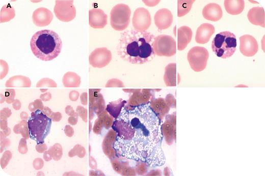
A 30-year-old woman with no significant medical history presented after 2 days of nonfebrile severe abdominal pain with vomiting and diarrhea. Initial laboratory tests revealed central anemia and thrombocytopenia (hemoglobin 8 g/dL, platelets 50 000/μL) without leukopenia (leukocytes 6400/μL), rhabdomyolysis (creatine kinase >30 000 U/L), severe coagulopathy (prothrombin time = 35.7 seconds), and hemophagocytic lymphohistiocytosis (ferritin >75 000 ng/mL, triglycerides 443 mg/dL, lactate dehydrogenase 3200 U/L) in the absence of hepatic or renal dysfunction. A gut-derived infection was ruled out based on negative blood and stool cultures and a negative procalcitonin test.
Peripheral blood smear analysis showed neutrophils with hyposegmented nuclei and pseudo-Pelger-Huët anomalies (panel A, May-Grünwald stain, original magnification ×100), containing abundant cytoplasmic vacuoles (panel B, May-Grünwald stain, original magnification ×100) and evidence of nuclear karyorrhexis (panel C, May-Grünwald stain, original magnification ×100). Bone marrow aspiration yielded a hypocellular specimen, with similar cytological abnormalities in hematopoietic precursors (panel D, May-Grünwald stain, original magnification ×100) and hemophagocytosis (panel E, May-Grünwald stain, original magnification ×100). Based on these findings, colchicine toxicity was suspected. Subsequent toxicological analysis confirmed colchicine poisoning due to self-administration, concealed by the patient during the initial interview (plasma colchicine level 2.5 ng/mL). This low level is, in this case, due to chronic intoxication, as the drug quickly leaves the bloodstream and accumulates in tissues where it remains toxic.
For additional images, visit the ASH Image Bank, a reference and teaching tool that is continually updated with new atlas and case study images. For more information, visit https://imagebank.hematology.org.


This feature is available to Subscribers Only
Sign In or Create an Account Close Modal