A 54-year-old man with a history of alcohol use disorder presented to our institution after a pulseless electrical activity arrest. Workup revealed severe anoxic brain injury and significant global cardiac hypokinesis. Cardiac catheterization, however, showed clean coronary arteries. An etiology could not definitively be determined, but cardiac sarcoidosis was considered. Unfortunately, there was no neurological recovery. He was palliatively extubated, and his organs were procured for donation.
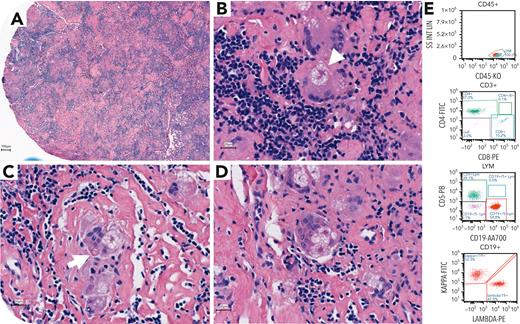
During collection, mediastinal lymphadenopathy and splenic nodules were noted. Histopathologic review revealed innumerable multinucleated giant cells with coalescing nonnecrotizing granulomas and hyalinized stroma effacing lymph node (panel A, 5× objective; hematoxylin and eosin stain) and splenic architecture. The multinucleated giant cells were particularly impressive at demonstrating classical, but often obfuscated, microscopic features of sarcoidosis. These included asteroid bodies (arrowhead), star-shaped eosinophilic intracytoplasmic collections of calcium, phosphorus, and lipids, and Hamazaki-Wesenberg inclusions (arrow), large ovoid yellow-brown lysosomes containing hemosiderin and lipofuscin (panels B-D, 200× objective; hematoxylin and eosin stain). Although these features have low sensitivity and specificity for sarcoidosis, they herald granulomatous diseases. As such, mycobacterial and fungal infections must be ruled out. Here, flow cytometry confirmed the absence of hematolymphoid neoplasms (panel E), and acid-fast bacilli (AFB), Fite, and Grocott special stains were negative for microorganisms, supporting a diagnosis of sarcoid lymphadenopathy.
For additional images, visit the ASH Image Bank, a reference and teaching tool that is continually updated with new atlas and case study images. For more information, visit https://imagebank.hematology.org.


This feature is available to Subscribers Only
Sign In or Create an Account Close Modal