In this issue of Blood, Sharma and colleagues present evidence that hematopoietic stem cell transplantation (HSCT) rapidly reverses biomarkers of cardiac fibrosis in young patients with sickle cell disease (SCD).1 Patients with SCD suffer from accelerated microvascular aging that afflicts all organs in the body, including the heart, kidney, lungs and brain. The resulting cardiac phenotype is known as heart failure with preserved ejection fraction, or HFpEF, characterized by normal systolic function, increased left ventricular mass, cardiac fibrosis, impaired diastolic function, left atrial dilation, impaired exercise performance, postcapillary pulmonary hypertension, QT prolongation, ventricular arrhythmias, and sudden death.2
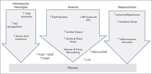
The progressive microvascular dysfunction results from sterile inflammation provoked by sickling as well as its downstream consequences including anemia, hypoxia, intravascular hemolysis, heme-mediated Toll-like receptor 4 (TLR4) activation, circulating microparticles, and other vasoactive cytokines.2,3 Resulting activation of the inflammasome, as well as overstimulation of the renin-angiotensin-aldosterone-endothelin axis and chronically elevated sympathetic nervous system tone, increase circulating interleukin-18 (IL-18) levels, a potent proinflammatory molecule.3,4 IL-18 also increases cardiac action potential duration and dispersion, predisposing to ventricular arrhythmias.4,5 Cardiac fibrosis increases ventricular passive stiffness, leading to atrial dilation and scarring, which are the strongest predictors of exercise intolerance.2,6 Elevated left ventricular filling pressures also trigger postcapillary pulmonary hypertension and represent an independent predictor of mortality.2
Cardiac fibrosis can be detected noninvasively using cardiac magnetic resonance imaging with or without the use of gadolinium contrast through a technique known as T1 mapping. Fibrosis increases the native T1 of left ventricular muscle, as well as the difference between the native T1 and the postcontrast T1. From both measurements, the fraction of extracellular water volume (ECV) may be estimated, with fibrosis increasing the measured ECV. Using contrast improves the diagnostic accuracy of fibrosis detection, albeit at a very small additive risk. ECV is markedly elevated in both animal models and patients with SCD and is correlated with disease severity.6-8
In the present study, the HFpEF phenotype was quite mild, consistent with the young age of the subjects. Diastolic dysfunction was not present, and no atrial dilation was noted. Nonetheless, ECV was elevated in 8 of 14 subjects to levels comparable to other cardiomyopathies suggesting ongoing vascular inflammation, microvascular damage, and fibrosis. HSCT normalized the ECV measurement within 1 month in most subjects, with sustained improvement at 1 year.
What is most striking about the present study is not that fibrosis reversed, but that changes were observed so quickly. Numerous animal models of myocardial fibrosis demonstrate fibrosis regression once the inciting stimulus is removed. The rapid ECV reduction suggests abrogation of the sterile inflammatory process but does not provide insight into the underlying mechanism. The lack of changes in myocardial T2 suggests the ECV changes could not be explained by water shifts (edema) alone; however, T1 and ECV are both elevated in myocardial inflammation as well as fibrosis. Given that inflammation and scarring go hand in hand, the specificity of the rapid ECV changes can always be questioned. However, the point is somewhat moot, as reversal of the primary stimulus is the ultimate goal of curative therapies, regardless.
In contrast, regression of myocardial mass was not observed until the 1-year time point. Normalization of left ventricular mass shortens oxygen diffusion distances from myocardial capillaries, which could contribute to continued normalization of the ECV over time. It is possible, perhaps even likely, that the observed ECV improvements result from multiple mechanisms with different time courses, but the study was underpowered to address this question. It is also possible that some residual myocardial fibrosis may remain after HSCT, particularly if HSCT is performed in older subjects. Nonetheless, the present data provide hope that older adults might derive cardiac protection from curative therapies. In a study of 12 adults, ages 19 to 51, who underwent haploidentical HSCT, native T1 normalized in most subjects by 12 months.9 The impact of gene therapies on cardiac fibrosis are also likely to be favorable; however, confirmatory studies will be required, particularly for agents that manipulate oxygen affinity.
So, what lessons can be drawn from the present study for those patients that must rely on disease-modifying, rather than curative, therapies? One key point is that complete success is possible and that we cannot be content with the ECV levels observed in “well-treated” patients with SCD.8,10 The second important lesson is that HFpEF in SCD begins at the red cell. Better red cell health will translate to better cardiac health. Hydroxyurea and transfusions do not reverse fibrosis because their target is incorrect, but because they incompletely ameliorate the primary problem. Additional therapies targeting iron restriction and pyruvate kinase activity are undergoing clinical trials; these could potentially improve red blood cell physiology either alone or in combination therapies, thereby slowing HFpEF progression. Transfusion strategies may also require critical reevaluation, particularly the pre- and posttransfusion hemoglobin levels. Anemia begets microvascular hypoxia in the brain and the heart because of their incessant oxygen demands, reinforcing oxidative stress and sterile inflammation. Thus, the ECV normalization demonstrated by HSCT must serve as the goalposts for palliative therapies as well. Drug development of antifibrotic agents also offers hope; however, many redundant pathways reinforce fibrosis in humans (see figure), potentially limiting efficacy of any single target. The importance of modifying traditional vascular risk factors (not shown in figure) also cannot be underestimated including dietary, weight, blood pressure, glucose, and lipid control coupled with regular physical activity. Although patients with SCD have many barriers to a healthy lifestyle, that does not free them or their practitioners from trying to achieve one.
A simple schematic illustrating the redundancy of pathways causally linking the primary red cell defect to cardiac fibrosis. Hemolysis, anemia, and vaso-occlusion represent the proximal stimuli, but so many interrelationships exist across these pathways that connecting arrows were suppressed for clarity. Some mediators clearly lie outside any given pathway, and other connections and mediators remain to be elucidated. Although IL-18 has shown promise as a convergent pathway for fibrosis and arrhythmias in mouse models,3,4 its suitability as a drug target in humans remains an open question. NO, nitric oxide; PLGF, placentally derived growth factor; RAS, renin-angiotensin-aldosterone system; SNS, sympathetic nervous system; VEGF, vascular endothelial growth factor.
A simple schematic illustrating the redundancy of pathways causally linking the primary red cell defect to cardiac fibrosis. Hemolysis, anemia, and vaso-occlusion represent the proximal stimuli, but so many interrelationships exist across these pathways that connecting arrows were suppressed for clarity. Some mediators clearly lie outside any given pathway, and other connections and mediators remain to be elucidated. Although IL-18 has shown promise as a convergent pathway for fibrosis and arrhythmias in mouse models,3,4 its suitability as a drug target in humans remains an open question. NO, nitric oxide; PLGF, placentally derived growth factor; RAS, renin-angiotensin-aldosterone system; SNS, sympathetic nervous system; VEGF, vascular endothelial growth factor.
Conflict-of-interest disclosure: J.C.W. declares no competing financial interests.


This feature is available to Subscribers Only
Sign In or Create an Account Close Modal