Background:
Platelets play a central role in thrombosis and hemostasis and immunity through their interaction with fibrinogen. Fibrinogen is a plasma protein that facilitates thrombosis and inflammation. Fibrinogen is the ligand for the major platelet receptor aIIbb3 and TREM- Like Transcript-1 (TLT-1), which platelets release from their a-granules, upon platelet activation.
TLT-1 binding fibrinogen was surprising since αIIbβ3, the most abundant platelet receptor, binds fibrinogen, facilitating platelet aggregation. Although TLT-1 may assist in clot formation and hemostasis with αIIbβ3, TLT-1's main association is with regulating inflammatory-derived bleeding. Little is known about the TLT-1-fibrinogen interaction. The goal of this study is to identify the binding sites for this interaction and determine whether it can regulate inflammatory hemostasis in an LPS inflammatory mouse model.
Aims:
Delineate the TLT-1-fibrinogen binding site and determine if, peptides representing the molecular interaction affect platelet function.
Methods:
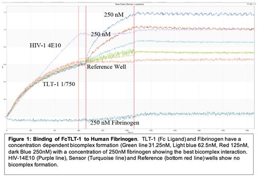
Using an Octet Qke Bio-layer Interferometry (BLI), we confirmed a binding interaction between TLT-1 and fibrinogen. Subsequently we digested fibrinogen using either trypsin or chymotrypsin and immunoprecipitated the peptides with a TLT-1-fc chimera followed by -Mass Spectrometry (LC-MS/MS) to identify potential interacting peptides. As an alternative method, we used a peptide microarray, where fluorescently labeled TLT-1 was incubated with 8-mers of α, β, γ chain fibrinogen peptides attached to a chip to determine which peptides bound TLT-1. Additionally, competitive ELISAs using peptides representing the TLT-1 extracellular domain loops (CDR-1, CDR2, CDR3 and LP17) were incubated with soluble TLT-1 in fibrinogen coated wells to determine the region of TLT-1 responsible for binding fibrinogen. To determine whether any of the selected peptides had inhibitory activity, we used two in vitro competitive inhibitory assays, first, we quantified FITC-labelled fibrinogen binding to activated human platelets and second, human platelet aggregometry incubated in the presence of these peptides at different concentrations (varying from 4-40 μM) using different agonists (ADP, thrombin, collagen, thromboxane A2).
Results:
We obtained an equilibrium dissociation constant (KD) of 3.02 ± 0.20 nM for the TLT-1 Fibrinogen interaction, suggesting a high affinity interaction. We identified four peptides that had consistent interaction without nonspecific binding in all three trials (trypsin and chymotrypsin IP, as well as the peptide microarray). The competitive platelet aggregometry and flow cytometry assays show decreased aggregation and significantly decreased fluorescent intensity of FITC-labelled fibrinogen in the presence of these peptides, suggesting two of these peptides are our potential binding sites (decrease of FITC-fluorescence intensity when compared to control: p1=20% decrease, p2=44% decrease, p3=21% decrease and P4=39% decrease, p=0.023). A HEK-293 transfected cell line expressing TLT-1 YFP is currently being used to access the ability of the identified peptides to mediate adhesion. In competitive ELISAs, TLT-1 loops LP17 and CDR3 show decreased absorbance when TLT-1 is incubated with fibrinogen in the presence of these peptides.
Conclusions:
Our TLT-1 data suggests that fibrinogen interacts with TLT-1 in the CD3 loop and in the region designated as LP17. With the aggregometry and flowcytometry competitive assays we have identified p2 as our most promising candidate for the TLT-1 interaction sight on fibrinogen. Currently we are adding the isolated peptides to our HEK/TLT-1 binding assay to determine if our selected peptide yields the same level of inhibition as seen in our aggregation and flow cytometric assays. We will then use an inflammatory in vivo mouse model to determine the effects of administering the selected peptide on platelet neutrophil conjugate formation and mortality. Our poster will report the current state of these studies .
Disclosures
No relevant conflicts of interest to declare.


This feature is available to Subscribers Only
Sign In or Create an Account Close Modal