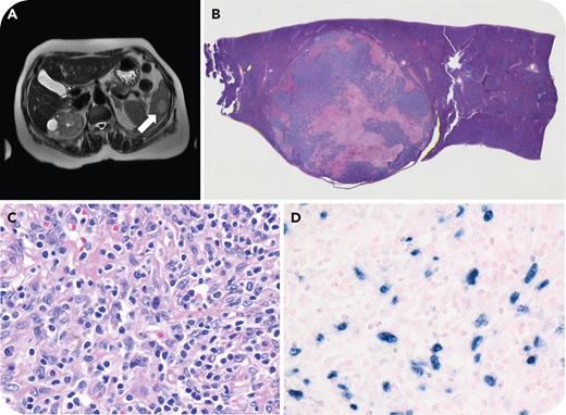
The findings are consistent with EBV-positive inflammatory follicular dendritic cell (FDC)/fibroblastic reticular cell (FRC) tumor, an indolent proliferation of stromal cells of mesenchymal origin not derived from hematopoietic stem cells. The proposed International Consensus Classification change in nomenclature from the 2016 World Health Organization entity inflammatory pseudotumor-like follicular/fibroblastic dendritic cell sarcoma to EBV-positive inflammatory FDC/FRC tumor acknowledges lineage heterogeneity and the indolent clinical behavior. EBER positivity in the spindle cells present in circumscribed mixed inflammatory splenic lesions is diagnostic for EBV-positive inflammatory FDC/FRC tumor even in the absence of FDC markers.
For additional images, visit the ASH Image Bank, a reference and teaching tool that is continually updated with new atlas and case study images. For more information, visithttp://imagebank.hematology.org.


This feature is available to Subscribers Only
Sign In or Create an Account Close Modal