Fanconi anemia (FA) is a predominantly autosomal recessive disorder resulting from mutations in one of >22 genes involved in the FA/BRCA DNA repair pathway. FA is characterized by multiple congenital abnormalities, progressive bone marrow failure (BMF) and cancer predisposition. Genetic heterogeneity and diverse clinical presentations challenge early diagnosis and optimal management. We previously reviewed the genotype-phenotype associations in FA from literature cases (Fiesco-Roa MO et al. Blood Rev. 2019). We now report the results from the NCI cohort.
We studied 147 patients with FA in the NCI inherited bone marrow failure syndromes Cohort Study (ClinicalTrials.gov, NCT00027274) to explore genotype phenotype associations by genes, location in the FA/BRCA pathway (upstream, ID complex, downstream), and compare information on the clinic cohort (CC) and field cohort (FC) patients. 57 patients (CC) were evaluated at the NIH Clinical Center between 2002 and 2020. Details on 90 patients in the FC were obtained from the review of medical records. The sex ratio (M:F) was similar (0.6:1 and 0.8:1). Patients in the FC were younger than in the CC (p=0.004) with median ages 27 (3-68) years for the CC and 19 (0-57) for the FC.
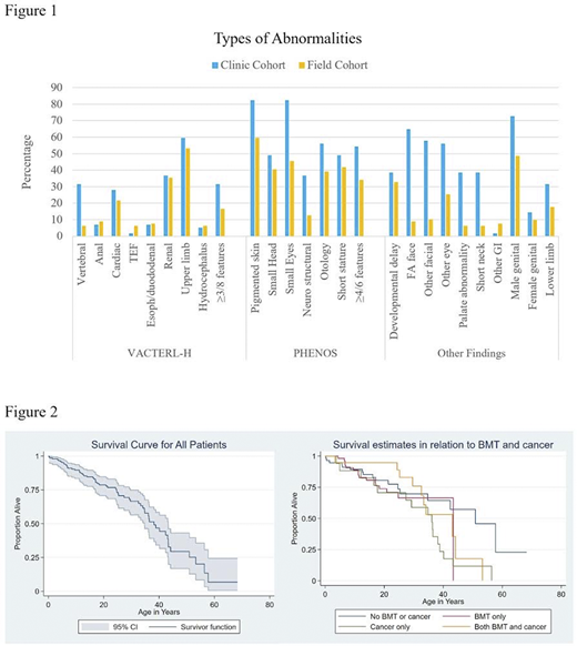
The main genotypes in the CC were 59% FANCA, 17% FANCC, 6% FANCI and in the FC were 60% FANCA, 13% FANCC and 8% FANCG. At least one FA type physical abnormality was present in all CC patients and 73/79 (92%) FC patients (phenotype data not reported on 11 FC patients). >3/8 VACTERL-H features (Vertebral, Anal, Cardiac, Tracheo-esophageal fistula (TEF), Esophageal or duodenal atresia, Renal, upper Limb (radial ray) and Hydrocephalus) were present in 32% of CC patients and 16% of FC (p=0.04). At least 4/6 PHENOS features (skin Pigmentation, small Head, small Eyes, other central Nervous system (CNS) anomalies, Otology and Short stature) were present in 54% of CC patients and 34% FC (p=0.02). The types and frequencies of phenotypic abnormalities are shown in figure 1. 17 patients in the CC (30%) and 10 in the FC (13%) had both VACTERL-H and PHENOS (p=0.01). We excluded patients with unknown genotype or phenotype from further analysis. In the CC, cardiac abnormalities were more common in patients with FANCI or ID complex gene variants than in all others (p=0.02 and 0.001, respectively) as were VACTERL-H and structural CNS abnormalities in patients with ID complex variants (p=0.03 and 0.006, respectively). In the FC, VACTERL-H, imperforate anus and hydrocephalus were more common in patients with FANCD1 genotype (p=0.03, 0.009 and 0.004, respectively) and downstream pathway gene variants (p=0.004, <0.001 and 0.03, respectively). PHENOS, renal and neurodevelopmental abnormalities were less common in patients with upstream genes variants (p=0.001, 0.009 and <0.001, respectively). Upper limb abnormalities were less common in patients with FANCC genotype (p=0.007).
BMF was present in 121/147 (88%) patients; 33% had been transfusion-dependent and 26% received androgen therapy. Clonal cytogenetic abnormalities were seen in 30%; 17% developed myelodysplastic syndrome at a median age of 17 (1.4-44) years and 6 patients developed acute myeloid leukemia at a median age of 19 (12-29) years. 72 (49%) patients underwent bone marrow transplant at a median age of 9.5 (1.5-44) years for BMF, MDS or leukemia. There was no significant difference between the FC and CC. The median survival age of our cohort is 38 (95% CI 34-43) years and at least 80% of our patients are >18 years of age. Kaplan-Meier survival estimates are presented in figure 2. Solid tumors developed in 30/135 (22%) patients with available data; median age at first cancer was 30 (2-44) years. The most common tumor was head and neck squamous cell carcinoma (n=15 patients), followed by skin (n=8) and anogenital cancers (n=6); many patients developed multiple cancers. Detailed hematologic, cancer, endocrine outcomes and survival analyses are ongoing.
Overall, renal and upper limb abnormalities were reported in most of the patients in both CC and FC, as shown previously (Alter BP et al. Mol Syndromol. 2013). Data from the CC were more complete than from the review of charts from the FC highlighting that the clinical in person evaluation of patients provides detailed characterization of FA phenotypes and more accurate assessment of genotype-phenotype associations. This will facilitate timely diagnosis, surveillance and clinical management of patients with FA.
No relevant conflicts of interest to declare.
Author notes
Asterisk with author names denotes non-ASH members.

This icon denotes a clinically relevant abstract


This feature is available to Subscribers Only
Sign In or Create an Account Close Modal