

Introduction: Sickle cell disease (SCD) affects multiple organs including the eye. Ophthalmologists have long relied on funduscopic exam to detect sickle cell retinopathy (SCR). Regular examinations to diagnose and monitor SCR permit timely initiation of treatment to prevent vision loss. Clinical guidelines recommending yearly retinal exam beginning at age 10 were based on data using conventional exam techniques. Optic coherence tomography (OCT) is a quick, noncontact test that provides images of distinctive retinal cell layers at high resolution. OCT has revolutionized the ability to examine retinal structure and provided new information on retinal damage in SCD (Bonanomi, 2013, Chow 2011). Our analysis using data from 69 patients shows that OCT has a higher detection rate for retinal changes in SCD, offering earlier diagnosis than fundoscopy alone (Jin 2018). In the pre-OCT era, patients with the SC genotype, although exhibiting less severe systemic SCD manifestations, were more frequently diagnosed with SCR and seemed to develop vision-threatening proliferative SCR more often than those with SS. In this study, we investigated the correlation between SCD genotype and retinal damage detected by OCT, using our expanded dataset to 97 patients.
We also investigated a potential link between hypoxic ischemic injuries in the retina and brain. Stroke is one of the most devastating complications of SCD. The retina and brain share the same embryonic origin, share blood supply from the internal carotid artery, have similar capillary structure and high sensitivity to hypoxia. Thus, improvements in detecting and monitoring retinal ischemia may advance both the surveillance of SCR and cerebral vascular disease (CVD), such as silent cerebral infarcts (SCI), in SCD. Early detection of CVD is critical since prompt intervention can halt progression. Currently, the only available means to detect SCI is MRI, which is costly and often requires sedation. The ability to identify children at high risk for SCI would enable targeted MRI screening.
Methods: In this prospective study, conducted over 4 years, 97 consecutive patients with SCD underwent complete ophthalmologic examination and Spectral-Domain OCT (SD-OCT) imaging of both eyes. The posterior pole volume scan involves a 30⁰× 25⁰ cuboid, with 31 raster lines separated by 240 μm. Because of the preponderance of retinal changes on the temporal side of macula, the scanning center was aimed at approximately 3 mm temporal to the foveal center by adjusting the internal or external fixation target.
Macular volume scans were assessed for areas of visible thinning. Areas of retinal thinning were determined using the circle grid function on the thickness map. Inner retinal thickness was measured from the internal limiting membrane to the external/outer limiting membrane, and outer retinal thickness was measured from the external limiting membrane to the Bruch's membrane. Retinal exam findings were compared with clinical data. Data analysis was performed using IBM SPSS Statistics 25 (IBM, Armonk, NY).
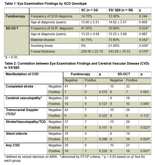
Results: 97 (48 male) patients aged 5-20 years (mean 12.19 ± 4.35) with SCD (34 SC, 53 SS, 5 Sβ+ thalassemia, 5 Sβ0 thalassemia) were examined. Visual acuity ranged from 20/20 to 20/40. On funduscopic exam, 14 of 97 (14.43%) showed signs of retinopathy whereas 59 of 97 (60.82%) showed inner retina thinning on SD-OCT.
By SD-OCT, patients with SS/Sβ0 showed a significantly higher frequency of detected SCR change than SC genotype (70.7% vs 47.1%) (Table 1). SS/Sβ0 was also associated with a higher frequency of bilateral SCR (75.6% vs 43.8%) and foveal involvement (22.0% vs 0%). Remarkably, all patients in our cohort with SS/Sβ0 genotype and documented CVD had evidence of SCR by SD-OCT (Table 2).
Conclusions: In our cohort, SD-OCT showed greater capabilities than fundoscopy in: 1) a higher detection rate for retinal changes consistent with SCR, offering earlier diagnosis; 2) a significantly higher frequency of SCR and more extensive retinal changes associated with the more severe SCD genotypes SS and Sβ0 as compared to SC; 3) demonstrated correlation between the presence of any form of CVD including stroke, cerebral vasculopathy by MRA, abnormal transcranial Doppler and SCI strongly suggests that retinal exam using SD-OCT may aid in detection and monitoring SCD related CVD. These important findings require further study in a larger patient population undergoing serial exams over time.
No relevant conflicts of interest to declare.

This feature is available to Subscribers Only
Sign In or Create an Account Close Modal