

Introduction:
Myelonecrosis is defined as necrosis of myeloid tissue within the hematopoietic medullary spaces with preservation of bony trabeculae. It is a rare finding on trephine which is reported to occur in 0.37% to 6.5% of all cases [1]. Most common cause of myelonecrosis is associated neoplasia [2]. Extensive myelonecrosis can make bone marrow aspiration and further evaluation difficult, leading to delay in diagnosis and management.
We describe five such interesting cases with varying degree of myelonecrosis where immune-histochemistry in the necrosed zone helped us clinch the diagnosis.
Cases and methods:
Clinicopathological characteristics, bone marrow morphology and Immuno-histochemistry findings are tabulated below. In all the cases trephine biopsies were fixed with 10% neutral buffered formalin and decalcification was done with 14% EDTA for 48-72hrs (as per ICSH 2008 Guidelines).
In all these cases, myelonecrosis was graded and ranged from mild to moderate. The procedure used was that of a semi-quantitative one.
Grade I (mild): <20% of the biopsy
Grade II (moderate-intermediate): 20-50% of the biopsy
Grade III (severe-extensive): >50% of the biopsy.
Gomori's technique was done for reticulin staining. Fibrosis was graded as per WHO protocols. Immuno-histochemistry was done by peroxidase - anti-peroxidase technique. Immuno-histochemical findings were recorded separately by two hemato-pathologists. Expression of each marker was graded as per strength and compared with the expression in the viable zones. The gradation of expression was done in a semi-quantitative manner.
Mild expression: +
Moderate expression: ++
Strong expression: +++
Discussion:
Out of the five cases, three were hematological malignancies and rest two are metastatic solid tumors. Myelonecrosis in all the cases was of coagulative type. Myelonecrosis was extensive in 1/5 cases, moderate in 3/5 cases and patchy or focal in 1/5 cases. We found that the necrosed tumor cells retained their antigenicity for most of the markers. It was observed that even the intensity of expression was at par with that of the viable zones.
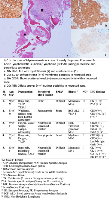
The necrosed zone in case of B-cell precursor- acute lymphoblastic leukemia showed diffuse and strong positivity for CD10, CD79a, TdT and dim positive for CD34, thus confirming the diagnosis. Similarly, in the case of Non-Hodgkin's lymphoma the expression of markers in the necrosed areas showed strong antigenic expression for CD20, CD79a and nuclear positivity for Bcl 2, Bcl 6 and Mib 1. In the myeloma case retention of CD138, CD56 and Lambda light chain restriction was noted.
Interestingly, both the cases with metastasis presented with bony pain and increased ALP (Alkaline Phosphatase). While in one case associated increase in PSA (Prostate specific antigen) in serum indicated the possibility of metastasis from a prostatic primary, the other case was evaluated in the line of myeloma. Immuno-histochemistry in the trephine suggested a possibility of metastasis from an occult primary. Later, on further evaluation a primary was found in the right breast of the patient. In both the metastatic cases, the expression of Cytokeratin was strong and diffuse. PSA was expressed in dot-like pattern.
Jinkala et. al(3). in their study found that malignancy was the commonest cause of myelonecrosis. They found that neoplastic aetiology in 91% cases of marrow necrosis, out of which primary hematologic malignancy constituted 60% and rest was due to metastatic solid tumours. Also, most of the studies have proved that myelonecrosis itself is a bad prognostic factor in patients.
Conclusion:
Thus, we conclude Immuno-histochemistry is an useful adjunct to demonstrate the retained antigenicity in myelo-necrosed tissues; and could be useful in these cases as they are usually associated with scant and difficult aspirate. It can be done easily leading to a faster diagnosis of the patient even in resource poor settings too. This will save the patient from undergoing a painful bone marrow procedure again.
References:
1. Markovic SN,. Pancytopenia due to bone marrow necrosis in acute myeloid leukemia: role of reactive CD8 cells. Am J Hematol 1998; 59:74.
2. Dunn Bone marrow necrosis in 38 adult cancer patients. J Famos Med Assoc 1993;92(12): 1107-10.
3. Jinkala S.R. Myelonecrosis: A Clinicopathological Study from a Tertiary Care Center in South India over a Twelve-Year Period. Bone Marrow Research 2014; 5
No relevant conflicts of interest to declare.

This feature is available to Subscribers Only
Sign In or Create an Account Close Modal